Tissue Inhibitor of Metalloproteases 3: Comparison
Please note this is a comparison between Version 5 by Vicky Zhou and Version 4 by Vicky Zhou.

The proteolytical cleavage of transmembrane proteins with subsequent release of their extracellular domain, so-called ectodomain shedding, is a post-translational modification that plays an essential role in several biological processes, such as cell communication, adhesion and migration. Metalloproteases are major proteases in ectodomain shedding, especially the disintegrin metalloproteases (ADAMs) and the membrane-type matrix metalloproteases (MT-MMPs), which are considered to be canonical sheddases for their membrane-anchored topology and for the large number of proteins that they can release. The unique ability of TIMP-3 to inhibit different families of metalloproteases, including the canonical sheddases (ADAMs and MT-MMPs), renders it a master regulator of ectodomain shedding. 

  • TIMPs
  • ADAMs
  • metalloproteases
  • ectodomain shedding
  • proteomics

1. Introduction

The human genome encodes for about 15,000 transmembrane proteins, encompassing 19% of the total genome. Ectodomain shedding is a post-translational modification by which membrane proteins are proteolytically cleaved and their ectodomain released. This biochemical mechanism is essential in a number of cellular processes, including cell communication, adhesion and migration. For instance, tissue bioavailability of certain cytokines and growth factors is controlled by ectodomain shedding, as it depends on membrane-level signalling receptors and therefore the ability of cells to respond to specific cues. Moreover, shedding controls cell-to-cell adhesion or adhesion of cells to the ECM, and it modulates cellular transport by controlling levels of endocytic receptors on the cell surface [1]. Members of several different classes of proteases have been shown to function as sheddases, with the metalloproteases playing a most prominent role for the number of substrates that they cleave and the subsequent pathophysiological processes in which they are involved [1][2]. Metalloproteases comprise three related families of proteases, the matrix metalloproteases (MMPs), the disintegrin metalloproteases (ADAMs) and the disintegrin metalloproteases with thrombospondin motifs (ADAMTSs). While most MMPs and ADAMTSs are mainly involved in the catabolism of extracellular matrix components, ADAMs and the membrane-type MMPs (MT-MMPs) act as canonical sheddases [3][4]. Nevertheless, this distinction is not always clear, as membrane-bound ADAMs can be shed themselves and cleave ECM components, and secreted metalloproteases can bind to components of the cell surface and function as sheddases [5][6][7].
Given the large number of substrates and biological processes in which they play a role, the activity of metalloprotease sheddases has to be finely modulated. When dysregulated, aberrant metalloprotease-dependent ectodomain shedding can lead to pathological conditions, such as cancer, arthritis, inflammatory diseases and Alzheimer’s [4][8]. Its regulation occurs, among other mechanisms, by formation of a 1:1 non-covalent complex with the tissue inhibitors of metalloproteases (TIMPs) [9]. Among the four mammalian TIMPs, TIMP-3 has the largest inhibitory profile being able to inhibit not only MMPs and ADAMTSs, but also all the canonical sheddases of the ADAM and MT-MMP families, thus rendering TIMP-3 a master regulator of ectodomain shedding.

2. Regulation of TIMP-3

Given its role in modulating ECM turnover and ectodomain shedding, the bioavailability of TIMP-3 must be finely regulated. This occurs at several levels, including transcriptional regulation, by growth factors and cytokines, and epigenetically by promoter methylation, post-transcriptional regulation by specific miRNAs and by receptor-mediated endocytosis (Figure 1).
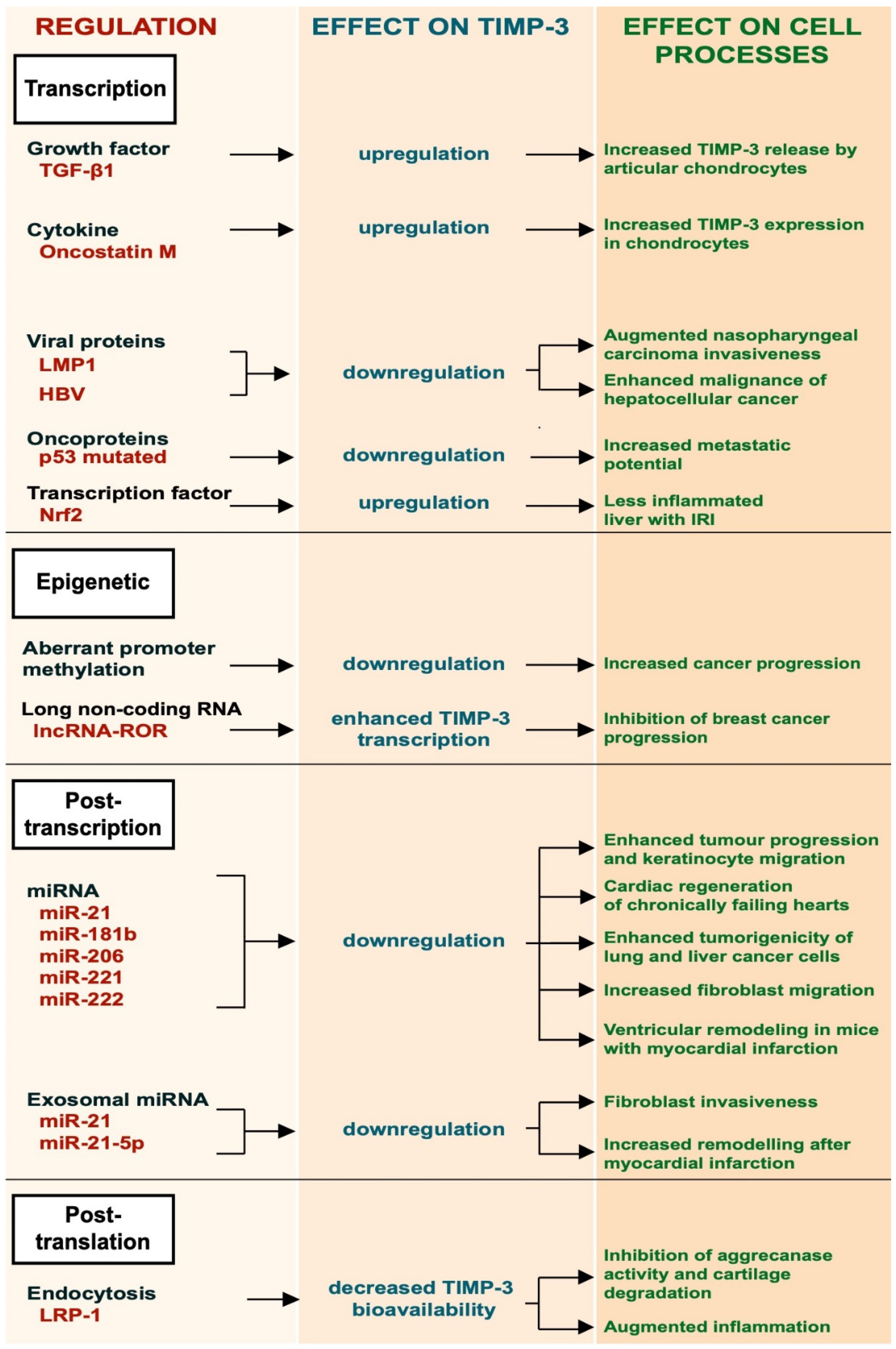
Figure 1. Schematic representation of the different regulatory mechanisms of TIMP-3.

2.1. Transcriptional Regulation

TGF-β1 is a multifunctional growth factor produced by different cell types, such as monocytes, platelets and chondrocytes, that promotes ECM deposition. It has been shown to induce the expression of TIMP-3 in articular chondrocytes [10]. This occurs by activation of the ERK/MAPK signaling pathway, leading to phosphorylation of the Sp-1 transcription factor and its subsequent binding to the TIMP3 promoter. Qureshi and colleagues also reported that TIMP-3 expression could be induced by TGF-β1 through different mechanisms involving Smad-2, -3 and -4 and the phosphatidylinositol 3-kinase/Akt signaling pathway [10][11]. Oncostatin M, a cytokine belonging to the IL-6 family, can also induce expression of TIMP-3 in human chondrocytes by activating the Janus Kinase (JAK)/STAT signaling pathway [12]. Epstein–Barr virus (EBV) latent protein 1 (LMP-1) is a viral protein essential for the capability of the Epstein–Barr virus to mediate growth transformation of B-cells. LMP-1 was shown to promote metastasis in EBV-negative nasopharyngeal carcinoma cells by suppressing TIMP-3 transcription [13]. Similarly, human hepatitis B virus suppresses TIMP-3 expression, although the molecular mechanism is not fully elucidated [14]. Missense mutations in the p53 gene inactivate its growth suppressing activity and are observed in the majority of human tumours. Thomas and colleagues showed that missense mutations in p53 created a mutant p53 that gained the ability to bind to the TIMP-3 promoter and suppress its expression [15]. Loss of TIMP-3 expression in cancer cells could lead to an increase of metalloprotease activity and therefore to their increased metastatic potential. Nuclear factor erythroid 2-related factor 2 (Nrf2), a transcription factor with a key role in inflammation, positively regulated the expression of TIMP-3, thereby attenuating inflammatory responses in the liver of mice challenged with ischemia/reperfusion injury (IRI) [16].

2.2. Epigenetic Regulation

The epigenetic regulation of TIMP-3 involves a number of mechanisms that are not dependent on its gene sequence. For example, aberrant methylation of the promoter region of TIMP-3 gene was found to be associated with primary cancer of the brain, kidney, colon, breast, etc. [17]. TIMP-3 plays a protective role in cancer. Epigenetic regulation of the TIMP3 gene has been reported to be a key mechanism in cancer progression [17][18][19]. Long non-coding RNAs (lncRNAs) are a new class of epigenetic regulators that play important roles in several diseases, including cancer. Long non-coding RNA reprogramming (lncRNA-ROR) was shown to recruit the transmethylase MLL1 to promote H3K4 trimethylation that enhanced TIMP3 transcription [20].

2.3. Post-Transcriptional Regulation

Post-transcriptional regulation of TIMP-3 expression has been investigated in both physiological processes and pathological conditions. Several miRNAs have been found to bind the 3′-UTR of the TIMP-3 mRNA, resulting in the degradation of TIMP-3 transcripts and its gene silencing [21][22]. miR-21 is one of the most expressed micro-RNAs in many different tumours, including glioblastoma, and its expression often correlates with tumour grade and poor prognosis [21]. miR-21 targets TIMP-3 mRNAs, leading to their degradation. Its ability to reduce TIMP-3 expression promotes tumour invasion [21][23][24]. Regulation of TIMP-3 by miR-21 has also been observed in physiological processes, including wound healing [25]. During skin wound healing, keratinocytes need to proliferate and migrate. Both keratinocyte proliferation and migration are favored by expression of miR-21, which attenuates TIMP-3 expression [25]. TIMP-3 mRNA is a validated target of miR-181b and miR-206 [22][26]. miR-181b is upregulated by TGF-β and its expression is markedly suppressed in hepatocarcinomas. These data suggest that TGF-β can modulate TIMP-3 expression by miR-181b [26]. miR-206-mediated downregulation of TIMP-3 promotes cardiac regeneration of chronically failing hearts [26]. miR-221 and miR-222 can downregulate the expression of TIMP-3. Garofalo and colleagues found that the hepatocyte growth factor (HGF)/scatter factor upregulates miR-221 and miR-222 expression. This enhances tumourigenicity of lung and liver cancer cells by downregulating the phosphatase and tensin homolog (PTEN) and TIMP3 [27].
Exosomes can be used as means for transferring miRNAs to target recipient cells. Few examples of exosomal miRNAs have been reported to regulate the expression of TIMP-3 and, in turn, cell migration. Melanoma-derived exosomes carried miR21, which promoted invasion of fibroblasts by downregulation of TIMP3 expression [28]. Similarly, macrophage-derived exosomes lowered the expression of TIMP-3 by miR-21-5p and favored ventricular remodeling in mice with myocardial infarction [29].

2.4. LRP-1-Mediated Endocytosis

Endocytosis has emerged as a major mechanism to regulate bioavailability of TIMP-3 in the tissues, with the low-density lipoprotein receptor-related protein-1 (LRP-1) playing a key role in this process [30][31]. TIMP-3 is constitutively internalized by cells through the receptor, either alone or in a complex with its target metalloproteases, and targeted to lysosomal degradation [30][32]. Shedding of LRP-1 and release of its soluble ectodomain negatively modulate TIMP-3 endocytosis, as soluble LRP-1 is able to bind to the inhibitor and act as a decoy receptor [30][33]. LRP-1 shedding, which is mediated by different metalloproteases, including ADAM17 and ADAM10, can function as a negative feedback loop to regulate ADAM-mediated shedding itself [34][35]. It has been reported that activation of ADAM17 by LPS promotes shedding of LRP-1 and subsequent extracellular accumulation of TIMP-3, which, in turn, dampens ADAM17 activity and TNF release [36]. Thus, LRP-1 shedding and accumulation of TIMP-3 may be a crucial mechanism for the resolution of ADAM-mediated cell responses, including inflammation.

3. Mass Spectrometry-Based Approaches to Investigate Functions of TIMP-3

The function of a protease strictly depends on the collection of substrates that it cleaves. Some proteases cleave a large number of substrates, while others have a very restricted activity towards a few proteins. Furthermore, the activity of proteases can be spatially and/or temporally confined, so that even proteases with a large spectrum of substrates may only access a limited number of them at specific locations and at specific times. In the last years, high-resolution mass spectrometry techniques have been developed to investigate the function of specific proteases by the systematic identification of their substrates in a specific cellular case [2][37]. These proteomics-based approaches have also been applied to TIMP-3, enabling the discovery of new functions of the inhibitor on the stabilization of transmembrane proteins and endocytosis of secreted proteins [38][39]. Here will provide a brief description of these mass spectrometry-based techniques that have been used to characterize the function of TIMP-3 and its target proteases and which may eventually be applied to other proteases and their inhibitors.

3.1. High-Resolution Secretome Analysis

High-resolution secretome analysis allows the determination of alterations of protein levels in the secretome of cultured cells. This includes the possibility to evaluate levels of proteins that are released by ectodomain shedding, whose levels are reduced when their sheddases are inhibited or genetically ablated (Figure 2). Conditioned media of specific cells (for instance, cells overexpressing TIMP-3) are applied to tryptic digestion, which transforms a complex protein sample containing secreted and shed proteins into a mixture of peptides that can be analysed by mass spectrometry. These peptides are usually separated by C18-reversed phase liquid chromatography (LC) prior to MS/MS analysis. MS/MS allows the search for each peptide against a protein database so that it can be linked to a protein and its relative quantification, for instance, by a label-free quantification method [37].
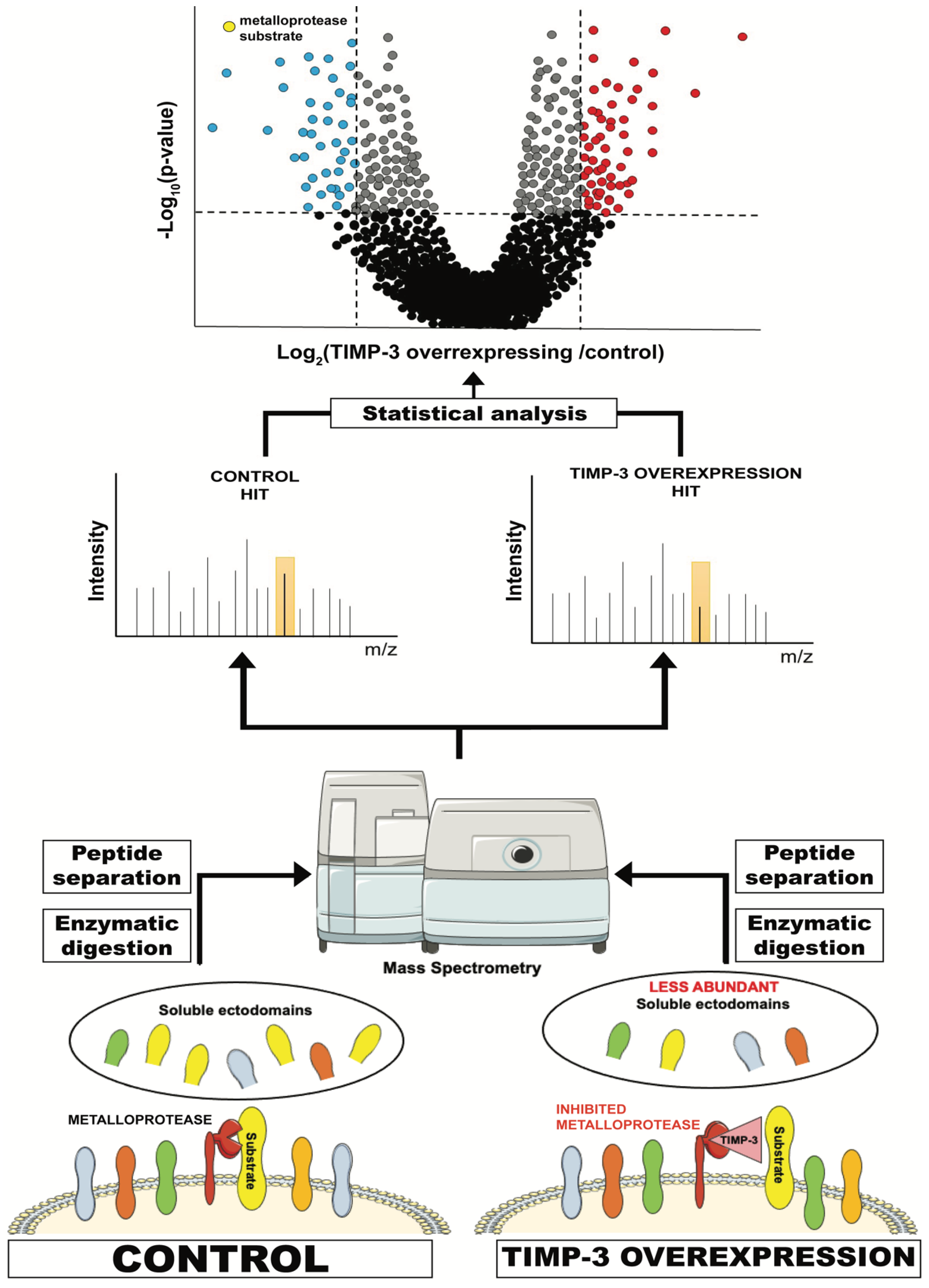
Figure 2. Schematic representation of a typical proteomic workflow to investigate ectodomain shedding. For instance, TIMP-3-overexpressing or control cells are cultured in serum-free media and conditioned media are collected. These contain, among other proteins secreted by the cell, the ectodomain of proteins that are shed by TIMP-3 target metalloproteases. Levels of these proteins in TIMP-3-overexpressing cells will be lower due to inhibition of their shedding by TIMP-3. Conditioned media from TIMP-3-overexpressing and control cells will be applied to tryptic digestion, C18 reversed phase liquid chromatography (LC) and, ultimately, MS/MS analysis. This will enable the identification of proteins contained in the conditioned media and the quantification of their levels in TIMP-overexpressing cells versus controls. Finally, a statistical analysis will show the levels of the proteins that are altered in the media of TIMP-3-overexpressing cells.
Secretome of TIMP-3-overexpressing HEK293 cells were analysed using a similar approach and revealed that TIMP-3 mainly inhibited shedding of ADAM10 substrates in these cells and under steady-state conditions [38]. A similar effect on ADAM10 shedding was noted when cells were treated with a molecule able to prevent TIMP-3 endocytosis and increase its extracellular levels [33]. These results complemented the phenotypical analysis of Timp3-null mice, whose most prominent phenotypes are related to a deregulated ADAM17 activation [9][40]. Altogether, proteomics and in vivo analysis suggest a dual role for TIMP-3 in regulating the homeostatic turnover of membrane proteins by ADAM10, and several biological processes, including cell-to-cell communication and adhesion, by controlling the stimulated activity of ADAM17.

3.2. Surfaceomics

As extensively discussed here, shedding regulates levels of transmembrane proteins and therefore biological responses. In turn, TIMP-3 has been reported to stabilize membrane proteins by inhibiting ectodomain shedding, such as death receptors and adhesion molecules, thereby modulating cellular processes. However, shedding can be a means to control the homeostatic turnover of transmembrane proteins. The effects of TIMP-3 on proteins that are shed at a low rate, as a consequence of their constitutive turnover at the cell surface, may be less pronounced and not lead to an evident increase of their levels at the cell surface and clear functional consequences on cell function. In order to discriminate which proteins are effectively stabilized on the cell surface by TIMP-3 (as well as by genetic ablation of specific sheddases of their pharmacological inhibition), quantitative proteomics can be used. Different approaches can be used to isolate cell surface proteins prior to being analysed by mass spectrometry, including enrichment of glycosylated membrane proteins by SPECS (discussed in the next section) and biotinylation of membrane proteins followed by streptavidin pulldown. The latter approach was used to identify proteins stabilized by TIMP-3 at the surface of HEK293 cells [39]. Carreca et al. demonstrated that only a group of shed proteins effectively accumulate on the cell surface when TIMP-3 is overexpressed and their shedding is inhibited. Such a proteomic approach can complement secretome analysis and may be useful to assess whether shedding of specific proteins can lead to functional consequences for cell behavior.

3.3. SPECS and SUSPECS

Secretome protein enrichment with click sugars (SPECS) is a proteomic method that has been proven useful to investigate shedding, especially in those cells that require serum and other additives to grow, such as primary neurons [41][42]. SPECS consists of metabolic labelling of cellular glycoproteins with azido sugars, followed by copper-free click chemistry-mediated biotinylation. This process allows one to label and purify only shed proteins, but not serum glycoproteins. Labelled ectodomains released by shedding will be pulled down from conditioned media using a streptavidin-conjugated resin and analysed by high-resolution mass spectrometry. Dibenzocyclooctyne (DBCO)-NHS is usually used as the click linker for biotin-conjugation in SPECS. This compound is not cell permeable, and therefore labelled ectodomains of cell surface transmembrane proteins can be biotinylated by click-chemistry and enriched by streptavidin pulldown in a similar manner to soluble ectodomains [43]. This method, namely, “surface-spanning protein enrichment with click sugars” (SUSPECS), in which the enrichment of cell membrane proteins by click chemistry is followed by mass spectrometry analysis, has been successfully used to investigate ectodomain shedding in neurons.

3.4. TAILS

Additional to ADAM-mediated ectodomain shedding, TIMP-3 modulates the activity of secreted metalloproteases, such as MMPs and ADAMTSs. These proteases are majorly involved in processing components of the ECM. The previously described proteomics methods, which have been used to investigate ectodomain shedding, are not suitable to identify soluble substrates of TIMP-3 target metalloproteases and therefore they are not appropriate to study the role of TIMP-3 in ECM turnover. In fact, these methods comprise a tryptic digestion of protein samples and evaluation of relative protein abundance. Relative abundance of an ECM component would not change upon cleavage by a protease, and therefore methods that are specifically targeted to identifying cleavages should be used. For instance, the terminal amine-based isotope labeling of substrates (TAILS) has been extensively characterized to investigate MMPs and their inhibitors ([44][45] and reviewed in [46]). Based on the TAILS method, primary amines of both the n-termini and lysine residues of proteins are chemically labeled with formaldehyde and isotopes are labeled for the following mass spectrometry analysis. Then, labeled proteins are digested by trypsin, which will cleave after arginines, but it will not be able to cleave after lysines, as they will be blocked after formaldehyde conjugation. Then, TAILS peptides are enriched and subjected to liquid chromatography and high-resolution mass spectrometry. The neo-n-termini peptides arising from cleavages of the protease of interest will appear with higher relative abundance, and from this the substrates of specific proteases can be deduced.

4. Conclusions and Perspectives

TIMP-3 is an endogenous inhibitor of metalloproteases that is able to inhibit all canonical sheddases of the ADAM family, in addition to MMPs and ADAMTSs. For this reason, TIMP-3 is considered a master regulator of ectodomain shedding. Its genetic ablation leads to several abnormalities related to unregulated shedding, while its overexpression has been proven as protective in a number of pathological conditions characterized by enhanced proteolysis, including cancer and arthritis. Functions of TIMP-3 have been characterized in a targeted manner, by analysis of phenotypes arising in mouse as a consequence of its genetic ablation. Nevertheless, some phenotypes become evident only when mice are challenged with specific stimuli, and bottom-up approaches may be required to uncover such functions of the inhibitor and its target proteases that do not lead to evident abnormalities in vivo under steady-state conditions. In the last few years, unbiased high-resolution proteomics has allowed the systematic identification of protease substrates, thereby providing key information to deduce new functions of proteases and their inhibitors, including TIMP-3. Indeed, proteomics has been used to identify transmembrane proteins that are regulated by TIMP-3 and revealed new molecular mechanisms in which TIMP-3 is involved.
Mutations in the TIMP3 gene lead to pathological variants of the inhibitor associated with the onset of Sorsby’s fundus dystrophy (SFD), a degenerative disease of the macula. Despite scientific efforts that have been made to understand how SFD mutations affect TIMP-3 function and cell behavior, the molecular mechanisms linking SFD mutations to the onset of the disease are still unknown [47]. The proteomic approaches that have been described may be used to evaluate effects of SFD TIMP-3 mutants on cell behavior that may be linked to the new pathological functions of the inhibitor. Interestingly, all mutations leading to development of SFD occur at the C-terminal domain of TIMP3, highlighting its involvement in pathophysiological processes that are yet to be elucidated. The C-terminal domain of TIMP-3 is dispensable for metalloprotease inhibition in most cases [9]. Nevertheless, similar to the SFD mutations, a truncated form of TIMP-3 lacking the C-terminal domain leads to different inflammatory responses than full-length TIMP-3 after myocardial infarction [47][48]. Methods of high-resolution proteomics, similar to those analyzed here, may serve for further characterization of the C-terminal domain of TIMP-3. Furthermore, while the substrate repertoire of ADAM10 and ADAM17 has been extensively characterized, the number of identified substrates of other TIMP-3 target sheddases, including ADAM12 and ADAM15, is still limited [8]. The function of such sheddases strictly depends on the array of their substrates. Thus, the identification of substrates may link the activity of sheddases and their inhibitors, including TIMP-3, to specific biological processes.
In conclusion, TIMP-3 is a major regulator of ectodomain shedding in vivo. Its ablation or overexpression in mice has led to the identification of several biological processes in which TIMP-3 plays a key role. Nevertheless, many aspects of TIMP-3 biology are not fully characterized yet. Proteomics represents a promising tool to uncover such TIMP-3 functions that have not been elucidated by in vivo analysis and targeted biochemical approaches, thus providing new insights into the biology of the inhibitor.

References

  1. Lichtenthaler, S.F.; Lemberg, M.K.; Fluhrer, R. Proteolytic ectodomain shedding of membrane proteins in mammals-hardware, concepts, and recent developments. EMBO J. 2018, 37, e99456.
  2. Muller, S.A.; Scilabra, S.D.; Lichtenthaler, S.F. Proteomic Substrate Identification for Membrane Proteases in the Brain. Front. Mol. Neurosci. 2016, 9, 96.
  3. Murphy, G.; Nagase, H. Localizing matrix metalloproteinase activities in the pericellular environment. FEBS J. 2011, 278, 2–15.
  4. Edwards, D.R.; Handsley, M.M.; Pennington, C.J. The ADAM metalloproteinases. Mol. Asp. Med. 2008, 29, 258–289.
  5. Fiore, E.; Fusco, C.; Romero, P.; Stamenkovic, I. Matrix metalloproteinase 9 (MMP-9/gelatinase B) proteolytically cleaves ICAM-1 and participates in tumor cell resistance to natural killer cell-mediated cytotoxicity. Oncogene 2002, 21, 5213–5223.
  6. Rodriguez-Manzaneque, J.C.; Carpizo, D.; Plaza-Calonge Mdel, C.; Torres-Collado, A.X.; Thai, S.N.; Simons, M.; Horowitz, A.; Iruela-Arispe, M.L. Cleavage of syndecan-4 by ADAMTS1 provokes defects in adhesion. Int. J. Biochem. Cell Biol. 2009, 41, 800–810.
  7. Scharfenberg, F.; Helbig, A.; Sammel, M.; Benzel, J.; Schlomann, U.; Peters, F.; Wichert, R.; Bettendorff, M.; Schmidt-Arras, D.; Rose-John, S.; et al. Degradome of soluble ADAM10 and ADAM17 metalloproteases. Cell. Mol. Life Sci. 2020, 77, 331–350.
  8. Hsia, H.E.; Tushaus, J.; Brummer, T.; Zheng, Y.; Scilabra, S.D.; Lichtenthaler, S.F. Functions of ‘A disintegrin and metalloproteases (ADAMs)’ in the mammalian nervous system. Cell. Mol. Life Sci. 2019, 76, 3055–3081.
  9. Brew, K.; Nagase, H. The tissue inhibitors of metalloproteinases (TIMPs): An ancient family with structural and functional diversity. Biochim. Biophys. Acta 2010, 1803, 55–71.
  10. Qureshi, H.Y.; Sylvester, J.; El Mabrouk, M.; Zafarullah, M. TGF-beta-induced expression of tissue inhibitor of metalloproteinases-3 gene in chondrocytes is mediated by extracellular signal-regulated kinase pathway and Sp1 transcription factor. J. Cell. Physiol. 2005, 203, 345–352.
  11. Qureshi, H.Y.; Ahmad, R.; Sylvester, J.; Zafarullah, M. Requirement of phosphatidylinositol 3-kinase/Akt signaling pathway for regulation of tissue inhibitor of metalloproteinases-3 gene expression by TGF-beta in human chondrocytes. Cell Signal. 2007, 19, 1643–1651.
  12. Li, W.Q.; Dehnade, F.; Zafarullah, M. Oncostatin M-induced matrix metalloproteinase and tissue inhibitor of metalloproteinase-3 genes expression in chondrocytes requires Janus kinase/STAT signaling pathway. J. Immunol. 2001, 166, 3491–3498.
  13. Chang, S.H.; Chang, H.C.; Hung, W.C. Transcriptional repression of tissue inhibitor of metalloproteinase-3 by Epstein-Barr virus latent membrane protein 1 enhances invasiveness of nasopharyngeal carcinoma cells. Oral Oncol. 2008, 44, 891–897.
  14. Kim, J.R.; Kim, C.H. Association of a high activity of matrix metalloproteinase-9 to low levels of tissue inhibitors of metalloproteinase-1 and -3 in human hepatitis B-viral hepatoma cells. Int. J. Biochem. Cell Biol. 2004, 36, 2293–2306.
  15. Thomas, S.; Reisman, D. Localization of a mutant p53 response element on the tissue inhibitor of metalloproteinase-3 promoter: Mutant p53 activities are distinct from wild-type. Cancer Lett. 2006, 240, 48–59.
  16. Rao, J.; Qiu, J.; Ni, M.; Wang, H.; Wang, P.; Zhang, L.; Wang, Z.; Liu, M.; Cheng, F.; Wang, X.; et al. Macrophage nuclear factor erythroid 2-related factor 2 deficiency promotes innate immune activation by tissue inhibitor of metalloproteinase 3-mediated RhoA/ROCK pathway in the ischemic liver. Hepatology 2021.
  17. Bachman, K.E.; Herman, J.G.; Corn, P.G.; Merlo, A.; Costello, J.F.; Cavenee, W.K.; Baylin, S.B.; Graff, J.R. Methylation-associated silencing of the tissue inhibitor of metalloproteinase-3 gene suggest a suppressor role in kidney, brain, and other human cancers. Cancer Res. 1999, 59, 798–802.
  18. Rohrs, S.; Dirks, W.G.; Meyer, C.; Marschalek, R.; Scherr, M.; Slany, R.; Wallace, A.; Drexler, H.G.; Quentmeier, H. Hypomethylation and expression of BEX2, IGSF4 and TIMP3 indicative of MLL translocations in acute myeloid leukemia. Mol. Cancer 2009, 8, 86.
  19. Eads, C.A.; Lord, R.V.; Wickramasinghe, K.; Long, T.I.; Kurumboor, S.K.; Bernstein, L.; Peters, J.H.; DeMeester, S.R.; DeMeester, T.R.; Skinner, K.A.; et al. Epigenetic patterns in the progression of esophageal adenocarcinoma. Cancer Res. 2001, 61, 3410–3418.
  20. Hu, A.; Hong, F.; Li, D.; Jin, Y.; Kon, L.; Xu, Z.; He, H.; Xie, Q. Long non-coding RNA ROR recruits histone transmethylase MLL1 to up-regulate TIMP3 expression and promote breast cancer progression. J. Transl. Med. 2021, 19, 95.
  21. Gabriely, G.; Wurdinger, T.; Kesari, S.; Esau, C.C.; Burchard, J.; Linsley, P.S.; Krichevsky, A.M. MicroRNA 21 promotes glioma invasion by targeting matrix metalloproteinase regulators. Mol. Cell. Biol. 2008, 28, 5369–5380.
  22. Wang, B.; Hsu, S.H.; Majumder, S.; Kutay, H.; Huang, W.; Jacob, S.T.; Ghoshal, K. TGFbeta-mediated upregulation of hepatic miR-181b promotes hepatocarcinogenesis by targeting TIMP3. Oncogene 2010, 29, 1787–1797.
  23. Song, B.; Wang, C.; Liu, J.; Wang, X.; Lv, L.; Wei, L.; Xie, L.; Zheng, Y.; Song, X. MicroRNA-21 regulates breast cancer invasion partly by targeting tissue inhibitor of metalloproteinase 3 expression. J. Exp. Clin. Cancer Res. 2010, 29, 29.
  24. Selaru, F.M.; Olaru, A.V.; Kan, T.; David, S.; Cheng, Y.; Mori, Y.; Yang, J.; Paun, B.; Jin, Z.; Agarwal, R.; et al. MicroRNA-21 is overexpressed in human cholangiocarcinoma and regulates programmed cell death 4 and tissue inhibitor of metalloproteinase 3. Hepatology 2009, 49, 1595–1601.
  25. Yang, X.; Wang, J.; Guo, S.L.; Fan, K.J.; Li, J.; Wang, Y.L.; Teng, Y.; Yang, X. miR-21 promotes keratinocyte migration and re-epithelialization during wound healing. Int. J. Biol. Sci. 2011, 7, 685–690.
  26. Limana, F.; Esposito, G.; D’Arcangelo, D.; Di Carlo, A.; Romani, S.; Melillo, G.; Mangoni, A.; Bertolami, C.; Pompilio, G.; Germani, A.; et al. HMGB1 attenuates cardiac remodelling in the failing heart via enhanced cardiac regeneration and miR-206-mediated inhibition of TIMP-3. PLoS ONE 2011, 6, e19845.
  27. Garofalo, M.; Di Leva, G.; Romano, G.; Nuovo, G.; Suh, S.S.; Ngankeu, A.; Taccioli, C.; Pichiorri, F.; Alder, H.; Secchiero, P.; et al. miR-221&222 regulate TRAIL resistance and enhance tumorigenicity through PTEN and TIMP3 downregulation. Cancer Cell 2009, 16, 498–509.
  28. Wang, C.; Wang, Y.; Chang, X.; Ba, X.; Hu, N.; Liu, Q.; Fang, L.; Wang, Z. Melanoma-Derived Exosomes Endow Fibroblasts with an Invasive Potential via miR-21 Target Signaling Pathway. Cancer Manag. Res. 2020, 12, 12965–12974.
  29. Dong, J.; Zhu, W.; Wan, D. Downregulation of microRNA-21-5p from macrophages-derived exosomes represses ventricular remodeling after myocardial infarction via inhibiting tissue inhibitors of metalloproteinase 3. Int. Immunopharmacol. 2021, 96, 107611.
  30. Scilabra, S.D.; Troeberg, L.; Yamamoto, K.; Emonard, H.; Thogersen, I.; Enghild, J.J.; Strickland, D.K.; Nagase, H. Differential regulation of extracellular tissue inhibitor of metalloproteinases-3 levels by cell membrane-bound and shed low density lipoprotein receptor-related protein 1. J. Biol. Chem. 2013, 288, 332–342.
  31. Troeberg, L.; Fushimi, K.; Khokha, R.; Emonard, H.; Ghosh, P.; Nagase, H. Calcium pentosan polysulfate is a multifaceted exosite inhibitor of aggrecanases. FASEB J. 2008, 22, 3515–3524.
  32. Carreca, A.P.; Pravata, V.M.; Markham, M.; Bonelli, S.; Murphy, G.; Nagase, H.; Troeberg, L.; Scilabra, S.D. TIMP-3 facilitates binding of target metalloproteinases to the endocytic receptor LRP-1 and promotes scavenging of MMP-1. Sci. Rep. 2020, 10, 12067.
  33. Scilabra, S.D.; Yamamoto, K.; Pigoni, M.; Sakamoto, K.; Muller, S.A.; Papadopoulou, A.; Lichtenthaler, S.F.; Troeberg, L.; Nagase, H.; Kadomatsu, K. Dissecting the interaction between tissue inhibitor of metalloproteinases-3 (TIMP-3) and low density lipoprotein receptor-related protein-1 (LRP-1): Development of a “TRAP” to increase levels of TIMP-3 in the tissue. Matrix Biol. 2017, 59, 69–79.
  34. Liu, Q.; Zhang, J.; Tran, H.; Verbeek, M.M.; Reiss, K.; Estus, S.; Bu, G. LRP1 shedding in human brain: Roles of ADAM10 and ADAM17. Mol. Neurodegener 2009, 4, 17.
  35. Yamamoto, K.; Santamaria, S.; Botkjaer, K.A.; Dudhia, J.; Troeberg, L.; Itoh, Y.; Murphy, G.; Nagase, H. Inhibition of Shedding of Low-Density Lipoprotein Receptor-Related Protein 1 Reverses Cartilage Matrix Degradation in Osteoarthritis. Arthritis Rheumatol. 2017, 69, 1246–1256.
  36. Schubert, K.; Collins, L.E.; Green, P.; Nagase, H.; Troeberg, L. LRP1 Controls TNF Release via the TIMP-3/ADAM17 Axis in Endotoxin-Activated Macrophages. J. Immunol. 2019, 202, 1501–1509.
  37. Yang, C.Y.; Troeberg, L.; Scilabra, S.D. Quantitative Mass Spectrometry-Based Secretome Analysis as a Tool to Investigate Metalloprotease and TIMP Activity. Methods Mol. Biol. 2020, 2043, 265–273.
  38. Scilabra, S.D.; Pigoni, M.; Pravata, V.; Schatzl, T.; Muller, S.A.; Troeberg, L.; Lichtenthaler, S.F. Increased TIMP-3 expression alters the cellular secretome through dual inhibition of the metalloprotease ADAM10 and ligand-binding of the LRP-1 receptor. Sci. Rep. 2018, 8, 14697.
  39. Carreca, A.P.; Pravata, V.M.; D’Apolito, D.; Bonelli, S.; Calligaris, M.; Monaca, E.; Muller, S.A.; Lichtenthaler, S.F.; Scilabra, S.D. Quantitative Proteomics Reveals Changes Induced by TIMP-3 on Cell Membrane Composition and Novel Metalloprotease Substrates. Int. J. Mol. Sci. 2021, 22, 2392.
  40. Fan, D.; Kassiri, Z. Biology of Tissue Inhibitor of Metalloproteinase 3 (TIMP3), and Its Therapeutic Implications in Cardiovascular Pathology. Front. Physiol. 2020, 11, 661.
  41. Tushaus, J.; Muller, S.A.; Kataka, E.S.; Zaucha, J.; Sebastian Monasor, L.; Su, M.; Guner, G.; Jocher, G.; Tahirovic, S.; Frishman, D.; et al. An optimized quantitative proteomics method establishes the cell type-resolved mouse brain secretome. EMBO J. 2020, 39, e105693.
  42. Kuhn, P.H.; Koroniak, K.; Hogl, S.; Colombo, A.; Zeitschel, U.; Willem, M.; Volbracht, C.; Schepers, U.; Imhof, A.; Hoffmeister, A.; et al. Secretome protein enrichment identifies physiological BACE1 protease substrates in neurons. EMBO J. 2012, 31, 3157–3168.
  43. Herber, J.; Njavro, J.; Feederle, R.; Schepers, U.; Muller, U.C.; Brase, S.; Muller, S.A.; Lichtenthaler, S.F. Click Chemistry-mediated Biotinylation Reveals a Function for the Protease BACE1 in Modulating the Neuronal Surface Glycoproteome. Mol. Cell. Proteom. 2018, 17, 1487–1501.
  44. Prudova, A.; Serrano, K.; Eckhard, U.; Fortelny, N.; Devine, D.V.; Overall, C.M. TAILS N-terminomics of human platelets reveals pervasive metalloproteinase-dependent proteolytic processing in storage. Blood 2014, 124, e49–e60.
  45. Doucet, A.; Kleifeld, O.; Kizhakkedathu, J.N.; Overall, C.M. Identification of proteolytic products and natural protein N-termini by Terminal Amine Isotopic Labeling of Substrates (TAILS). Methods Mol. Biol. 2011, 753, 273–287.
  46. Das, N.; Benko, C.; Gill, S.E.; Dufour, A. The Pharmacological TAILS of Matrix Metalloproteinases and Their Inhibitors. Pharmaceuticals 2020, 14, 31.
  47. Anand-Apte, B.; Chao, J.R.; Singh, R.; Stohr, H. Sorsby fundus dystrophy: Insights from the past and looking to the future. J. Neurosci. Res. 2019, 97, 88–97.
  48. Lobb, D.C.; Doviak, H.; Brower, G.L.; Romito, E.; O’Neill, J.W.; Smith, S.; Shuman, J.A.; Freels, P.D.; Zellars, K.N.; Freeburg, L.A.; et al. Targeted Injection of a Truncated Form of Tissue Inhibitor of Metalloproteinase 3 Alters Post-Myocardial Infarction Remodeling. J. Pharmacol. Exp. Ther. 2020, 375, 296–307.
More
ScholarVision Creations