The CB is a membrane-less perinuclear organelle present in male germ cells which serve as storehouse for mRNAs transported by RNA binding and transport proteins like GRTH/DDX25. It also serves as a processing center of mRNAs awaiting translation during later stages of spermatogenesis. These CBs are involved in diverse pathways like RNA transport, decay, surveillance and regulate the stability of mRNAs to secure the correct timing of protein expression at different stages of spermiogenesis.
- phospho-GRTH
- chromatoid bodies
- spermatogenesis
- small RNAs
- siRNAs
- miRNAs
- PiRNAs
Spermatogenesis is ongoing differentiation process that occurs in the seminiferous tubules of testis. It’s a highly specialized process by which immature male germ cells divide, undergo meiosis and differentiate into highly specialized haploid cells resulting in the production of functional sperm [1,2,3]. The process is regulated by synchronous expression of a set of genes in a precise order that produces genetically unique spermatozoa [2,3,4]. During spermatogenesis, the haploid round spermatids formed have highly complex transcriptomes which require perfect quality control mechanisms necessary to produce functional germ cells [5,6,7]. There is active transcription occurring at the initial stages of spermatogenesis and the mRNAs are transported and stored transiently in large cytoplasmic ribonucleoprotein (RNP) granules called “chromatoid bodies” (CBs) [8,9].
1. Introduction
The functions of CBs overlap with those of P-bodies and stress granules of somatic cells. Inside CBs, there are several proteins that are involved in different steps of RNA metabolism with different classes of RNAs, including microRNAs (miRNAs) and Piwi-interacting RNAs (piRNAs) [9,10]. CBs are involved in diverse pathways through which it regulates a wide variety of processes, including RNA transport, regulation, decay, surveillance, translational arrest, store of translation machinery components. Thus, the CB functions as an RNA processing center. Mature mRNAs are transported from nucleus to the cytoplasm and then to CBs during the early stages of spermatogenesis (active transcription stages) by RNA binding/transport proteins like gonadotropin-regulated testicular RNA helicase (GRTH/DDX25) [9,11,12].
- Role of RNPs and CBs
RNP complexes and granules are unique composite structures formed under physiological conditions and stress (heat, starvation, oxidative stress). These granules are dynamic structures with changes in shape, location, and composition and these can exist in condensed or diffused states. Specific mutations which affect their assembly or disassembly result in several pathologies like fragile X-syndrome and amyotrophic lateral sclerosis (a progressive neurodegenerative disease affecting brain and the spinal cord nerve cells) etc. The association of RNA-binding proteins such as DDX4 and DDX25 with RNA in the germ line of several organisms gives rise to non-membranous structures called germ granules. Several terminologies are used to describe them in different organisms like Nuage, Pole plasm, piNG body (piRNA nuage giant body) in Drosophila, Polar granule in C elegans, and chromatoid body, Balbiani body, and Cajal body in mice and zebrafish [13]. One of the more voluminous RNP granules is the CB, which consists primarily of RNA and other RNPs involved in the RNA processing of male germ cells.
The CB is a membrane-less, filamentous-lobular, perinuclear organelle present in the cytoplasm of male germ cells [9,14]. It acts as a repository of important mRNAs associated as mRNPs waiting for translation during later stages of spermatogenesis. CBs dynamically changes shape and size and also move between cells. Their biochemical composition include small RNAs like miRNAs, piRNAs, endogenous small interfering RNAs (endo-siRNAs) and mRNAs, long non-coding RNAs, RNA-binding proteins, members of the small interfering RNA pathway such as MIWI, Argonaute protein, Dicer endonuclease, decapping enzyme, and proteins involved in RNA post-transcriptional regulation and translational initiation etc [8,9,15,16]. CBs also contain GRTH [9], phospho-GRTH [10] and MVH/DDX4, a mouse homolog of VASA, a germ cell marker, and these proteins are the major constituents of CBs with few others [16]. CBs also contains other proteins which are involved in the piRNA pathway, nonsense-mediated RNA decay (NMD) pathway, and in RNA post-transcriptional and translational regulation etc. Specifically, other CB proteins are MILI, MIWI, TDRD6, TDRD7, D1PAS1, PABP1, HSPA2 constitute with DDX25 and DDX4 around 70% of the CB proteome [9,10,17,18].
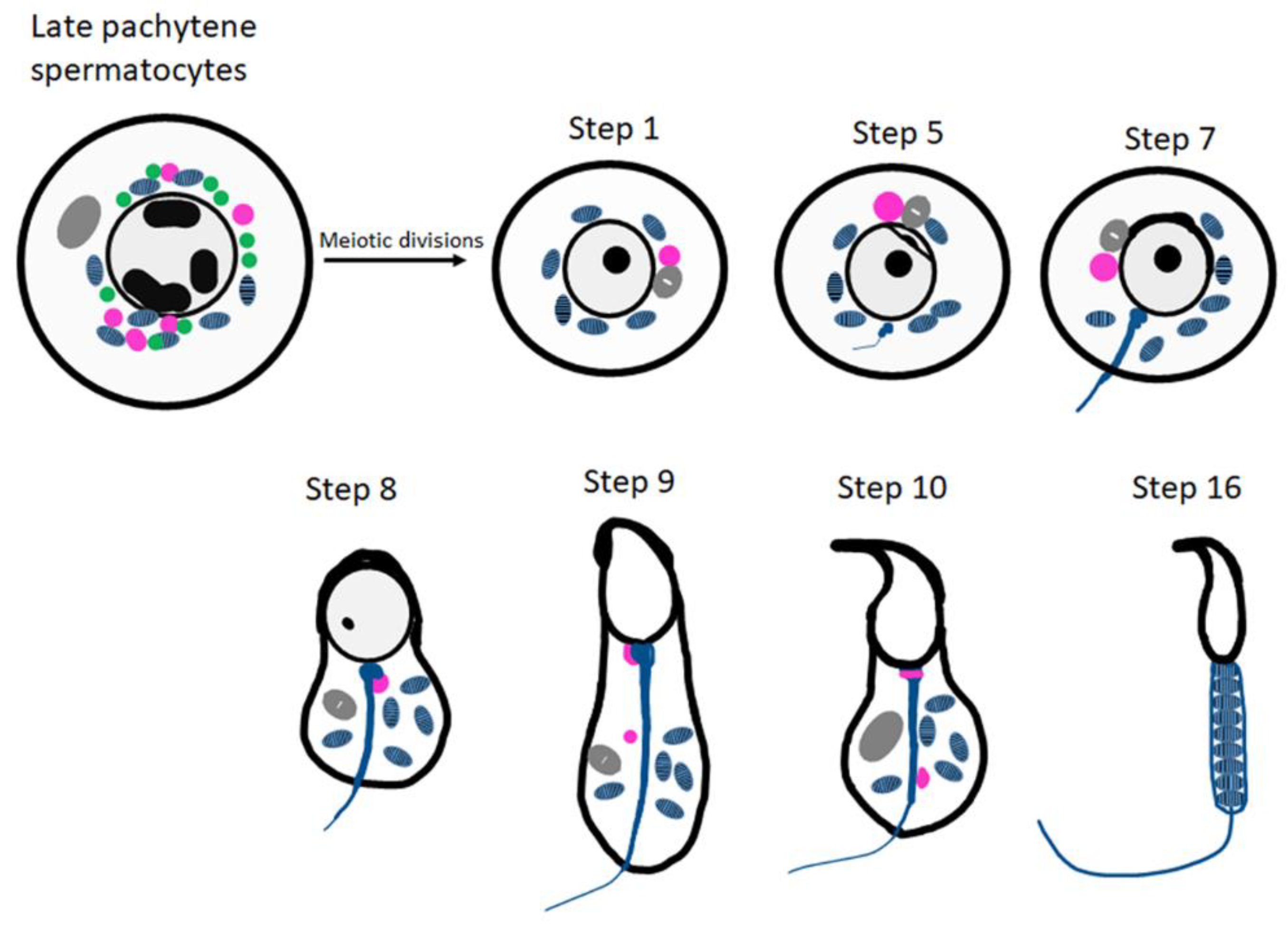
The origin of CBs is not clearly understood and more consensus view being their emergence from small granules (precursors of CBs) which are associated with the nuclear envelope present near the electron-dense inter-mitochondrial cement in the late pachytene spermatocytes (
2. RNPs Are Critical for Cellular and Organism Function: Role of CBs
Figure 1). The CB observed during all steps of round spermatid differentiation (steps 1–8 of spermiogenesis). The largest CBs are observed in step four, five and six of spermiogenesis [18]. At later stages of spermatid elongation, CBs move caudally to the neck region and split into two separate structures; one is discarded along with residual cytoplasm, and the other forms a ring around the base of the flagellum (
Figure 1).

Figure 1.
Schematic representation of CB organization and fate during spermatiogenesis The inter-mitochondrial cement (IMC; green) intermixed with mitochondria (blue) and the CB precursors (pink) co-exist in late pachytene spermatocytes. The Golgi complex is depicted in gray. The CB (pink) is condensed to its final single form in the early round spermatids. At step 8 of spermiogenesis, the CB is found at the basis of the flagellum. Later, it splits into two separate structures (step 9 onwards) and eventually disappears.
- CBs Are Analogous to Stress Granules and P-Bodies
RNPs lack demarcating membrane which gets formed under physiological conditions in male germ cells. CBs’ overall functions fall between P-bodies and stress granules and are in concert with the maintenance of RNA regulation. Stress granules and processing bodies are also membrane-less RNA granules that sequester translationally inactive messenger ribonucleoprotein particles (mRNPs) into compartments distinct from the surrounding cytoplasm [19,20]. Like P-bodies, stress granule assembly is dependent on the pool of non-translating mRNAs. Stress granules and P-bodies can physically interact to facilitate the shuttling of RNA and protein between them. The key variation between P-bodies and stress granules is that P-bodies assemble around the key enzymes of cytoplasmic RNA degradation (physiological conditions), and stress granules assemble around the essential components of the translation machinery (stress conditions) under heat, glucose deprivation, viral or bacterial infection, hypoxia, and oxidative stress [19,21].
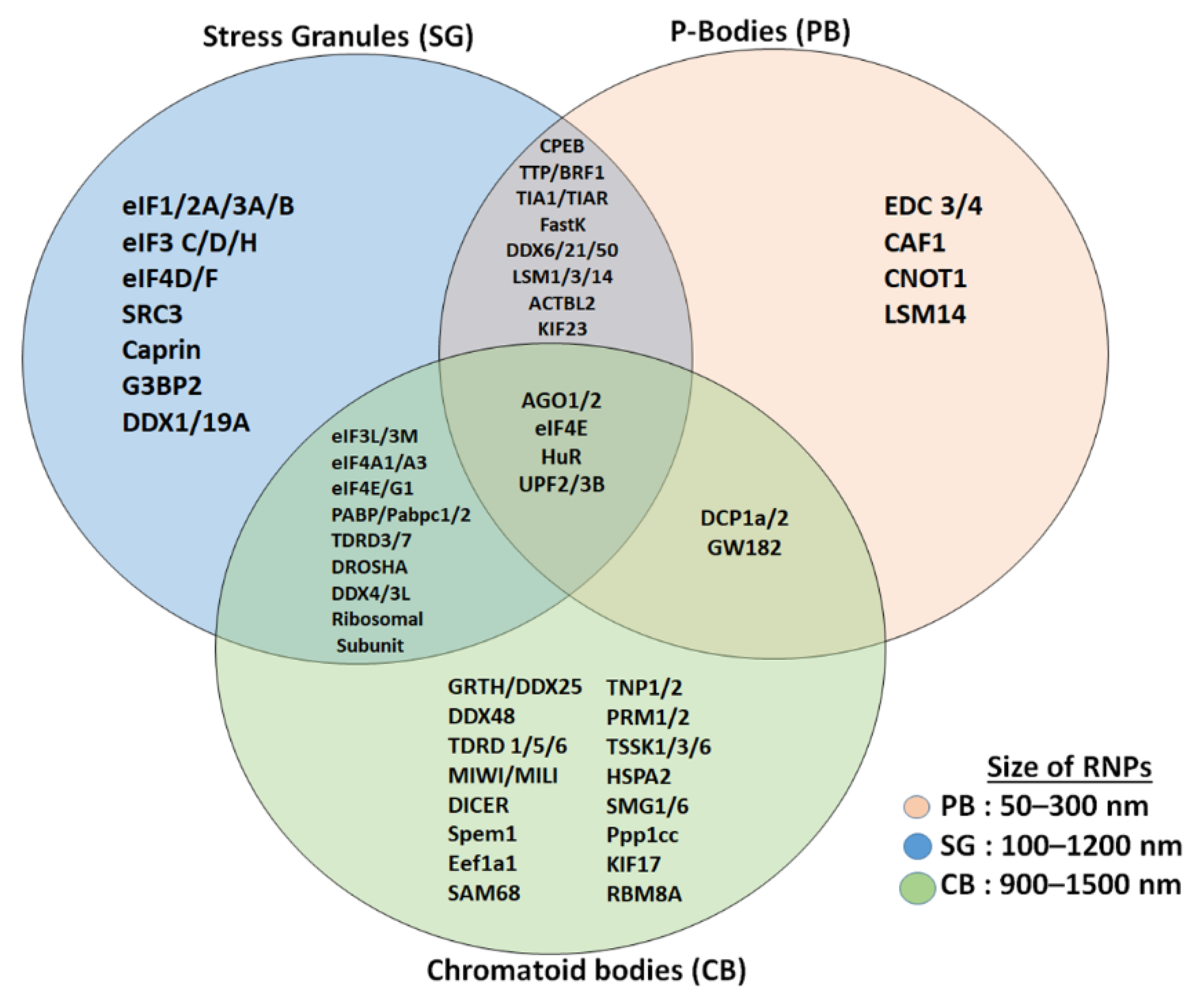
Unlike CBs and stress granules, P-bodies are not associated with the regulation of translation initiation instead it serve as a site for mRNA degradation, translation repression and storage of non-translating mRNAs (
3. CBs Are Analogous to Stress Granules and P-Bodies
Figure 2) [10,20,22]. P-bodies are uniquely enriched with factors related to mRNA decay and the NMD pathway, such as members of mRNA decapping machinery like decapping enzymes DCP1/2; UPF1/2, the activators of decapping EDC3, Dhh1/RCK/p54, Pat1, and Scd6/RAP55; the LSM1-7 complex; and the exonuclease XRN1 (
Figure 2) [20-24]. P-bodies are independent of initiation factors or translational assembly, while CBs seem to regulate mRNA storage and decay, as well as translational machinery, all in one compartment effectively [10,20]. Recent studies have implicated post-translational modifications of RNP granule proteins as the driver for the assembly and disassembly of the RNP granule [13,19]. The role the RNPs plays is unique and irreplaceable and has a critical role in the survival of the cell. More like stress granules, CBs have proteins or mRNAs that are involved in the translation process and other associated factors, and mRNA regulatory factors such as PABPC1, UPF2, and others [10,17,21]. Detailed studies on the interaction between these RNPs inside the cell provide vital clues on the molecular mechanisms of gene regulation.

Figure 2.
Comparison of proteins (mRNA decay and translation machinery) present in CBs with stress granules and P-bodies. Stress granules and CBs share several initiation factors and translation initiation assembly proteins, while P-bodies and CBs share few of the proteins. CBs are more related to stress granules than to P-bodies and CBs are bigger in size compared to stress granules and P-bodies.
- Small RNAs and Regulation of Spermatogenesis
During early spermatogenesis, small regulatory RNAs such as microRNAs control the expression of an array of genes at transcriptional or post-transcriptional levels. The role of CBs in small RNA-mediated gene control are not clearly understood at present. The repertoires of microRNAs and piRNAs have been identified inside CBs and both serve as important regulators of male germ cell differentiation.
miRNAs (21–23 nucleotides) belong to the class of noncoding RNAs which participate in a wide array of biological functions, by promoting target mRNA degradation and inhibition of translation [28-30]. miRNAs recognize their target mRNAs by sequence-specific bp pairing in the RNA-induced silencing complex together with Argonaute proteins (AGO) [9]. Most miRNAs are derived from primary miRNA transcripts which are processed by the Drosha-DGCR8 complex in the nucleus. The resulting precursor miRNAs (pre-miRNAs) get transported to the cytoplasm where mature miRNAs are generated via a Dicer-dependent or independent route. Another crucial component of the miRNA and siRNA pathways is the cytoplasmic endonuclease Dicer, which is critical for male fertility [31]. Dicer interacts with the germ-cell-specific RNA helicase MVH (mouse VASA homolog, DDX4). Sertoli cell-specific deletion of Dicer in mice results in spermatogenic malfunction, defective maturation, and infertility [31]. Transcripts of AGO proteins, Drosha, and Dicer have been demonstrated to be present in germ cells and Sertoli cells [29,32]. GRTH regulates proteins of the microprocessor complex, Drosha and DGCR8 (miRNA biogenesis) at the levels of mRNA and protein [29]. miRNA pathway proteins have been demonstrated to accumulate in the CBs in haploid round spermatids, suggesting that the CB and GRTH have a role in miRNA-dependent gene regulation [28,29].
Several testis-specific miRNAs such as miR-469, testis-preferred miR-34c, miR-470 and other let-7 family members (let-7a/d, b, and e-g), and miR203 were upregulated in round spermatids of GRTH KO mice. In addition, the enzyme complex (Drosha-DGCR8) required for the processing of miRNA was also upregulated in the GRTH KO mice. The miRNA, miR-469 target Tnp2 and Prm2 mRNAs by binding to their coding regions and thereby preventing translation of these essential mRNAs to proteins [29]. GRTH negatively regulates the overall miRNA biogenesis via Drosha/DGCR8 microprocessor complex, to generate mature miR-469 and other miRNAs which could play critical roles during spermatid elongation and spermiogenesis. miRNAs such as miR-469, through their inhibitory action on Tnp2/Prm2 mRNA, control the timely expression of genes for chromatin compaction and the progression of spermatogenesis [29]. The other small RNA, piRNAs comprise the largest and most complex class of small non-coding RNAs that are enriched in the germline tissues. Unlike miRNAs (22 nt long), piRNAs are slightly longer (26–32 nt long), and the biosynthesis of piRNAs is different from that of miRNAs and siRNAs [33,34,35]. During post-meiotic germ cell differentiation, the CB accumulates piRNAs and proteins of piRNA machinery. The CBs provide platforms for the piRNA pathway and appear to be involved both in piRNA biogenesis and piRNA-targeted RNA degradation.
The piRNAs play significant roles in suppressing transposons, maintaining genome integrity, formation of heterochromatin, and epigenetic regulation of sex determination, epigenetic inheritance etc [35]. Mature piRNA sequences are surprisingly diverse between different organisms,
even between closely related species. The piRNA pathway relies on the cooperation between 26-30 nucleotide long piRNAs and Piwi-clade Argonaute proteins to silence targets both at the co-transcriptional and posttranscriptional levels [35]. The embryonic and post-natal male germ cells express high levels of piRNAs where it regulates post-transcriptional regulation of mRNAs, maintenance of germline DNA integrity, silencing of transposon transcription, suppression of translation [35]. piRNAs have been found within the nucleus, cytoplasm, and the CBs.
In mice, piRNAs are produced in two waves: the first occurs during fetal and perinatal stages, generating piRNAs (pre-pachytene piRNAs) enriched in transposon element (TE) sequences, while the second occurs during postnatal spermatogenesis and produces piRNAs unrelated to TE sequences but mostly from intergenic regions called ‘pachytene piRNAs’. Pre-pachytene piRNAs bind to MILI and MIWI2 and participate in silencing of transposable elements both at epigenetic and posttranscriptional level in fetal and neonatal germ cells [36]. Pachytene piRNAs account for more than 95% of piRNAs in the adult mouse testes. The pachytene piRNAs do not show strong complementarity to any genomic sequence, except the loci that produce them. Recent findings indicated that pachytene piRNAs associated with MILI and MIWI and were shown to mediate global mRNA decay in spermatocytes and spermatids [37, 38, 39]. Pachytene piRNAs together with MIWI (mouse homolog of Aub) target spermiogenic mRNAs with imperfect base pairing and induce their decay either through MIWI-dependent cleavage or by assembling a complex containing CAF1, a deadenylase in the CCR4-NOT complex [40]. The other class of small RNAs are endo-siRNAs (siRNAs) or silencing RNAs. They are a class of 20-25 nucleotide-long double-stranded RNA molecules which play critical role in regulation of gene expression specifically reducing the translation of specific mRNAs [16]. Recent studies have shown that its also involved in DNA repair and transcriptional regulation within the nucleus [41]. Biosynthesis of endo-siRNAs is more similar to miRNAs derived from double stranded intrinsic transcripts and are processed by Dicer and Argonaute proteins. Any transcript that are capable of forming double-stranded structures, either inter- or intramolecularly, has the potential to be processed into an siRNA. Due to this property, siRNAs are only rarely conserved in sequence and to differentiate between siRNAs possessing functional relevance are major challenges in the small RNA field. The siRNA binding to the target sequence always results in mRNA cleavage/degradation (Endonucleolytic cleavage of mRNA) while miRNAs binding is involved in translational repression in addition to mRNA cleavage/degradation [42]. There are other major differences exists as well. To elicit RNAi, the siRNA must be fully complementary to its target mRNA. In contrast, the target recognition of miRNAs is more complex with multiple binding sites (mRNA CDS and UTR) and different degree of complementarity [42]. The miRNAs only need partially complementary to its target mRNA to be functional unlike siRNAs which require complimentary to full mRNAs. In addition to their role in the RNAi pathway, siRNAs are also involved in RNAi-related pathway which play critical role in shaping the chromatin structure of a genome.
References:
- Steger, K. Haploid spermatids exhibit translationally repressed mRNAs. Embryol. 2001, 203, 323–334.
- Kimmins, S.; Sassone-Corsi, P. Chromatin remodelling and epigenetic features of germ cells. Nature 2005, 434, 583–589.
- Griswold, M.D. Spermatogenesis: The Commitment to Meiosis. Rev. 2016, 96, 1–17.
- Kavarthapu, R.; Anbazhagan, R.; Raju, M.; Morris, C.T.; Pickel, J.; Dufau, M.L. Targeted knock-in mice with a human mutation in GRTH/DDX25 reveals the essential role of phosphorylated GRTH in spermatid development during spermatogenesis. Mol. Genet. 2019, 28, 2561–2572.
- Shima, J.E.; McLean, D.J.; McCarrey, J.R., Griswold, M.D. The murine testicular transcriptome: characterizing gene expression in the testis during the progression of spermatogenesis. Reprod. 2004, 71, 319–330.
- Tsai-Morris, C.H.; Sheng, Y.; Gutti, R.; Tang, P.Z.; Dufau, M.L. Gonadotropin-regulated testicular RNA helicase (GRTH/DDX25): a multifunctional protein essential for spermatogenesis. Androl. 2010, 31, 45–52.
- Maclean, J.A.; Wilkinson, M.F. Gene regulation in spermatogenesis. Top. Dev. Biol. 2005, 71, 131–197.
- Kotaja, N.; Sassone-Corsi, P. The chromatoid body: a germ-cell-specific RNA-processing centre. Rev. Mol. Cell Biol. 2007, 8, 85–90.
- Sato, H.; Tsai-Morris, C.H.; Dufau, M.L. Relevance of gonadotropin-regulated testicular RNA helicase (GRTH/DDX25) in the structural integrity of the chromatoid body during spermatogenesis. Biophys. Acta 2010, 1803, 534–543.
- Anbazhagan, R.; Kavarthapu, R.; Dufau, M.L. Role of phosphorylated gonadotropin-regulated testicular RNA helicase (GRTH/DDX25) in the regulation of germ cell specific mRNAs in chromatoid bodies during spermatogenesis. Cell Dev. Biol. 2020, 8, 580019.
- Sheng, Y.; Tsai-Morris, C.H.; Gutti, R.; Maeda, Y.; Dufau, M.L. Gonadotropin-regulated testicular RNA helicase (GRTH/Ddx25) is a transport protein involved in gene-specific mRNA export and protein translation during spermatogenesis. Biol. Chem. 2006, 281, 35048–35056.
- Dufau, M.L.; Tsai-Morris, C.H. Gonadotropin-regulated testicular helicase (GRTH/DDX25): an essential regulator of spermatogenesis. Trends Endocrinol. Metab. 2007, 18, 314–320.
- Schisa, J.A.; Elaswad, M.T. An emerging role for post-translational modifications in regulating rnp condensates in the germ line. Mol. Biosci. 2021, 8, 658020.
- Peruquetti, RL. Perspectives on mammalian chromatoid body research. Reprod. Sci. 2015, 159, 8–16.
- Eulalio, A.; Behm-Ansmant, I.; Izaurralde, E. P bodies: at the crossroads of post-transcriptional pathways. Rev. Mol. Cell Biol. 2007, 8, 9–22.
- Meikar, O.; Da Ros, M.; Korhonen, H.; Kotaja, N. Chromatoid body and small RNAs in male germ cells. Reproduction 2011, 142, 195–209.
- Meikar, O.; Vagin, V.V.; Chalmel, F.; Sõstar, K.; Lardenois, A.; Hammell, M.; Jin, Y.; Da Ros, M.; Wasik, K.A.; Toppari, J.; et al. An atlas of chromatoid body components. RNA 2014, 20, 1–3.
- Lehtiniemi, T.; Kotaja, N. Germ granule-mediated RNA regulation in male germ cells. Reproduction 2018, 155, 77–91.
- Kedersha, N.L.; Gupta, M.; Li, W.; Miller, I.; Anderson, P. RNA-binding proteins TIA-1 and TIAR link the phosphorylation of eIF-2 α to the assembly of mammalian stress granules. Cell. Biol. 1999, 147, 1431–1442.
- Riggs, C.L.; Kedersha, N.; Ivanov, P.; Anderson, P. Mammalian stress granules and P bodies at a glance. Cell Sci. 2020, 133, jcs242487.
- Kedersha, N.; Stoecklin, G.; Ayodele, M.; Yacono, P.; Lykke-Andersen, J.; Fritzler, M.J.; Scheuner, D.; Kaufman, R.J.; Golan, D.E.; Anderson, P. Stress granules and processing bodies are dynamically linked sites of mRNP remodeling. Cell Biol. 2005, 16, 871–884.
- Ivanov, P.; Kedersha, N.; Anderson, P. Stress Granules and Processing Bodies in Translational Control. Cold Spring Harb. Perspect. Biol. 2019, 11, a032813.
- Parker, R.; Sheth, U. P Bodies and the Control of mRNA Translation and Degradation. Cell 2007, 25, 635–646.
- Decker, C.J.; Parker R P-bodies and stress granules: possible roles in the control of translation and mRNA degradation. Cold Spring Harb. Perspect. Biol. 2012, 4, a012286.
- Guo, L.; Shorter, J. It’s raining liquids: RNA tunes viscoelasticity and dynamics of membraneless organelles. Cell 2015, 60, 189–192.
- Kim, B., Cooke, H.J.; Rhee, K. DAZL is essential for stress granule formation implicated in germ cell survival upon heat stress. Development 2012, 139, 568–578.
- Ma, W.; Mayr, C. A membraneless organelle associated with the endoplasmic reticulum enables 3’utr-mediated protein-protein interactions. Cell 2018, 175, 1492–1506.
- Kotaja, N.; Bhattacharyya, S.N.; Jaskiewicz, L.; Kimmins, S.; Parvinen, M.; Filipowicz, W.; Sassone-Corsi, P. The chromatoid body of male germ cells: similarity with processing bodies and presence of Dicer and microRNA pathway components. Natl. Acad. Sci. USA 2006, 103, 2647–2652.
- Dai, L.; Tsai-Morris, C.H.; Sato, H.; Villar, J.; Kang, J.H.; Zhang, J.; Dufau, M.L. Testis-specific miRNA-469 up-regulated in gonadotropin-regulated testicular RNA helicase (GRTH/DDX25)-null mice silences transition protein 2 and protamine 2 messages at sites within coding region: implications of its role in germ cell development. Biol. Chem. 2011, 286, 44306–44318.
- Krol, J.; Loedige, I.; Filipowicz, W. The widespread regulation of microRNA biogenesis, function and decay. Rev. Genet. 2010, 11, 597–610.
- Papaioannou, M.D.; Pitetti, J.L.; Ro, S.; Park, C.; Aubry, F.; Schaad, O.; Vejnar, C.E.; Kühne, F.; Descombes, P.; Zdobnov, E.M. et al. Sertoli cell Dicer is essential for spermatogenesis in mice. Dev Biol. 2009, 326, 250–259.
- Gonzalez-Gonzalez, E.; Lopez-Casa,s P.P.; del Mazo, J. The expression patterns of genes involved in the RNAi pathways are tissue-dependent and differ in the germ and somatic cells of mouse testis. Biophys. Acta 2008, 1779, 306–311.
- Aravin, A.A.; Sachidanandam, R.; Girard, A.; Fejes-Toth, K.; Hannon, G.J. Developmentally regulated piRNA clusters implicate MILI in transposon control. Science 2007, 316,744-747.
- Thomson, T.; Lin, H. The biogenesis and function of PIWI proteins and piRNAs: progress and prospect. Rev. Cell Dev. Biol. 2009, 25, 355–376.
- Chuma, S.; Nakano, T.; piRNA and spermatogenesis in mice. Philos Trans R Soc Lond B Biol Sci. 2013, 368:0110338.
- Simonelig, M. piRNAs, master regulators of gene expression. Cell Res 2014, 24, 779–780.
- Gou, LT., Dai, P., Yang, JH. et al. Pachytene piRNAs instruct massive mRNA elimination during late spermiogenesis. Res. 2014, 24, 680–700.
- Zhang, P., Kang, JY., Gou, LT. et al. MIWI and piRNA-mediated cleavage of messenger RNAs in mouse testes. Res. 2015, 25, 193–207.
- Wang, C.; Lin. H. Roles of piRNAs in transposon and pseudogene regulation of germline mRNAs and lncRNAs. Genome Biol. 2021,22:27.
- Barckmann, B.; Pierson, S.; Dufourt, J.; Papin, C.; Armenise, C.; Port, F.; Grentzinger, T.; Chambeyron, S.; Baronian, G.; Desvignes, J.P. Aubergine iCLIP Reveals piRNA-Dependent Decay of mRNAs Involved in Germ Cell Development in the Early Embryo. Cell Rep. 2015, 12, 1205–1216.
- Hilz, S.; Modzelewski, A.J.; Cohen, P.E.; Grimson, A. The roles of microRNAs and siRNAs in mammalian spermatogenesis. Development 2016, 143, 3061-3073.
- Lam, J.K.W.; Chow, M.Y.T; Zhang, Y.; Leung, SWS. siRNA Versus miRNA as Therapeutics for Gene Silencing. Ther. Nucleic Acids 2015, 4, e252.
4. Small RNAs of CBs and Regulation of Spermatogenesis
References
- Steger, K. Haploid spermatids exhibit translationally repressed mRNAs. Anat. Embryol. 2001, 203, 323–334.
- Kimmins, S.; Sassone-Corsi, P. Chromatin remodelling and epigenetic features of germ cells. Nature 2005, 434, 583–589.
- Griswold, M.D. Spermatogenesis: The Commitment to Meiosis. Physiol. Rev. 2016, 96, 1–17.
- Kavarthapu, R.; Anbazhagan, R.; Raju, M.; Morris, C.T.; Pickel, J.; Dufau, M.L. Targeted knock-in mice with a human mutation in GRTH/DDX25 reveals the essential role of phosphorylated GRTH in spermatid development during spermatogenesis. Hum. Mol. Genet. 2019, 28, 2561–2572.
- Shima, J.E.; McLean, D.J.; McCarrey, J.R.; Griswold, M.D. The murine testicular transcriptome: Characterizing gene expression in the testis during the progression of spermatogenesis. Biol. Reprod. 2004, 71, 319–330.
- Tsai-Morris, C.H.; Sheng, Y.; Gutti, R.; Tang, P.Z.; Dufau, M.L. Gonadotropin-regulated testicular RNA helicase (GRTH/DDX25): A multifunctional protein essential for spermatogenesis. J. Androl. 2010, 31, 45–52.
- Maclean, J.A.; Wilkinson, M.F. Gene regulation in spermatogenesis. Curr. Top. Dev. Biol. 2005, 71, 131–197.
- Kotaja, N.; Sassone-Corsi, P. The chromatoid body: A germ-cell-specific RNA-processing centre. Nat. Rev. Mol. Cell Biol. 2007, 8, 85–90.
- Sato, H.; Tsai-Morris, C.H.; Dufau, M.L. Relevance of gonadotropin-regulated testicular RNA helicase (GRTH/DDX25) in the structural integrity of the chromatoid body during spermatogenesis. Biochim. Biophys. Acta 2010, 1803, 534–543.
- Anbazhagan, R.; Kavarthapu, R.; Dufau, M.L. Role of phosphorylated gonadotropin-regulated testicular RNA helicase (GRTH/DDX25) in the regulation of germ cell specific mRNAs in chromatoid bodies during spermatogenesis. Front. Cell Dev. Biol. 2020, 8, 580019.
- Sheng, Y.; Tsai-Morris, C.H.; Gutti, R.; Maeda, Y.; Dufau, M.L. Gonadotropin-regulated testicular RNA helicase (GRTH/Ddx25) is a transport protein involved in gene-specific mRNA export and protein translation during spermatogenesis. J. Biol. Chem. 2006, 281, 35048–35056.
- Dufau, M.L.; Tsai-Morris, C.H. Gonadotropin-regulated testicular helicase (GRTH/DDX25): An essential regulator of spermatogenesis. Trends Endocrinol. Metab. 2007, 18, 314–320.
- Schisa, J.A.; Elaswad, M.T. An emerging role for post-translational modifications in regulating rnp condensates in the germ line. Front. Mol. Biosci. 2021, 8, 658020.
- Peruquetti, R.L. Perspectives on mammalian chromatoid body research. Anim. Reprod. Sci. 2015, 159, 8–16.
- Eulalio, A.; Behm-Ansmant, I.; Izaurralde, E. P bodies: At the crossroads of post-transcriptional pathways. Nat. Rev. Mol. Cell Biol. 2007, 8, 9–22.
- Meikar, O.; Da Ros, M.; Korhonen, H.; Kotaja, N. Chromatoid body and small RNAs in male germ cells. Reproduction 2011, 142, 195–209.
- Meikar, O.; Vagin, V.V.; Chalmel, F.; Sõstar, K.; Lardenois, A.; Hammell, M.; Jin, Y.; Da Ros, M.; Wasik, K.A.; Toppari, J.; et al. An atlas of chromatoid body components. RNA 2014, 20, 1–3.
- Lehtiniemi, T.; Kotaja, N. Germ granule-mediated RNA regulation in male germ cells. Reproduction 2018, 155, 77–91.
- Kedersha, N.L.; Gupta, M.; Li, W.; Miller, I.; Anderson, P. RNA-binding proteins TIA-1 and TIAR link the phosphorylation of eIF-2 α to the assembly of mammalian stress granules. J. Cell. Biol. 1999, 147, 1431–1442.
- Riggs, C.L.; Kedersha, N.; Ivanov, P.; Anderson, P. Mammalian stress granules and P bodies at a glance. J. Cell Sci. 2020, 133, jcs242487.
- Kedersha, N.; Stoecklin, G.; Ayodele, M.; Yacono, P.; Lykke-Andersen, J.; Fritzler, M.J.; Scheuner, D.; Kaufman, R.J.; Golan, D.E.; Anderson, P. Stress granules and processing bodies are dynamically linked sites of mRNP remodeling. J. Cell Biol. 2005, 16, 871–884.
- Ivanov, P.; Kedersha, N.; Anderson, P. Stress Granules and Processing Bodies in Translational Control. Cold Spring Harb. Perspect. Biol. 2019, 11, a032813.
- Parker, R.; Sheth, U. P Bodies and the Control of mRNA Translation and Degradation. Mol. Cell 2007, 25, 635–646.
- Decker, C.J.; Parker, R. P-bodies and stress granules: Possible roles in the control of translation and mRNA degradation. Cold Spring Harb. Perspect. Biol. 2012, 4, a012286.
- Guo, L.; Shorter, J. It’s raining liquids: RNA tunes viscoelasticity and dynamics of membraneless organelles. Mol. Cell 2015, 60, 189–192.
- Kim, B.; Cooke, H.J.; Rhee, K. DAZL is essential for stress granule formation implicated in germ cell survival upon heat stress. Development 2012, 139, 568–578.
- Ma, W.; Mayr, C. A membraneless organelle associated with the endoplasmic reticulum enables 3’utr-mediated protein-protein interactions. Cell 2018, 175, 1492–1506.
- Kotaja, N.; Bhattacharyya, S.N.; Jaskiewicz, L.; Kimmins, S.; Parvinen, M.; Filipowicz, W.; Sassone-Corsi, P. The chromatoid body of male germ cells: Similarity with processing bodies and presence of Dicer and microRNA pathway components. Proc. Natl. Acad. Sci. USA 2006, 103, 2647–2652.
- Dai, L.; Tsai-Morris, C.H.; Sato, H.; Villar, J.; Kang, J.H.; Zhang, J.; Dufau, M.L. Testis-specific miRNA-469 up-regulated in gonadotropin-regulated testicular RNA helicase (GRTH/DDX25)-null mice silences transition protein 2 and protamine 2 messages at sites within coding region: Implications of its role in germ cell development. J. Biol. Chem. 2011, 286, 44306–44318.
- Krol, J.; Loedige, I.; Filipowicz, W. The widespread regulation of microRNA biogenesis, function and decay. Nat. Rev. Genet. 2010, 11, 597–610.
- Papaioannou, M.D.; Pitetti, J.L.; Ro, S.; Park, C.; Aubry, F.; Schaad, O.; Vejnar, C.E.; Kühne, F.; Descombes, P.; Zdobnov, E.M.; et al. Sertoli cell Dicer is essential for spermatogenesis in mice. Dev Biol. 2009, 326, 250–259.
- Gonzalez-Gonzalez, E.; Lopez-Casas, P.P.; del Mazo, J. The expression patterns of genes involved in the RNAi pathways are tissue-dependent and differ in the germ and somatic cells of mouse testis. Biochim. Biophys. Acta 2008, 1779, 306–311.
- Aravin, A.A.; Sachidanandam, R.; Girard, A.; Fejes-Toth, K.; Hannon, G.J. Developmentally regulated piRNA clusters implicate MILI in transposon control. Science 2007, 316, 744–747.
- Thomson, T.; Lin, H. The biogenesis and function of PIWI proteins and piRNAs: Progress and prospect. Annu. Rev. Cell Dev. Biol. 2009, 25, 355–376.
