Therapeutic Efficacies of Berberine against Neurological Disorders: Comparison
Please note this is a comparison between Version 1 by P.C. Shaw and Version 3 by Beatrix Zheng.

Neurological disorders refer to any dysfunctions of the nervous system, and mainly include Alzheimer’s disease (AD), Parkinson’s disease (PD), Huntington’s disease (HD), dementia, schizophrenia, anxiety, depression, epilepsy, traumatic brain injury (TBI), and brain tumor. The burden of deaths and disabilities caused by neurological disorders has been increasing dramatically, ranking it as the leading cause of disability and the second leading cause of death worldwide. The World Health Organization predicts that by 2040, as many developed countries’ populations age, neurological disorders will overtake cancer to become the second leading cause of death worldwide. Nevertheless, there is no treatment that can cure neurological disorders, and the current treatments mainly target the amelioration of symptoms. Berberine, a natural alkaloid, is mainly isolated from Coptis chinensis, Berberis vulgaris, Hydrastis canadensis, and Phellodendron amurense. For over a thousand years, these herbs have been used for treating diarrhea without any obvious side effects in patients. With the advances of pharmacological research, BBR has been considered as a promising multitarget drug (MTD) for treating neurological disorders.

  • berberine
  • neurological disorders

1. BBR on Alzheimer’s Disease

Alzheimer’s disease (AD) most often develops in people over 65 years of age and is characterized by memory loss and handicapped daily functions [1][22]. To date, the exact cause of AD has not been fully discovered, but it is believed that AD results from multiple contributing factors. Thus, there is no direct and effective treatment for AD. There are two main strategies for treatment. Firstly, inhibiting the activity of cholinesterase (ChE), an enzyme to catalyze the breakdown of acetylcholine (ACh) and other choline esters that function as neurotransmitters, is one of the potential therapeutic strategies based on the cholinergic hypothesis [2][3][23,24]. Secondly, it is important to reduce amyloid beta (Aβ) and Tau protein plaques, which may lead to neurofibrillary tangle formation, oxidation, inflammation, and excitotoxicity [4][5][25,26].
Due to its multifaceted nature, BBR has been shown to address AD mainly in two aspects: anti-ChE and anti-Aβ/Tau pathways (Figure 12).
Figure 12.
Effects of BBR against AD and PD.

1.1. Inhibitory Effect of BBR on ChE

The cholinergic hypothesis states that a deficit in central cholinergic neurotransmission resulting from a loss of ACh contributes to pathological development [6][27]. ChE, including acetylcholinesterase (AChE) and butyrylcholinesterase (BChE), is responsible for hydrolyzing ACh into choline and acetic acid [7][28]. ChE inhibitors are effective medication for AD as they enhance central cholinergic function by inhibiting ChE activities, thereby increasing the availability of ACh to stimulate memory and learning ability in the brain [6][27]. In the streptozotocin-induced sporadic AD model and the heavy-metals-induced AD-like disease model, BBR maintained the ACh level by inhibiting AChE activity [8][9][29,30]. BBR has a large hydrophobic surface and a cation; thus, hydrophobic residues in AChE interacted with BBR to form a binding pocket, which accounts for the interaction between AChE and BBR [10][31]. However, there is no publication discussing the effect of BBR on BChE in vivo.

1.2. Anti-Aβ and Tau Effects of BBR

The Aβ peptide, consisting of 39–43 amino acids, is derived from the abnormal processing of the amyloid precursor protein (APP), and the accumulation of Aβ peptide has been considered as a hallmark of AD pathogenetic development [11][32]. The enzymes α-secretase, β-secretase (also called BACE), and γ-secretase take active roles in the processing of APP [12][33]. Tau proteins within the brain cells of AD brains are misfolded and abnormally shaped, deposits of which form tangles within the neural cells [13][34]. In AD, it is common to find tau hyperphosphorylation and aggregation, thus losing its ability to maintain the microtubule tracks; as a result, tau dysfunction could lead to the retraction of neuronal processes and thus cell death [13][34].
The oral administration of BBR significantly ameliorated learning deficits and spatial memory retention in transgenic mouse models of AD (TgCRND8 mice, APP/PS1 mice, and 3×Tg AD mice) [14][15][16][17][35,36,37,38]. A mechanistic study showed that BBR significantly decreased the levels of C-terminal fragments of APP and the hyperphosphorylation of APP via the protein kinase B/glycogen synthase kinase 3 (AKT/GSK3) signaling pathway [14][35]. BBR also inhibited the activity of β/γ-secretases or suppressed PRKR-like endoplasmic reticulum kinase/eukaryotic translation initiation factor-2 α (PERK/EIF2α) signaling-mediated BACE1 translation to downregulate the Aβ level in the AD mouse hippocampus [15][16][18][36,37,39]. In addition, promoting the clearance of Aβ is another mechanistic aspect of BBR. To promote Aβ clearance, BBR activated the autophagic process through initiating the phosphoinositide 3-kinase (PI3K)/Beclin-1 pathway [17][38] or by inhibiting the mammalian target of rapamycin/P70 S6 kinase (mTOR/p70S6K) signaling [19][40]. Additionally, Aβ is toxic to neural cells, as it can cause pore formation resulting in ion leakage, disturb cellular calcium balance, and destroy membrane potential, thus leading to apoptosis, synaptic loss, and cytoskeleton disruption [20][41]. BBR is effective in preventing Aβ-induced damage to neural cells. Bilaterally injecting rats with Aβ induced learning and memory impairments, while BBR administration ameliorated Aβ-induced toxicity [21][42]. BBR showed this beneficial effect via modulating the Ca2+-activated K+ channel to maintain the optimal level of Ca2+ entry [21][42]. Moreover, BBR reduced Aβ-related oxidative and inflammatory damage. The antioxidant effect of BBR was exerted via downregulating reactive oxygen species (ROS) level, promoting the activity of glutathione (GSH), and inhibiting lipid peroxidation [22][43]. BBR also normalized the production of cytokines such as tumor necrosis factor α (TNFα), interleukin 12 (IL-12), IL-6, and IL-1β to retard inflammation [9][30]. In addition, exposure to Aβ could potentially lead to microglial activation, thereby triggering a detrimental neural response [23][44]. No in vivo function of BBR regarding microglial activation has been revealed; only an in vitro study indicated that BBR could inhibit Aβ-induced microglial activation via a silencing of cytokine signaling factor 1 (SOCS1)-dependent modulation of the microglial M1/M2 activated state [24][45].
Targeting Tau, BBR can reduce its hyperphosphorylation and increase its degradation. In 3×Tg AD mice, BBR improved the spatial learning capacity, memory retention, and the mechanism involved in reducing tau hyperphosphorylation via modulation of the AKT/glycogen synthase kinase 3β (GSK3β) pathway, enhancing autophagic flux, and increasing tau clearance through the PI3K/Beclin-1/B-cell lymphoma 2 (Bcl-2) pathway [25][46]. In APP/PS1 mice, BBR was found to suppress nuclear factor kappa-light-chain enhancer of the activated B cells (NF-κB) signaling pathway to limit tau hyperphosphorylation [22][43].
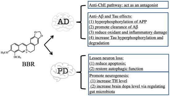
In conclusion, BBR exhibits therapeutic efficacy on PD mainly through the inhibition of ChE activity and suppression of Aβ- and Tau-induced toxicity. The downregulation of ChE activity by BBR contributes to increased ACh availability in the brain [8][9][29,30]. Both Aβ and Tau are toxic to neural cells via triggering oxidant, inflammatory, and even death signals, while BBR can degrade Aβ and Tau to ameliorate their toxicity [15][16][17][18][19][21][22][25][36,37,38,39,40,42,43,46] (Figure 12).

2. BBR on Parkinson’s Disease

Parkinson’s disease (PD) is a progressive neurodegenerative disorder characterized by the degeneration of dopamine (DA) and non-DA neurons, which could lead to tremors, rigidity, bradykinesia, and gait disturbance [26][47]. No cure has been discovered for treating PD and the current therapy mainly focuses on lessening neuron loss [27][8]. BBR showed beneficial effects against the chemical-induced PD model (Figure 12).
BBR protected neurons from apoptosis induced by 1-methyl-4-phenyl-1,2,3,6-tetrahydropyridine/probenecid (MPTP/P) through the downregulation of the Bcl2/Bcl-2-associated X protein (BAX) ratio [28][48], through AMP-activated protein kinase (AMPK)-dependent enhancement of autophagy [29][30][49,50], or by preventing NLRP3 inflammasome activation [30][50]. In the 6-hydroxydopamine-induced PD model, BBR reduced ROS production, caspase-3 activation, and subsequent neuronal death [31][32][51,52] BBR also increased the expression of tyrosine hydroxylase (TH), a rate-limiting enzyme for dopamine synthesis, to promote neurogenesis [28][30][48,50]. Additionally, a recent study demonstrated that BBR could ameliorate PD by regulating gut microbiota. BBR enhanced TH to produce L-dopa by triggering the biosynthesis of tetrahydrobiopterin in the gut microbiota and subsequently led to an increased brain dopa level, therefore improving brain function in MPTP-induced PD mice [33][53].
In addition, rotenone is also widely used to establish PD models [34][54], whereas the effect of BBR on the rotenone model is less well understood and controversial. There is no in vivo study of BBR on the rotenone-induced PD model. For the in vitro efficacy, Kysenius and colleagues claimed that the subtoxic nanomolar concentration (30 nM) of BBR could sensitize neurons to rotenone injury [35][55], while Han and colleagues found that BBR protected SH-SY5Y cells from rotenone injury by activating the antioxidant and PI3K/AKT signaling pathway [36][56].
Collectively, BBR maintains neural viability in PD models. BBR not only lessens neuron loss [28][29][30][31][32][33][48,49,50,51,52,53], but also promotes neurogenesis [28][30][31][48,50,51] (Figure 12).

3. BBR on Stroke

Stroke, also defined as a cerebrovascular accident, is one of the major causes of mortality and long-term disability, and it is induced by either inadequate focal blood flow or hemorrhage into the brain tissue or the surrounding subarachnoid space [37][57]. The current prevention or treatment of stroke includes primary prevention, recanalization and thrombolysis, neuroprotection, secondary prevention, and neurorepair [38][39][58,59]. Both pretreatment and post-treatment of BBR have shown prominent efficacies for stroke (Figure 23).
Figure 23.
Effects of BBR against stroke.
BBR was found to be a thrombin inhibitor and had the ability to inhibit thrombin-induced platelet aggregation in washed platelet samples in vitro [40][60]; however, there is no research exploring the thrombolysis effect of BBR in vivo.
Middle cerebral artery occlusion (MCAO) surgery has been widely used to establish a successful murine stroke model [41][61]. After cerebral infarction occurs, oxidative factors and proinflammatory cytokines are released, leading to ischemic neuronal death including apoptosis and necrosis [42][62]. Following ischemia and reperfusion, a cascade of inflammatory responses is triggered. The high-mobility group box 1 (HMGB1) protein is released from necrotic and dying neural cells, subsequently activating the NF-κB pathway, which is commonly used as an indicator of inflammation in stroke studies [43][44][63,64]. Then, TNFα, IL-1β, and IL-6 are activated [45][46][65,66]. Seven-day pretreatment of BBR prevented the translocation of NF-κB into the nucleus and the transcription of proinflammatory cytokines; consequently, the expression of proinflammatory factors such as TNFα, IL-1β, and IL-6 was downregulated and the expression of anti-inflammatory cytokines, including IL-10, was upregulated [47][67]. Inflammation in stroke could lead to the production of ROS [48][68]. Excessive ROS may cause severe damage to neural cells, and then cell death by either necrosis or apoptosis may be initiated [43][63]. BBR pretreatment lowered the increased level of MDA and enhanced the activities of antioxidases such as superoxide dismutase (SOD), catalase (CAT), peroxiredoxin, and NAD(P)H dehydrogenase quinone 1 (NQO1) [47][49][67,69]; in addition, the preadministration of BBR lessened neural cell apoptosis via decreasing caspase cascades (caspase-3 and caspase-9) and increasing Bcl-2 expression [50][51][52][70,71,72], and promoting the cell-survival-related pathways such as the phosphor activation of AKT and increase of ERK1/2 [50][53][70,73]. Moreover, BBR bound to the poly (A) tail on retinoblastoma mRNA to antagonize the mRNA degradation and upregulation of the retinoblastoma protein during ischemia/reperfusion, which in turn inhibited apoptosis and facilitated cell survival in the injured brain [54][74].
Additionally, post-treatment of BBR results in effects similar to pretreatment. BBR administered after MCAO surgery reduced the infarction volume in mice and rats [55][56][57][75,76,77]. BBR functioned as a potent anti-inflammatory agent for ameliorating focal cerebral ischemia injury by enhancing the IL-10 level [55][75] and downregulating NF-κB nuclear transposition [56][76]; BBR was also able to activate the upregulation of claudin-5 expression to reduce access to the blood–brain barrier [56][76]. Scavenging ROS also contributed to the effect of post-treatment with BBR. The reseaOurchers' previous study found that BBR acted as a potent agonist of peroxisome proliferator-activated receptor delta (PPARδ) to increase nuclear factor (erythroid-derived 2)-like 1/2 (NRF1/2) and NQO1 to lower the ROS content in MCAO mice brains, thus exhibiting the neuroprotective effect of BBR [57][77]. Moreover, BBR is also beneficial for facilitating angiogenesis by modulating AMP-activated protein kinase (AMPK)-dependent M2 macrophage/microglial polarization, which promoted functionary recovery against ischemic stroke [58][78].
Stroke occurrence results in a detrimental impact on the brain. The most prominent efficacy of BBR on stroke is to reduce brain infarct damage, which is achieved by promoting thrombolysis [40][60], decreasing oxidative and inflammatory damage [47][49][55][56][57][67,69,75,76,77], reducing neural cell death [47][50][51][52][53][54][67,70,71,72,73,74], and facilitating angiogenesis [58][78] (Figure 23).

4. BBR on Huntington’s Disease

Huntington’s disease (HD), also known as Huntington’s chorea, is mostly inherited and mainly characterized by chorea, dystonia, loss of motor coordination, and mental deterioration [59][79]. HD results from an expanded CAG repeat in the huntingtin gene, which encodes an abnormally long polyglutamine repeat in the huntingtin protein [60][80]. BBR effectively improved motor function and prolonged the survival rate of transgenic N171-82Q HD mice by increasing autophagic function to reduce mutant huntingtin accumulation [61][81].

5. BBR on Dementia

Dementia describes a group of symptoms regarding memory loss and thinking disability. It is not a specific disease, but brain disorders and aging have been confirmed to give rise to dementia [62][63][82,83]. The most common cause of dementia is AD, which accounts for 60–70% of dementia cases worldwide [62][82]. Vascular dementia accounts for at least 20% of dementia cases, making it the second most common type [64][84].
BBR treated AD-related dementia mainly via targeting AD symptoms (shown in Section 3.1). Moreover, BBR is also effective in treating vascular dementia, which is usually caused by reduced blood flow to the brain [64][84]. In the chronic cerebral hypoperfusion (CCH)-induced vascular dementia model, BBR treatment prevented cognitive deficits and reversed CCH-induced neuronal cell death [65][85]. In the diabetes-related vascular dementia model, BBR increased the blood supply from the posterior cerebral artery, which was achieved by the inhibition of miR-133a ectopic expression in the vascular endothelium and by the normalization of vascular bioactivity in the cerebral middle artery [66][86]. Neurotoxic chemicals, such as doxorubicin, d-galactose, and lipopolysaccharide also led to cognitive impairment. BBR significantly improved cognitive disability in doxorubicin- or lipopolysaccharide-treated rats, and also improved the mechanism of the antioxidant and anti-inflammatory effect [67][68][87,88]. BBR diminished oxidative stress through enhancing glutathione peroxidase (GPx), SOD, CAT, and GSH; additionally, BBR attenuated inflammation, as evidenced by the downregulation of cyclooxygenase 2 (COX-2), NF-κB, TLR4, TNFα, and IL-6 levels [67][68][87,88]. In d-galactose-induced dementia rats, BBR ameliorated memory loss by restoring the Arc expression level, which is a pivotal mediator in maintaining normal synaptic plasticity [69][89].
In conclusion, BBR restores normal brain functions in dementia subjects by increasing the brain blood supply [66][86], reducing oxidative and inflammatory damage [67][68][87,88], and maintaining normal synaptic plasticity [69][89].

6. BBR on Psychiatric Disorders and Epilepsy

Psychiatric disorders are mental illnesses that greatly disturb thinking, moods, and behaviors, which may increase the risk of disability, pain, and even death [70][71][90,91]. Major psychiatric disorders include schizophrenia, anxiety, and depression [72][92]. Moreover, these disorders are considered as comorbidities in epilepsy patients, as clinical evidence has shown a much higher rate of psychiatric disorders in epilepsy patients than in the healthy control group [73][74][93,94]. BBR produces prominent effects on psychiatric disorders and epilepsy (Figure 34).
Figure 34.
Effects of BBR against psychiatric disorders and epilepsy.

7. BBR on Traumatic Brain Injury

Traumatic brain injury (TBI) is an injury to the brain caused by an external force, and it can result in bruising, torn tissues, bleeding, and other physical damage to the brain, which might subsequently cause long-term complications or death [75][117]. TBI leads to neurological disability due to primary and secondary injury mechanisms [76][118]. The primary injury occurs during the initial insult, while the secondary injury is due to the pathological changes that follow the insults [76][118]. The secondary injury affects the recovery outcome post-TBI, and the post-treatment of BBR has shown good efficacy in attenuating secondary injury. BBR reduced cortical lesion size and neuronal death by inhibiting microglia and astrocyte activation in both the cortical lesion border zone (LBZ) and the ipsilateral hippocampal CA1 region, and by inhibiting inducible nitric oxide synthase (iNOS) and COX-2 expression, thus suppressing the following oxidative and inflammatory injury [77][119]. In addition, the post-injury administration of BBR was found to be related to the inhibition of the TLR4/MyD88/NF-κB signaling pathway, which suppressed the inflammatory cascade in glial cells to ameliorate TBI [78][120]. Thus, the antioxidant and anti-inflammatory properties of BBR [77][78][119,120] contribute to its efficacies.

8. BBR on Tumor

Brain tumors occur due to a mass or growth of abnormal cells in the brain. Brain tumors can begin in the brain (primary brain tumors), or cancer in other body parts may spread to the brain as secondary (metastatic) brain tumors [79][121]. BBR can suppress various kinds of tumors, including brain tumors (Figure 45).
Figure 45.
Effects of BBR against brain tumor.
Gliomas account for nearly 70% of malignant primary brain tumors in adults, and the prognosis is quite poor [80][122]. BBR has emerged as a promising antiglioma medication via promoting cell death, senescence, and inhibiting angiogenesis and drug resistance.
BBR induced glioblastoma cell apoptosis through autophagy activation, which was achieved by the inhibition of the AMPK/mTOR/unc-51-like kinase 1 (ULK1) pathway [81][123]. In addition, BBR treatment could lead to glioblastoma cell oncosis [82][124], which is a noncanonical form of programmed cell death resulting from a rapid decrease in intracellular adenosine triphosphate (ATP) and mitochondrial dysfunction [83][125]. BBR reduced the oxygen consumption rate and inhibited mitochondrial aerobic respiration by repressing phosphorylated ERK1/2 (p-ERK1/2), thereby triggering oncosis-like cell death [82][124]. The induction of cellular senescence is another antiglioma mechanism of BBR, which is likely mediated by the downregulation of the epidermal growth factor receptor (EGFR)/ Raf-1 Proto-Oncogene (RAF)/mitogen-activated protein kinase (MEK)/ERK pathway [84][126].
Angiogenesis refers to the formation of new blood vessels, and it does not cause malignancy itself but can promote tumor progression and metastasis [85][127]. The antiangiogenesis effect of BBR was evidenced by the decreased level of hemoglobin and cluster of differentiation 31 (CD31) mRNA, proving that BBR reduced vascular density in glioma; this occurred by inhibiting the phosphorylation of vascular endothelial growth factor receptor-2 (VEGFR2) and ERK [86][128].
The efficacy of chemotherapy might be hampered by the development of therapeutic resistance in glioma [87][129]. BBR enhanced the sensitization of glioma against temozolomide (a chemotherapeutic agent) [88][130]. BBR efficiently increased glioma responses to temozolomide treatment, with a profound effect on the activation of the ERK1/2 pathway, triggering the autophagy and apoptosis processes [88][130].
In sum, the anti-brain-tumor action of BBR is mainly due to the inhibition of tumor growth, such as inducing cell death [81][82][84][123,124,126] and suppressing angiogenesis [86][128]. Additionally, BBR shows a synergic effect by enhancing chemotherapy efficacy [88][130].
Video Production Service