Bacteria and fungi are known to be inactivated not only by ultraviolet radiation but also by visible light. Viruses appear to be sensitive to visible (violet/blue) light.
1. Introduction
Conventional disinfection techniques such as chemical disinfection or disinfection with ultraviolet radiation are very effective [1][2][3][4][5][2,3,4,5,6], but are not applicable in many everyday situations because, for example, ultraviolet radiation damages not only viruses but also human cells and various materials.
Blue and violet light are especially able to inactivate pathogenic bacteria and fungi [6][7][8][9][10][11][12][13][14][7,8,9,10,11,12,13,14,15] if the applied irradiation doses are high enough. Endogenous photosensitizers, such as porphyrins or flavins, naturally present in bacteria and fungi, absorb this visible light and subsequently generate reactive oxygen species such as 1O2, OH or H2O2. These intracellular reactive oxygen species attack DNA, proteins or membranes and, if the produced damage becomes too great, the cell dies. Human cells also contain such photosensitizers, but they have nevertheless proven to be very resistant to visible light [15][16][17][18][19][20][21][22][23][16,17,18,19,20,21,22,23,24].
So far, it is assumed that viruses do not contain photosensitizers as bacteria or fungi do. Nevertheless, it is obvious to ask whether visible light might also have an inactivating effect on SARS-CoV-2 or other viruses. In fact, previous studies on the effect of light on viruses have actually observed a significant coronavirus reduction with the help of visible light.
However, much of the experiments to date have been conducted with viruses in cell culture media, which may themselves contain photosensitizers such as riboflavin, that generate reactive oxygen species under illumination as reported by Grzelak et al.
[24][25]. Riboflavin is of particular importance in this respect, as it’s known strong antiviral effect is used in the disinfection of blood products. This is mostly undertaken in combination with UVB or UVA irradiation
[25][26][27][28][29][30][26,27,28,29,30,31]—also against SARS-CoV-2
[31][32]—but virus inactivation with riboflavin also works with visible light
[32][33].
Therefore, it cannot be excluded that media components influence virus photoinactivation results. Regardless, there are already commercial illumination devices that advertise white light, with partially increased short-wavelength (violet/blue) components, as safe measures against coronaviruses under a variety of different conditions in the air and on surfaces, based on currently published studies performed using viruses in media.
2. Virus Experiments Performed in Liquids
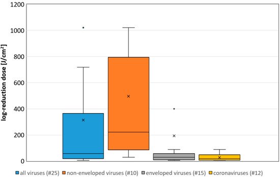
The median log-reduction doses of all virus experiments performed in liquids is 58 J/cm2. For the non-enveloped, enveloped and coronaviruses only, they are 222, 29 and 19 J/cm2, respectively (results on surfaces or at riboflavin concentrations above 1 mg/L were ignored). The differences between these groups is also illustrated in the boxplot in Figure 1.
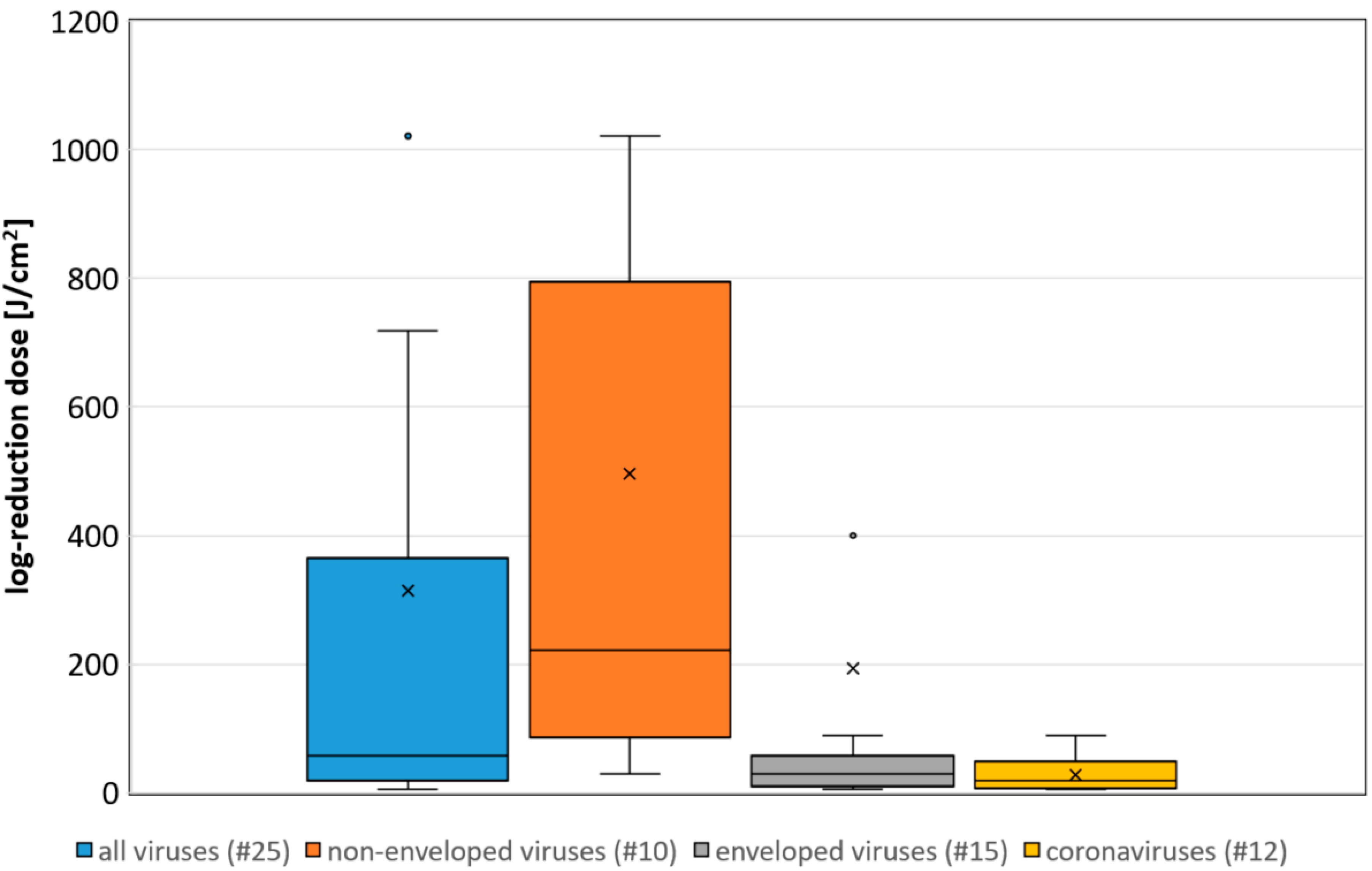
Figure 1. Boxplot of the determined necessary log-reduction dose for all, non-enveloped, enveloped and coronaviruses. Illustrated are the medians, and the quantiles of the result distribution. The number in brackets give the quantity of data sets for each virus category (not all outliers are depicted).
3. A correlation between the Assumed Riboflavin Concentration and the Sensitivity
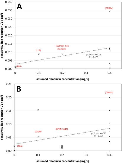
A correlation between the assumed riboflavin concentration (or the applied media) and the sensitivity (inverse of the log-reduction dose) is illustrated in Figure 2A,B for all non-enveloped and enveloped virus data, respectively. The results for both groups, with those of enveloped viruses including those for all coronaviruses, show that sensitivity increases with higher riboflavin concentration. However, this tendency appears to be less pronounced for non-enveloped viruses.
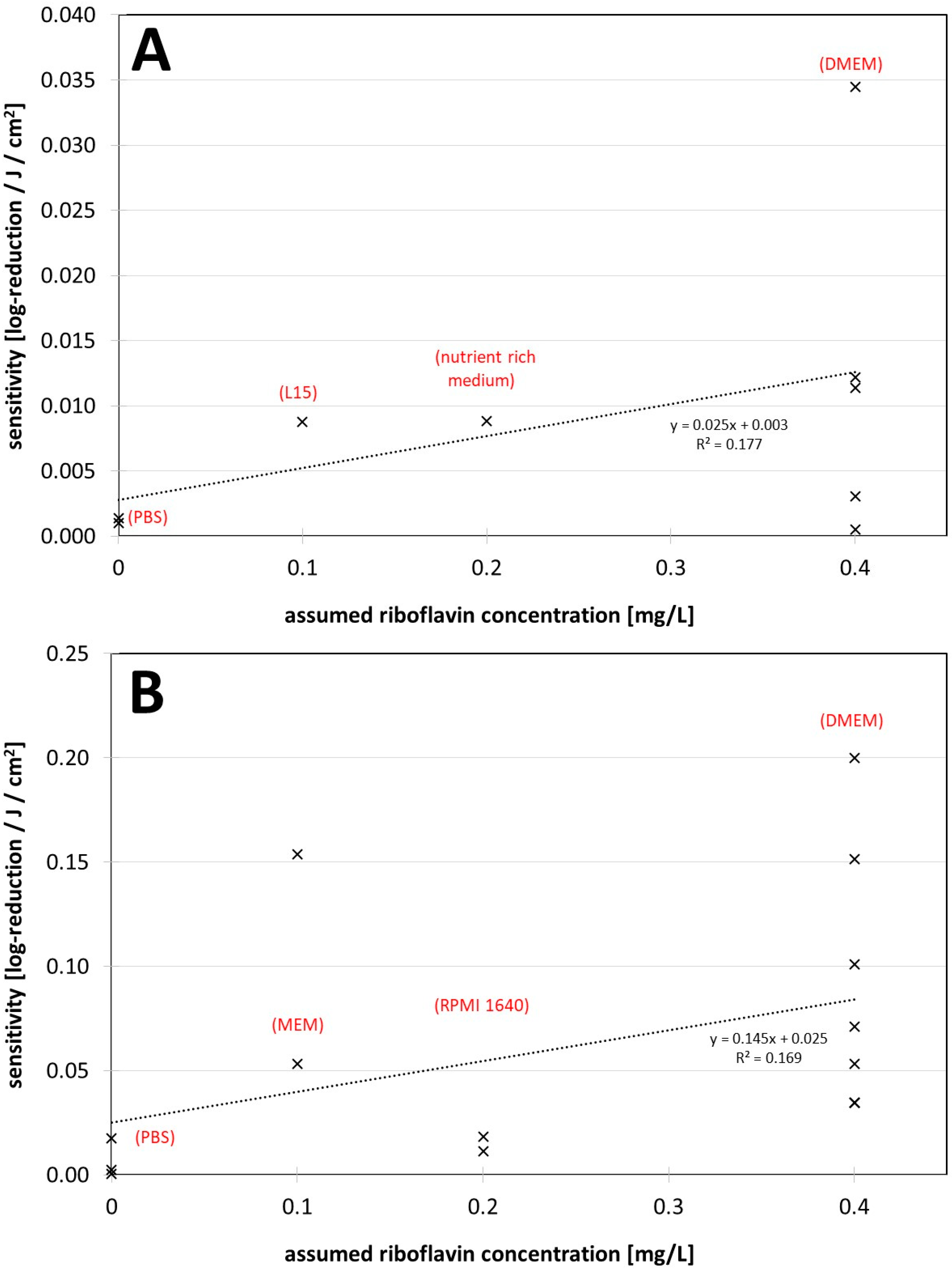
Figure 2. (A) Visible light sensitivity (inverse of the log-reduction dose) for viruses in different assumed riboflavin concentrations (media) for the non-enveloped viruses. (B) Visible light sensitivity for viruses in different assumed riboflavin concentrations (media) for enveloped viruses including coronaviruses. The dotted lines were derived by linear regression and are meant to visualize the dependence between riboflavin concentration (medium) and observed virus sensitivity.
4. The Findings regarding Virus Inactivation by Visible Light
The overall impression from Figure 1 is that, although there are large differences between the various viruses and experimental conditions, viruses can be inactivated by visible violet or blue light. Enveloped viruses such as coronaviruses appear to be particularly sensitive and non-enveloped less sensitive.
The median log-reduction doses are roughly in the same order of magnitude as previously observed for bacteria and fungi
[13][14][14,15], although it is as yet unclear whether the mechanisms or photosensitizers are comparable.
However, there is a major difference in how the virus experiments are conducted. Irradiations of bacterial and fungal solutions occur predominantly in media such as PBS, which do not contain photosensitizers. In particular, irradiation experiments with animal or human viruses usually take place in cell culture media with additives such as FCS or even phenol red. The latter may lead to a lower irradiance than expected due to optical absorption in the violet/blue spectral region and should always be considered as Tomb et al. and Biasin et al.
[33][34][39,51] did.
Another aspect may be even more critical. Many of the recent (corona-) virus investigations consider the observed photoinactivation to be an intrinsic property of the viruses studied. However, the applied media contain at least one photosensitizer, riboflavin, which can make an appreciable contribution to inactivation. Most of the new coronavirus studies do not discuss this possibility. Rathnasinghe et al. have mentioned a comparative study with a riboflavin solution but published no results
[35][42].
Riboflavin is known to act as a photosensitizer in combination with short wavelength visible light or with UV radiation. In photoinactivation experiments performed 50 years ago, it has already been found that the medium has an effect on virus reduction and that riboflavin concentrations, as found in common culture media, enhanced virus reduction
[36][37][38][39][40][44,47,49,62,63]. This has been reconfirmed in more recent studies by Tomb et al.
[33][41][39,40] and Kingsley
[42][43][43,45] and also fits the results of Grezlak et al.
[24][25] according to which irradiated media generate reactive oxygen species, for which riboflavin is mainly responsible.
The riboflavin concentrations in Figure 2 are only very roughly estimated and the data are sparse and scattered, but it already appears that irradiation experiments performed in DMEM with 0.4 mg/L riboflavin in the starting medium lead to higher photoinactivation sensitivities or smaller log-reduction doses compared with experiments in other media.
The question of the cause of the viral photosensitivity and the underlying mechanism of action remains unresolved. If exogenous photosensitizers in the medium, such as riboflavin, are responsible for the virus inactivation then it is a kind of photodynamic therapy (PDT), in which virus nucleotides, proteins, or the virus envelope are destroyed by reactive oxygen species
[44][45][67,68].
However, in saline solutions such as PBS, no external photosensitizers are expected and yet many authors have observed photoinactivation of viruses. This effect also still appears to be oxygen dependent
[40][46][63,65], which could be indicative of a mechanism based on endogenous photosensitizers and ROS generation. However, there are no known endogenous virus photosensitizers and no microbiological reason why viruses might require such photosensitizers.
Nonetheless, it has already been observed that typical PDT dyes can attach to viral envelopes
[47][61]. This allows the speculation that viruses might inadvertently carry photosensitizers from their host cells, which contain mainly porphyrins and flavins. This would explain the particularly efficient effect of violet or blue light on viruses—especially enveloped viruses—and for example, the relationship between the antiviral effects of 405 and 455 nm as observed by Vatter et al.
[48][59] for the phage phi6.
53. Conclusions
It appears that riboflavin, and also possibly other media components
[24][33][40][25,39,63] may have a major impact on photoinactivation results. DMEM especially might lead to higher virus sensitivities and should be used with caution in such experiments. Authors of future studies should consider this.
As long as it cannot be excluded that the medium has an influence on photoinactivation of (corona-) viruses, it should be mentioned in publications that virus reduction with visible light may be quite different under other conditions, such as in air. This is especially important in the ongoing corona pandemic, where frightened citizens seek protective measures and companies might offer deceptive security based on misunderstood studies.