You're using an outdated browser. Please upgrade to a modern browser for the best experience.
Mitochondrial Processing Peptidases: Comparison
Please note this is a comparison between Version 1 by Nina Kunová and Version 2 by Beatrix Zheng.

Mitochondrial proteins are encoded by both nuclear and mitochondrial DNA. While some of the essential subunits of the oxidative phosphorylation (OXPHOS) complexes responsible for cellular ATP production are synthesized directly in the mitochondria, most mitochondrial proteins are first translated in the cytosol and then imported into the organelle using a sophisticated transport system. These proteins are directed mainly by targeting presequences at their N-termini. These presequences need to be cleaved to allow the proper folding and assembly of the pre-proteins into functional protein complexes. In the mitochondria, the presequences are removed by several processing peptidases, including the mitochondrial processing peptidase (MPP), the inner membrane processing peptidase (IMP), the inter-membrane processing peptidase (MIP), and the mitochondrial rhomboid protease (Pcp1/PARL). Their proper functioning is essential for mitochondrial homeostasis as the disruption of any of them is lethal in yeast and severely impacts the lifespan and survival in humans.

  • mitochondrial processing peptidases
  • MPP
  • MIP
  • IMP
  • mitochondrial rhomboid protease
  • mitochondrial disease

1. Introduction

Mitochondria are vital components of all eukaryotes. They supply their cells with energy, maintain calcium homeostasis and biosynthesize heme and steroid molecules. They are also the main junction point of several metabolic and signaling pathways. Rather unsurprisingly, therefore, mitochondrial dysfunction is connected with an enormous range of diseases including myopathies (e.g., Kearns-Sayre syndrome), neurodegenerative diseases (e.g., Alzheimer’s disease (AD) and Parkinson’s disease (PD)), processes involved in ageing, and various types of cancers.
Generally, proper mitochondrial function depends on two sets of proteins. The smaller set is synthesized directly inside the mitochondria, while the larger set is synthesized on the cytosolic ribosomes. Cytosolically synthesized proteins have a presequence on their N-terminus that causes them to be imported into mitochondria using a complex transportation system [1][2][3][4][5][6][7][8][1,2,3,4,5,6,7,8]. After translation, cytosolic heat shock proteins chaperone the unfolded polypeptides to the outer mitochondrial membrane. Here, they are recognized by the receptors of the TOM (translocase of the outer membrane) complex and cross the outer membrane. In the intermembrane space, mitochondrial pre-proteins are immediately directed to the TIM23 (translocase of the inner membrane) complex for further translocation into the mitochondrial matrix (Figure 1) [9].
/media/item_content/202202/620c5e58d1b98ijms-23-01297-g001.png
Figure 1. Protein transport and processing from cytosol to mitochondria. MTS (mitochondrial targeting sequence)-carrying pre-proteins are imported through the TOM and TIM23 complexes. Proteins containing hydrophobic sorting signal are embedded into the inner membrane (IMM), while hydrophilic proteins are sent into the mitochondrial matrix through the PAM (protein import motor) complex. Cysteine-rich proteins are imported by the TOM and MIA protein translocation systems. The precursors of β-barrel proteins are translocated through the TOM and TIM9/10 complexes and sorted and assembled by the SAM complex. Metabolite carrier precursors are imported via TOM, TIM9/10 and TIM22, and several α-helical outer mitochondrial membrane (OMM) proteins are imported by the MIM complex. Pre-proteins imported into the mitochondria are processed by the mitochondrial processing peptidase (MPP) and later by the mitochondrial intermembrane peptidase (IMP) or mitochondrial intermediate peptidase (MIP). Proteins synthesized inside the mitochondria themselves are processed by IMP. Some inner membrane proteins are processed by rhomboid protease Pcp1/PARL. OMM, mitochondrial outer membrane; IMM, mitochondrial inner membrane; IMS, intermembrane space.
Protein transport and processing from cytosol to mitochondria. MTS (mitochondrial targeting sequence)-carrying pre-proteins are imported through the TOM and TIM23 complexes. Proteins containing hydrophobic sorting signal are embedded into the inner membrane (IMM), while hydrophilic proteins are sent into the mitochondrial matrix through the PAM (protein import motor) complex. Cysteine-rich proteins are imported by the TOM and MIA protein translocation systems. The precursors of β-barrel proteins are translocated through the TOM and TIM9/10 complexes and sorted and assembled by the SAM complex. Metabolite carrier precursors are imported via TOM, TIM9/10 and TIM22, and several α-helical outer mitochondrial membrane (OMM) proteins are imported by the MIM complex. Pre-proteins imported into the mitochondria are processed by the mitochondrial processing peptidase (MPP) and later by the mitochondrial intermembrane peptidase (IMP) or mitochondrial intermediate peptidase (MIP). Proteins synthesized inside the mitochondria themselves are processed by IMP. Some inner membrane proteins are processed by rhomboid protease Pcp1/PARL. OMM, mitochondrial outer membrane; IMM, mitochondrial inner membrane; IMS, intermembrane space.
Outer mitochondrial membrane proteins and inner mitochondrial membrane carrier proteins possess an N-terminal, C-terminal and/or internal signal sequences instead of a cleavable presequence [10]. Upon mitochondrial localization, the N-terminal presequence must be removed to avoid problems with further sorting and protein folding and assembly. For this task, specialized metalloproteinases, mitochondrial processing peptidases, have evolved.
The mitochondrial processing peptidase (MPP) plays an essential role in this process: MPP deletion was shown to be lethal [11][12][11,12]. MPP is responsible for processing the protein precursors that are fully translocated in the mitochondrial matrix as well as the precursors in transit to the inner membrane or inter-membrane space (Figure 1). Several inter-membrane proteins possess a so called bipartite presequence consisting of both a mitochondrial targeting sequence (MTS) cleaved by MPP and also of an intermembrane space-sorting signal that is subsequently eliminated by the inner membrane peptidase (IMP) residing in the inner mitochondrial membrane (Figure 1) [11].
In addition, some proteins possess a typical octapeptide (Phe/Leu/Ile-XX-Ser/Thr/Gly-XXXX) in their targeting presequence and after cleavage by MPP, are further processed by the mitochondrial intermediate peptidase (MIP) located in the mitochondrial matrix [11]. Moreover, a rhomboid-type serine protease, Pcp1 in yeast and PARL in humans, was also shown to act in the processing of several mitochondrial inner membrane proteins [13]. The peptidases involved in presequence cleavage are quite conserved in eukaryotes, including yeast and humans, which reflects their vital role in mitochondrial biogenesis. In humans, mutations in these peptidases were reported to have an impact on the development of several serious diseases including neuropsychiatric disorders [14][15][16][14,15,16], Friedreich’s ataxia (FRDA) [17], autosomal recessive spinocerebellar ataxia type 2 (SCAR2) [18], and type 2 diabetes (T2D) [19] (see Table 1 below).

2. Mitochondrial Processing Peptidase

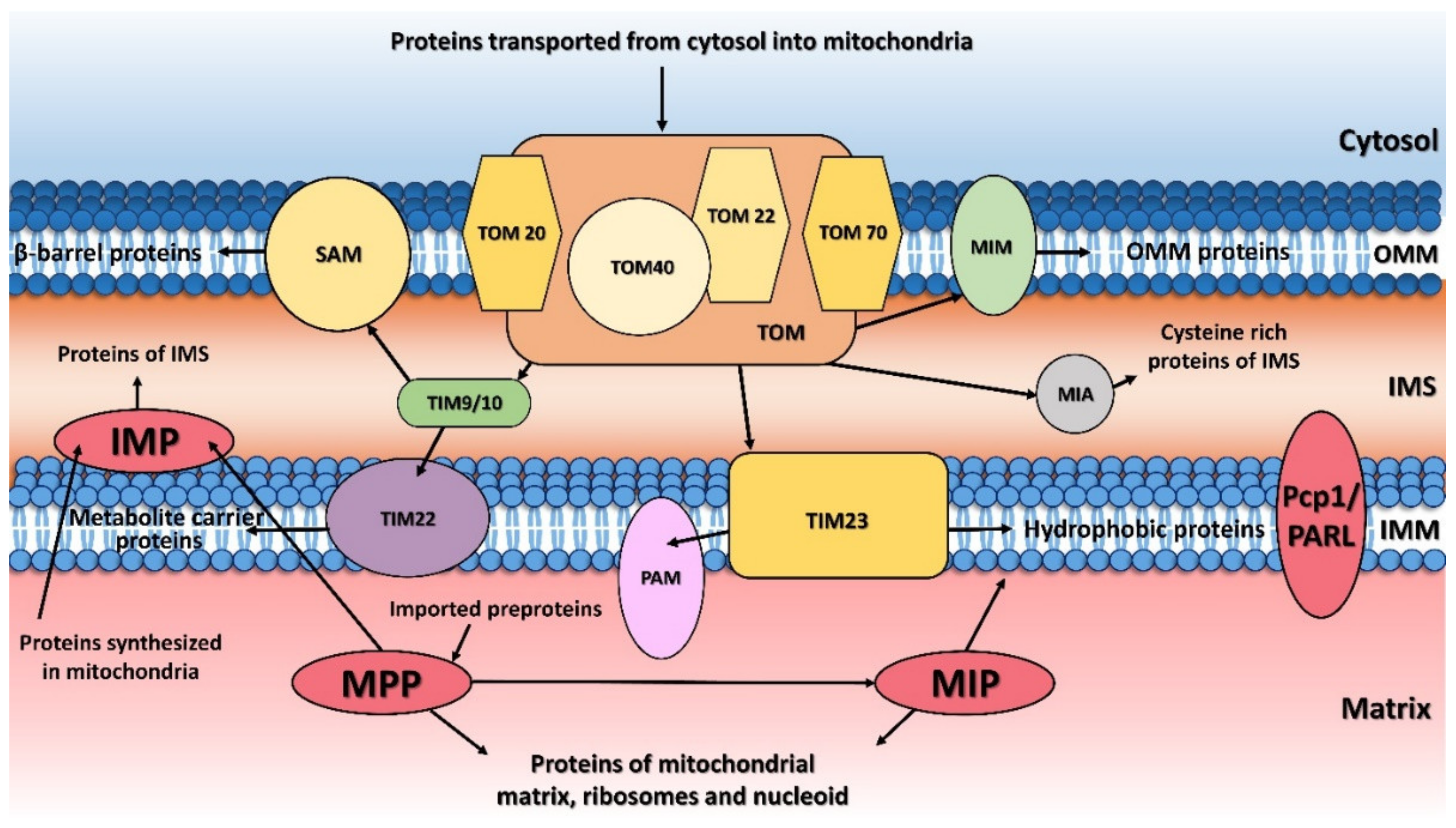
The principal responsibility of the mitochondrial processing peptidase is to remove the N-terminal targeting presequences of proteins imported into the mitochondria (Figure 1). Although the presequences can vary in length and amino-acid composition, they have several common properties. They are all predicted to form an amphiphilic α-helix [20][21][21,22], have an overall positive charge, and have an arginine residue at position -2 or -3 from the cleavage site [22][23].

MPP is a hetero-dimeric protein consisting of two subunits, α and β, which are referred to as PMPCA and PMPCB in humans [11][12][7,8]. These subunits together create a large substrate-binding cavity with a Zn2+-binding site on the MPPβ subunit. The site itself is created by a conserved HxxEHx76E motif in the MPPβ subunit; the mutation of any of these residues eliminates Zn2+ binding and blocks the peptidase activity. Although the β subunit contains the entirety of the catalytic site, the cooperation of action of both MPP subunits is required for proper processing of pre-proteins. The most conserved part of all known MPPα subunits is a glycine-rich loop (GRL; residues G284GGGSFSAGGPGKGMYS300 in yeast MPPα), which is essential for substrate binding [23][24][17,18] and which moves the precursor protein towards the active site through a multistep process [25][19]. An electrostatic analysis of MPP complexed to a peptide substrate showed that the binding cavity was strongly negatively-charged while the substrate peptide is positively charged.

Deletion of both MPP encoding genes (MPPA and MPPB) is incompatible with the viability of S. cerevisiae under any and all growth conditions, including even anaerobic growth [26][27][24,25]. In humans, mutations in either PMPCA or PMPCB cause mitochondrial diseases that are characterized by neurological disorder with an early childhood onset and a severe neurodegenerative course [28][29][30][26-28] (Table 1).

Table 1. The overview of human mitochondrial processing peptidase mutations and their involvement in human diseases.
Processing Peptidase Protein Variant Disease, Symptoms Ref.
PMPCA Homozygous mutation: c.1129G>A (p.Ala377Thr)

Heterozygous mutations: c.287C>T (p.Ser96Leu) with c.1543G>A (p.Gly515Arg)
SCAR2 with non- or slowly progressive cerebellar ataxia and developmental delay [31][38]
Homozygous mutation: c.766G>A (p.Val256Met) slowly progressive SCAR2 without intellectual disability [32][47]
Heterozygous mutation: c.677C>T (p.Arg223Cys) with c.853del (p.Asp285Ilefs*16) SCAR2 with progressive cerebellar ataxia and onset in infancy [18]
Heterozygous mutations: c.1066G>A (p.Gly356Ser) with c.1129G>A (p.Ala377Thr) SCAR2 with progressive, extensive brain atrophy, muscle weakness, visual impairment, respiratory defects [33][48]
Homozygous mutation: c.553C>T (p.Arg185Thr) SCAR2 with psychomotor delay [34][46]
PMPCB Heterozygous mutations: c.523C>T (p.Arg175Cys) with c.601G>C (p.Ala201Pro); c.524G>A (p.Arg175His) with c.530T>G (p.Val177Gly)

Homozygous mutation: c.1265T>C (p.Ile422Thr)
Prominent cerebellar atrophy in early childhood [35][50]
IMMP2L Duplication: 46,XY,dup(7)(q22.1-q31.1) GTS/TS [15]
Deletions ranged from ~49 kb to ~337 kb Neurological disorders (ADHD, GTS/TS, OCD, ASD, Asperger′s syndrome, schizophrenia and developmental delay) [36][37][38][39][53,54,55,56]
Base pair change Autism [40][57]
Copy number variation Alzheimer′s disease [41][58]
Downregulation Prostate cancer [42][59]
MIP Homozygous SNV: p.K343E

Heterozygous SNVs: p.L582R with p.L71Q; p.E602* with p.L306 and p.H512D with 1.4-Mb deletion of 13q12.12
LVNC and developmental delay, seizures, hypotonia [43][60]
Heterozygous mutation: c.916C > T (p.Leu306Phe) with c.1970 + 2 T>A (p.Ala658Lysfs*38) Developmental delay, hypotonia and intellectual disability [44][61]
Hypomethylation Metabolic syndrome [45][62]
Downregulation Prostate cancer [42][59]
PARL Reduced levels Type 2 diabetes [19]
Leu262Val polymorphism Increased plasma insulin concentration [46][63]
Mutation: c.230G>A (p.Ser77Asn) Parkinson′s disease [47][64]

3. Mitochondrial Inner Membrane Peptidase

The mitochondrial inner membrane peptidase is responsible for the maturation of proteins transported into the mitochondrial inter-membrane space (Figure 1) [48][49][50][51][65–68]. These include mature proteins synthesized both within the mitochondria (e.g., yeast mitochondrially encoded subunit 2 of cytochrome c oxidase, Cox2), or nuclear-encoded proteins synthesized in the cytosol and then transported into the mitochondria (e.g., yeast cytochrome b2, Cyb2, cytochrome c1, Cyt1, and NADH cytochrome b5 reductase, Mcr1).

Structurally, IMP consists of two subunits; in humans, these are IMMP1L (inner membrane mitochondrial peptidase 1-like) and IMMP2L (inner membrane mitochondrial peptidase 2-like) [49][66], and in S. cerevisiae there are three subunits, Imp1, Imp2, and Som1 [50][67]. Although the sequence identities between the individual yeast and human IMP homologues are relatively low (between 25-37%), their tertiary structures share a number of common features. All four IMP homologues are predicted to have a membrane-anchored α-helical N-terminal domain and a catalytic C-terminal domain. The yeast Imp1 and Imp2 subunits share 31% amino-acid sequence identity and both possess catalytic activity and are bound to the inner mitochondrial membrane [52][53][54][70–72]. The catalytic domain possesses a catalytic Ser/Lys dyad, which is present in all four proteins and is structurally located in the C-terminal region [51][55][68,73]. The third yeast subunit, Som1, most likely serves to recognize substrates and was shown to physically interact with Imp1 [51][56][68,74]. Surprisingly, Som1 seems to be important for the Imp1-mediated proteolytic processing of Cox2 and Mcr1, but not for the maturation of the Cyb2 and Cyt1 cytochromes processed by the Imp2 subunit [51][56][68,74].

The currently known natural Imp1 substrates all possess a characteristic [I/V][H/D/F/M][N](↓)[D/E] amino-acid motif surrounding the cleavage site (indicated by ↓) [67]. Although the substrate specificities for Imp1 and Imp2 do not overlap, there are recognizable similarities between the protein precursors that they cleave. These include a hydrophobic residue at position -3 from the cleavage site and, for the nucleus-encoded substrates, the distances between the transmembrane segment and the cleavage site are also preserved. The accessibility of the cleavage site to the peptidase is also a prerequisite for cleavage by IMP [50][67].

In humans, the IMP homolog, IMMP2L has a 41% similarity to the yeast Imp2 subunit and a 90% similarity to the mouse IMMP2L [49][66]. It is composed of 175 amino acids with a gene of 860 kb located on chromosome 7q (AUTS1 locus), whose integrity has been shown to be critical for the development of autism spectrum disorders (ASDs). IMMP2L is expressed at a basal level in all human tissues except for the lungs and liver of adults [15][36][15,53]. Mutations associated with the gene encoding IMMP2L have been observed in several neurodegenerative diseases, including Gills de la Tourette syndrome or Tourette′s syndrome (GTS/TS), attention-deficit hyperactivity disorder (ADHD), ASD, and schizophrenia [36][37][39][57][58][53,54,56,75,76] (see Table 1 above).

4. Mitochondrial Intermediate Peptidase

The mitochondrial intermediate peptidase is important for the maturation of a subgroup of precursor proteins imported into the mitochondrial matrix or embedded into the mitochondrial inner membrane [59][43]. These pre-proteins are first processed by MPP and only afterwards by MIP, which cleaves an additional octapeptide following MPP cleavage. The cleavage site targeted by MIP is characterized by an RX(↓)(F/L/I)XX(T/S/G)XXXX(↓) motif [48][65] and is located at the C-terminus of a leader peptide (↓). Active MIP is a soluble monomer of 75 kDa in yeast and 81 kDa in humans. Its proteolytic activity is stimulated by manganese, magnesium and calcium ions while 1 mM Co2+, Fe2+ or Zn2+ completely inhibits it. Unlike MPP, MIP is also sensitive to N-ethylmaleimide (NEM) and other sulfhydryl reagents [60][88].

Positioning at the substrate N-terminus and a large hydrophobic residue (phenylalanine, leucine and isoleucine) at position -8 from the cleavage site are both essential features for cleavage by MIP; this type of substrate specificity is not shared by any other known peptidase [48][65].

In S. cerevisiae, mitochondrial oxidative phosphorylation is severely affected when mip1 is missing. Branda et al. [48][65] showed that at least three vital components of the yeast mitochondrial gene expression machinery—mitochondrial small ribosomal subunit protein MrpS28, single-stranded DNA-binding protein Rim1, and elongation factor Tuf1—are processed by MIP. These proteins are essential for maintaining mitochondrial protein synthesis and mitochondrial DNA replication, which explains why the loss of mip1 impairs the mitochondrially encoded OXPHOS subunits. MIP1 disruption also results in the failure of at least two yeast nuclear-encoded respiratory chain components, the cytochrome c oxidase subunit 4 (Cox4) and the Rieske iron-sulfur protein of cytochrome bc1 catalytic subunit, to be cleaved [59][43].

In humans, MIP is encoded by the MIPEP gene, which contains 19 exons and is located on chromosome 13q12.12 [61][89]. MIPEP is expressed at high levels in energy-dependent tissues, such as the heart, brain, skeletal muscles, and pancreas [61][62][63][89–91]. Previously, some patients were reported with mutations in MIPEP which may have been linked to their diagnoses, but the first study showing that MIPEP is truly involved in a human disease was published in 2016 by Eldomery et al. [43][60] (see Table 1 above). They identified several single nucleotide variants (SNVs) in the MIPEP gene that caused a loss of MIP function in four unrelated patients suffering from an oxidative phosphorylation deficiency. Further studies in human fibroblasts showed that MIP has an important role in OXPHOS function since its loss impaired the processing of several OXPHOS subunits, including the OXPHOS complexes I, IV and V [44][61].

5. Mitochondrial Rhomboid Protease

The mitochondrial rhomboid protease—Pcp1 (processing of cytochrome c peroxidase protein 1) in yeast and PARL (presenilin-associated rhomboid-like) in humans—plays an essential role in mitochondrial quality control and steady-state maintenance [13][64][65][13,95,96]. Pcp1/PARL is a member of the rhomboid family of intramembrane serine proteases, which have a core consisting of transmembrane helices. Preliminary cellular localization studies demonstrated that the N-terminal part of PARL is localized in the mitochondrial matrix, while its C-terminus extends into the mitochondrial intermembrane space. Conserved serine and histidine catalytic residues (Ser256/His313 in Pcp1, and Ser277/His335 in PARL) are responsible for its intramembrane proteolytic activity [66][97].
While there is no significant conservation in the N-terminal regions of yeast Pcp1 and human PARL, this region is strongly conserved among vertebrates, especially in mammals [67][98]. The N-terminal domain of PARL is processed by two cleavages [68][99]. The first is mediated by MPP, which removes the mitochondrial targeting sequence, while the second is developmentally regulated and depends on PARL’s own intramembrane-cleaving protease activity. The PARLΔ53 truncated form (without its MTS) is considered to be its mature form and is predominantly present in lung, brain, heart and muscle tissues [66][97]. The second cleavage occurs at Ser77 and releases the so-called Pβ peptide, whose sequence is conserved only in mammals. This Pβ peptide is exported into the nucleus and contributes to mammalian-specific, developmentally regulated signaling between the mitochondria and nucleus [68][99]. Phosphorylation of PARL has also been proposed to play a key role in regulating its activity. Ser65, Thr69, and Ser70 have all been identified as possible PARL phosphorylation sites: phosphomimetic mutations at these sites dramatically reduced Pβ-cleavage and the PARLΔ77 production [69][100]. As a result of that, phosphorylation increases PARLΔ53 levels, which enhances its activity towards substrates [70][101].
The C-terminal parts of Pcp1 and PARL have higher sequence identity, where several transmembrane α-helices are located. Six of them are present in both Pcp1 and PARL, while PARL has an additional transmembrane α-helix. Both proteins possess endopeptidase activity and their catalytic dyads, Ser256 and His313 in Pcp1 and Ser277 and His335 in PARL, are highly conserved and both occur in the transmembrane helices in their respective proteases.
Yeast Pcp1 is known to process two mitochondrial substrates, cytochrome c peroxidase (Ccp1), and mitochondrial genome maintenance protein 1 (Mgm1), a mitochondrial outer membrane dynamin-like GTPase [71][102]. A Pcp1-deficient yeast strain was unable to grow in a glycerol medium and displayed striking mitochondrial morphological abnormalities, such as mitochondrial fragmentation, aggregation, and loss of mitochondrial DNA [72][105].
Mammalian PARL is responsible for the processing of several mitochondrial substrates, e.g., the serine protease HTRA2 (high-temperature requirement factor A2), the serine/threonine-protein kinase PINK1, the serine/threonine-protein phosphatase PGAM5 (phosphoglycerate mutase family member 5), and OPA1 (optic atrophy protein 1), the human homolog of yeast Mgm1 [13]. Recent proteomic analyses identified several additional PARL substrates including a subunit of mitochondrial respiratory chain complex III, TTC19 (tetratricopeptide repeat domain 19), the pro-apoptotic protein Smac (second mitochondrial-derived activator of caspases), the mitochondrial lipid transferase STARD7 (StAR-related lipid transfer protein 7), and CLPB (caseinolytic peptidase B), a putative mitochondrial chaperone. The identification of these substrates further supports the role of PARL in mitochondrial homeostasis [70][73][74][75][101,115,116,117].
In humans, reduced PARL levels have been connected with type 2 diabetes (T2D) and ageing (see Table 1 above). Civitarese et al. [19] showed that lowering PARL levels in human muscle cells resulted in lower mitochondrial oxidative capacity, reduced mitochondrial mass, increased protein oxidation and ROS production and impaired insulin signaling, all of which are known metabolic defects in T2D and ageing. Moreover, the presence of a Leu262Val polymorphism in PARL was associated with increased plasma insulin concentration, making it a possible risk factor for diabetes. Interestingly, two other groups [46][76][63,120] were unable to confirm this observation, though one of them, Hatunic et al. [46][63] did find that the Leu262Val PARL genetic variation is associated with an earlier onset of diabetes and may be a marker of increased susceptibility to nephropathy and cardiovascular complications in patients with diabetes.
An additional PARL missense mutation, Ser77Asn, was detected in two patients diagnosed with Parkinson’s disease, which also implicates PARL dysregulation in PD pathogenesis [47][64] (see Table 1 above). This mutation is located in the highly conserved N-terminus of the protein, in a position that is crucial for the second maturation of PARL and the release of the Pβ peptide (see above). Unfortunately, further independent studies failed to detect this mutation or any other pathogenic mutation in the PARL gene. This suggests that if PARL mutations are a genetic cause of Parkinson′s disease, then they are likely to be extremely rare [77][78][121,122].

6. Conclusions

Mitochondria are semiautonomous organelles that use a sophisticated system for importing pre-proteins synthetized in the cytosol and directing them to their final destination within mitochondria. This system is necessary for maintaining proper mitochondrial function and homeostasis. Mitochondrial processing peptidases are important components of the mitochondrial import machinery; their responsibility lies in removing the signal presequences of peptides and pre-proteins transported into the mitochondria. Most of these pre-proteins are processed by the mitochondrial processing peptidase, MPP, while other processing peptidases, including the mitochondrial inner membrane peptidase, IMP, the mitochondrial intermediate peptidase, MIP, and the mitochondrial rhomboid proteases, Pcp1/PARL, are responsible for the further processing of specific protein substrates. Disruption of any of these proteases is lethal, and their malfunctions are connected with a number of severe human diseases. These diseases are characterized by an early onset and patients often exhibit neurodegeneration. The short peptides produced by these proteases might also accumulate inside the organelle, causing further problems; consequently, functional coupling between the processing of precursor proteins and presequence degradation is crucial for maintaining a functional organellar proteome [79][123]. The mitochondria eliminate these peptides in two ways. Either the free presequences can be exported into the cytosol, like the Pβ peptide of PARL, which is further translocated into the nucleus where it associates with chromatin and contributes to developmentally regulated mitochondria-to-nuclei signaling, or, like the majority, they can be degraded by a peptidase to generate amino acids that can be used for the synthesis of new proteins within the mitochondria. In humans, the mutation of these peptidases may also lead to severe diseases.
Academic Video Service