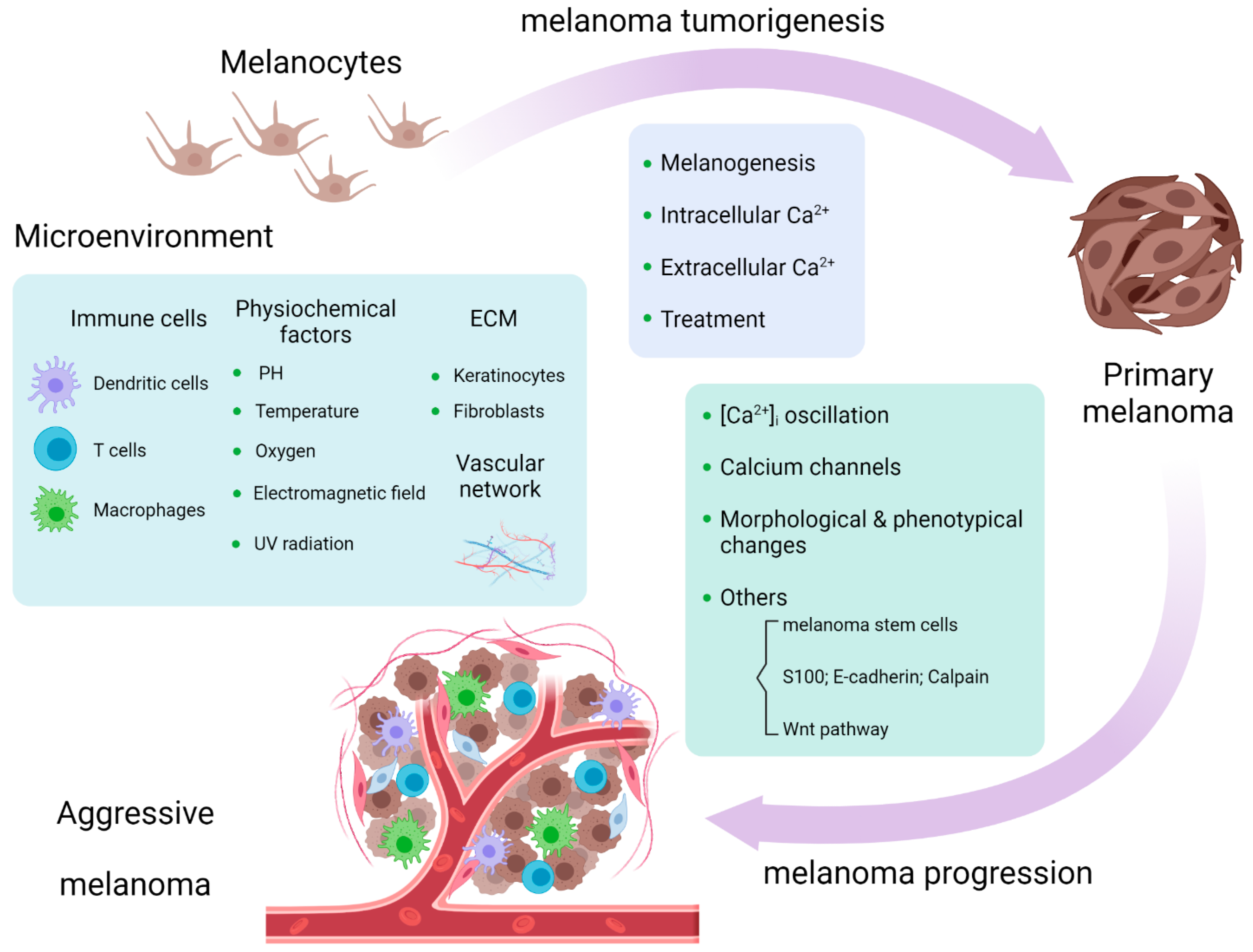
Calcium Signaling in Melanoma: Comparison
Please note this is a comparison between Version 1 by Haoran Zhang and Version 2 by Beatrix Zheng.

Calcium signaling plays important roles in physiological and pathological conditions, including cutaneous melanoma, the most lethal type of skin cancer. Intracellular calcium concentration ([Ca2+]i), cell membrane calcium channels, calcium related proteins (S100 family, E-cadherin, and calpain), and Wnt/Ca2+ pathways are related to melanogenesis and melanoma tumorigenesis and progression. Calcium signaling influences the melanoma microenvironment, including immune cells, extracellular matrix (ECM), the vascular network, and chemical and physical surroundings. Other ionic channels, such as sodium and potassium channels, are engaged in calcium-mediated pathways in melanoma. Calcium signaling serves as a promising pharmacological target in melanoma treatment, and its dysregulation might serve as a marker for melanoma prediction. 

  • calcium
  • melanoma
  • progression
  • melanoma microenvironment
  • mitochondria

1. Calcium Signaling in Melanoma Progression

Melanoma progression happens when the primary melanoma progresses to a metastatic melanoma with a migrating and invading capacity. Intracellular calcium concentration ([Ca2+]i) and its multiple channels function as regulators of melanoma progression that serve as mechanistic targets for control of melanoma growth and management of metastasis.

1.1. [Ca

2+

]i Oscillation Influences Melanoma Progression

Evidence documents that increased intracellular calcium stores are associated with highly metastatic melanoma cells [1][28]. Calcium released from ER facilitates melanoma cell migration. Epac1 activated by cAMP induces calcium elevation from ER via the PLC/IP3 receptor pathway and facilitates cell migration with the involvement of actin assembly, which is inhibited by mSIRK, a Gβγ-activating peptide, activating calcium influx from the extracellular space [2][3][29,30]. The expression of cGMP phosphodiesterase PDE5A is downregulated by oncogenic BRAF in BRAFV600E mutated melanoma by the extracellular-signal-regulated kinase (ERK) pathway, which induces an increase in [Ca2+]i, stimulating melanoma cell invasion and short-term and long-term lung colonization [4][31]. Y-box binding protein 1 is an unfavorable prognostic marker secreted from melanoma depending on [Ca2+]i and ATP levels, the expression of which increases in primary and metastatic melanoma, compared to benign melanocytic nevi. Conversely, elevated Y-box binding protein 1 secretion stimulates melanoma cell migration, invasion, and tumorigenicity [5][32]. Paradoxically, increased [Ca2+]i was reported to decrease melanoma progression. Olfactory receptor 51E2 activated by its ligand β-ionone suppresses the migration of vertical-growth phase melanoma cells by increasing [Ca2+]i [6][33].

1.2. Calcium Channels Are Involved in Melanoma Progression

Since [Ca2+]i plays an important mechanistic role in melanoma progression, the role of calcium channels cannot be neglected. Basically, NMDAR calcium channel function is weak in melanoma cells but strongly contributes to cell proliferation and invasion when its encoding gene GRIN2A is mutated at certain sites, such as G762E, with less glutamate supplementation [7][34]. Another glutamate receptor calcium channel mGluR5 was proved to have a profound effect on melanoma progression in vivo by triggering the phosphorylation of ERK [8][5]. The ERK pathway is also implicated in SOCE-mediated melanoma progression. Inhibition of SOCE by knockdown of STIM1 or Orai or by SOCE inhibitors suppresses melanoma cell proliferation and migration, while induction of SOCE activates ERK, which is inhibited by calmodulin kinase II or Raf-1 inhibitors [9][35]. TPC2 influences melanoma progression via SOCE. Downregulation of TPC2 expression in metastatic melanoma leads to a decrease of Orai1 expression and an increase of YAP/TAZ activity, which is responsible for melanoma’s aggressive property [10][36]. In BRAF mutant melanoma—the BRAFV600E mutation in particular—the expression of Ca2+-ATPase isoform 4b (PMCA4b) on the plasma membrane is low compared with benign nevi and is markedly elevated by vemurafenib (BRAF inhibitor) or selumetinib (MEK inhibitor) treatment, which indicates crosstalk between PMCA4b and the MAPK pathway. Activation of p38 MAPK induces the degradation of PMCA4b, while suppression of p38 MAPK by increasing the abundance of PMCA4b promotes the [Ca2+]i clearance and inhibits the migration of melanoma cells [11][12][37,38]. Moreover, SERCA on the ER membrane, controlled by the interaction between calcium-modulating cyclophilin ligand and basigin, was reported to have an effect on invasion and metastasis by regulating [Ca2+]i and matrix metalloproteinase (MMP)-9 activity in A375 cells [13][39]. Unlike Ca2+-ATPase, T-type VDCCs drive migration and invasion in BRAF mutant melanoma cells depending on Snail1 levels, suggesting therapeutic strategies by blocking T-type VDCCs to inhibit progression of melanoma [14][40]. Other ion channels are implicated in melanoma progression through calcium signaling. Nav1.6 sodium channel promotes melanoma cell (WM266 and WM115) invasion and proliferation by mTOR-mediated Na+/Ca2+ exchange [15][41]. KCa3.1 potassium channel was reported to promote melanoma cell migration by controlling the secretion of melanoma inhibitory activity proteins depending on [Ca2+]i [16][42].

1.3. Ca

2+

Signaling Influences Melanoma Progression through the Change of Morphological and Phenotypical Changes

Ca2+ signaling also leads to cell morphological and phenotypical changes, including the elongated cell axonal- and mesenchymal-like shape, formulation of invadopodia, and altered cytoskeleton structure, making cancer cells become more deformable and more invasive. Except for the role in melanogenesis, synaptotagmin-4 is thought to have a relationship with the growth and metastasis of melanoma by influencing axonal elongation [17][43]. Orai- and STIM1-mediated Ca2+ oscillation signals were reported to facilitate invadopodium assembly and thus promote melanoma invasion by regulating the recycling of membrane-bound MT1-MMP and extracellular matrix (ECM) remodeling [18][19][18,44]. The effect of the β2-adrenergic–Ca2+–actin axis on cancer invasion was reported in melanoma and other cancer types. β-adrenergic receptor (βAR) signaling triggers actin remodeling and reorganization to enhance cell contractility and promote cell invasion. β-adrenergic receptor-induced Ca2+ acts as a regulator of cytoskeletal actin by directly binding to actin or binding to filamin, the crosslinker of actin [20][45]. Meghnani et al. reported the upregulated expression of receptor for advanced glycation end products (RAGE) in melanoma patients in late metastatic stages. Overexpression of RAGE induced melanoma cells to become more metastatic by triggering cells into mesenchymal-like morphologies, which is associated with the upregulation of its ligand S100B, a calcium-binding protein [21][46].

1.4. Calcium-Related Pathways Participate in Melanoma Progression

Other factors (melanoma stem cells), other proteins (S100 family, E-cadherin, and calpain), and the Wnt/Ca2+ pathway influence melanoma progression through calcium signaling. Ca2+ released through IP3R in melanoma cells is crucial for the function of cancer stem cells. IP3R impairment leads to a diminution in the population of melanoma stem cells and reduced melanoma growth [22][47]. A network analysis of the expression of Ca2+ signaling and stem cell pluripotency-related genes (e.g., GSTP1, SMAD4, CTNNB1, MAPK3, GNAQ, PPP1CC, GSK3B, and PRKACA) showed some candidates that may contribute to the melanoma metastatic transformation and potential therapeutic biomarkers for metastatic melanoma [23][48].
S100A4 is a metastasis-promoting protein in melanoma cells which acts by targeting metabolic reprogramming, that is, the suppression of mitochondrial respiration and the activation of aerobic glycolysis [24][49]. Upregulation of S100P, ezrin, and RAGE improves the malignancy of melanoma [25][50]. E-cadherin has extracellular Ca2+-binding domains whose functions are dependent on Ca2+ and is essential for melanogenesis and melanoma suppression. E-cadherin silencing is related to melanoma metastatic dissemination and poor prognosis [26][27][51,52]. The decreasing expression of E-cadherin by overexpression of T-box transcription factors Tbx2 and Tbx3 is associated with enhanced melanoma invasiveness [28][53]. Promoter methylation by activating E-cadherin expression represents its therapeutic role in the treatment of melanoma [26][51]. Evidence in vitro and in vivo showed that inhibition of calpain, whose activity is promoted by calcium signaling, blunts melanoma growth, allows melanoma cells to escape from anti-tumor immunity, and increases metastatic dissemination by accelerating the migration process and reducing apoptosis [29][54].
Wnt5a was found to be expressed in highly aggressive melanoma and was able to increase melanoma invasive potential by activating PKC and raising [Ca2+]i in a transfected model [30][55]. Interestingly, Wnt5a signaling was engaged into melanoma cell movement, rendering them more aggressive. Wnt5a leads to the remodeling of the cytoskeleton and increases melanoma motility by activating calpain-1, leading to the cleavage of filamin A [31][56]. The assembly of the “Wnt-receptor-actin-myosin-polarity” structure, which is promoted by Wnt5a, promotes actomyosin contractility and substrate detachment for membrane retraction, mediated by the recruitment of cortical ER and elevation of Ca2+ [32][57]. (Figure 12).
/media/item_content/202202/62046a121e497ijms-23-01010-g002.png
Figure 12. Calcium signaling is involved in melanoma tumorigenesis and progression and melanoma microenvironment [33].
Calcium signaling is involved in melanoma tumorigenesis and progression and melanoma microenvironment [58].

2. Calcium Signaling in Melanoma Microenvironment

The tumor microenvironment, including surrounding immune cells and other cells, signaling molecules, blood vessels, and ECM, is closely related to and constantly interactive with melanoma cells, playing pivotal roles in melanoma generation, progress, and prognosis. Calcium signaling influences the altered microenvironment to change the fate of the melanoma by influencing the function of innate and adaptive immune cells, regulating ECM and tumor vascularization, and adapting to different physical and chemical surroundings.

2.1. Immune Cells

In T cell-based tumor immunosurveillance, cytotoxic T lymphocytes (CTLs) kill tumor cells by recognizing their specific T cell receptor. It was proved that CTLs-mediated cytotoxic function in melanoma and other cancers depends on a SOCE-mediated [Ca2+]I rise by regulating the degranulation of CTLs, the production of TNFα and IFNγ, and the expression of Fas ligand both in vivo and in vitro [34][59]. CD4+CD25+Foxp3+ regulatory T cells cause effector T cell death and suppress activation of T cells to induce immunosuppression through TGFβ-induced inhibition of IP3 production with a decrease in intracellular Ca2+ flux. Accordingly, Kim et al. increased IFNγ production and activated T cells in vitro and reduced melanoma growth in vivo through highly selective optical control of Ca2+ signaling in CTLs [35][60]. EGR4, a member of the zinc finger transcription factor family, was reported as a key regulator of T cell differentiation. Knocking out EGR4 in T cells triggers an enhanced Ca2+ response and increased IFNγ production in vitro and leads to regulatory T cells loss, Th1 bias, and CTL generation in a mouse melanoma lung colonization model [36][61]. Histamine and its H4 receptor induce the chemotaxis and migratory properties of γδ T cells through Gi protein-dependent [Ca2+]i increase in the microenvironment of melanoma cells [37][62].
Moreover, Ca2+ flux was involved in the NK cell-mediated innate immune response to melanoma cells. Although no difference in the formation of metastatic lung lesions was observed, NK cells are hyporesponsive to MHC class I-deficient target cells, with NK cells continuously activating by the Ly49H receptor [38][63]. Tumor-associated macrophages, especially CD163+ M2 macrophages, are related to immune escape, supporting cancer development [39][64]. Secreted flavoprotein renalase enhances the function of M2 macrophages to promote melanoma growth through the PMCA4b calcium channel by activating the MAPK and PI3K/AKT pathways [40][65]. Recently, mesencephalic astrocyte-derived neurotrophic factor, a novel immunoregulator basically secreted from pancreatic beta cells, was found to be secreted from melanoma and other cancer cell lines upon IFNγ-induced ER calcium depletion, which was proved to activate M2 macrophages and promote melanoma growth [41][42][66,67]. In addition, macrophages in the melanoma microenvironment are less susceptible to calcium electroporation compared with melanoma cells, but calcium electroporation stimulates the immunogenic capacity of melanoma-conditioned macrophages [43][68]. Calcium electroporation is a promising method in anti-cancer treatment under clinical trial which utilizes high-voltage electric pulses to introduce calcium flux into cells [44][69]. Recently, a near-infrared-stimulable optogenetic platform was established to remotely and selectively control Ca2+ oscillations and Ca2+-related gene expression and to modulate immunoinflammatory responses by regulating the functions of T lymphocytes, macrophages, and dendritic cells [45][70]. What is more, bone marrow-derived mast cells prefer to locate in hypoxic zones of the melanoma microenvironment, inducing CCL-2 synthesis and calcium rise by activating LVDCCs [46][71].

2.2. ECM and Vascular Network

In melanoma, ECM, molecules, proteins, and stromal cells interacting with Ca2+ signaling influence melanoma development. As we discussed above, Orai1- and STIM1-mediated Ca2+ oscillations regulate melanoma ECM degradation by MT1-MMP [18][19][18,44]. Attenuated [Ca2+]i enhances the chemotaxis of melanoma cells to type IV collagen, a member of the ECM proteins, depending on CD47 and integrins α2β1 and ανβ3 [47][48][72,73]. Thrombomodulin, an integral membrane glycoprotein on endothelial cells, acts as a Ca2+-dependent molecule controlling melanoma cell adhesion [49][74]. Kallikrein-related peptidase 6 is detected in neighboring stromal cells and keratinocytes and displays a paracrine function to accelerate melanoma migration and invasion which was proved to depend on protease-activated receptor 1-induced intracellular Ca2+ flux [50][24]. Skin keratinocytes and fibroblasts in melanoma ECM play important roles in melanoma development. Keratinocytes reduce the expression of TRPC1, 3, and 6 to decrease [Ca2+]i and negatively regulate the N-cadherin levels, a progressive factor in melanoma cells [51][75]. Keratinocytes can lead to cutaneous malignant lesions, dependent on the loss of calcium channel P2X1–3 and P2Y2 receptors and E-cadherin [52][76]. N-cadherin can promote melanoma cell migration and metastasis by facilitating the adhesion of melanoma cells to dermal fibroblasts and vascular endothelial cells [53][77].
The vascular network in the melanoma microenvironment, tightly interacting with ECM, provides nutrients and advantageous conditions for proliferation and metastasis. As we discussed above, the positive effects of Wnt5a on melanoma metastasis also include Ca2+-dependent exosome release, containing the pro-angiogenic and immunosuppressive factors (VEGF, IL-6, and MMP-2), which suppresses endothelial cell branching. Wnt5a expression has a potential relationship with the angiogenesis marker ESAM [54][78]. Nicotinic acid adenine dinucleotide phosphate, which is capable of triggering Ca2+ release from endosomes and lysosomes by targeting TPCs, was reported to control VEGF-induced angiogenesis in melanoma cells [55][79]. Moreover, vasculogenic mimicry is specific in less vascularized areas of the tumor microenvironment, providing nutrients and oxygen to facilitate tumor metastasis. Zhang et al. reported the role of the calcium/phospholipid-binding protein myoferlin in the inhibition of vasculogenic mimicry formation in melanoma by inducing mesenchymal-to-epithelial transition and decreasing MMP-2 expression [56][80]. The reconstitution of vascular mimicry with the combination of VEGFA signaling in ECM contributes to the formation of capillary-like structures in the melanoma microenvironment which is regulated by intracellular and extracellular Ca2+ levels and ανβ3 and ανβ5 integrins [57][81]. Studies displayed some anti-vascular methods in anti-tumor treatment by targeting Ca2+ signaling. Carboxyamido-triazole, an inhibitor of non-VDCCs, displayed inhibitory effects on melanoma invasion and angiogenesis, disrupting the signaling between melanoma and its microenvironment by suppressing VEGF production and endothelial cell response to VEGF [58][82]. Calcium electroporation not only directly induced melanoma necrosis and indirectly affected macrophages in the melanoma microenvironment but recently was found to suppress the formation of capillary-like structures in vitro and damage melanoma blood vessels in vivo [59][60][83,84]. Particularly, vascular endothelial cadherin is basically specific to endothelia but also presented in some melanomas [61][85]. Vascular endothelial cadherin-mediated interaction between melanoma and adjacent endothelium plays an important role in tumor metastasis properties. Inhibition of the PLC/IP3 pathway disrupts the melanoma–endothelium junctions by diminishing endothelial [Ca2+]i response [62][63][86,87].

2.3. Physical and Chemical Surroundings

The extracellular pH in melanoma is acidic because of the excess amount of anaerobic glucose metabolites [46][71]. Acidic extracellular pH enhances Ca2+ influx through VDCCs [64][88]. Noguchi et al. demonstrated therapeutic roles of mitochondrial inhibitors against melanoma accompanied by increasing [Ca2+]i at acidic extracellular pH, but a neutral or alkaline microenvironment enhanced melanoma growth and lung metastasis under the treatment of mitochondrial inhibitors [65][89]. Consequently, the tumor microenvironment was utilized to improve the treatment of melanoma. Cold atmospheric plasma induced Ca2+ influx in melanoma cells and acidification in the tumor microenvironment, which was thought to be the reason for its anti-cancer effects [66][90]. Except for low pH in the melanoma microenvironment, hypoxic conditions in melanoma lead to increased adenosine levels and high production of ROS [46][71]. Physical microenvironment changes, such as confinement, are able to elevate [Ca2+]i and suppress PKA activity via a PDE1-dependent pathway in melanoma cells which affects cell stiffness and locomotion [67][91]. Exposing melanoma cells to low-intensity, frequency-modulated electromagnetic fields for more than 15 min exhibits cytotoxic effects, with the involvement of VDCCs in an in vitro study [68][92]. Yu et al. reported the “cold/hot” properties of traditional Chinese medicine, which changes the temperature in A375 cells by TRPV4-mediated intracellular calcium influx [69][93]. UV radiation is a risk factor of melanoma. The roles of UV radiation in melanoma with calcium signaling involvement occur mainly by influencing vitamin D signaling, mitochondria-related Ca2+ influx, and ORAI1 channel-mediated melanogenesis [70][71][72][94,95,96] (Figure 12).
Video Production Service