Triterpenoids in Momordica charantia: Comparison
Please note this is a comparison between Version 2 by Vivi Li and Version 1 by Hsueh-ling Cheng.

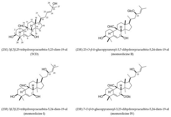
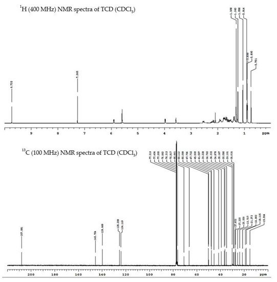
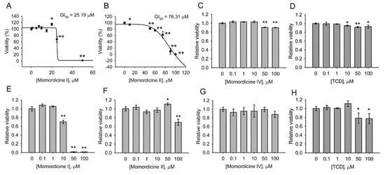
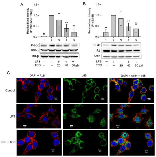
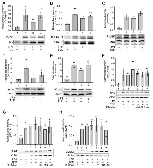
The vines and leaves of Momordica charantia L. are used as herbal medicines to treat inflammation-related disorders. However, their safety profile remains uncharacterized, and the constituents in their extracts that exert anti-inflammatory and adverse effects remain unclear. This study isolated the characteristic cucurbitane-type triterpenoid species in the vines and leaves of M. charantia L. and analyzed their cytotoxicity, anti-inflammatory effects, and underlying mechanisms. Four structurally related triterpenoids—momordicines I, II, IV, and (23E) 3β,7β,25-trihydroxycucurbita-5,23-dien-19-al (TCD)—were isolated from the triterpenoid-rich fractions of extracts from the vines and leaves of M. charantia. Momordicine I was cytotoxic on normal cells, momordicine II exerted milder cytotoxicity, and momordicine IV and TCD had no obvious adverse effects on cell growth. TCD had anti-inflammatory activity both in vivo and in vitro. In lipopolysaccharide-stimulated RAW 264.7 cells, TCD inhibited the inhibitor kappa B kinase/nuclear factor-κB pathway and enhanced the expression of nuclear factor erythroid 2-related factor 2, heme oxygenase-1, and glutamate-cysteine ligase modifier subunit through the extracellular signal-regulated kinase1/2 and p38. Thus, the vines and leaves of M. charantia should be used with caution. An extraction protocol that can enrich TCD but remove momordicine I would likely enhance the safety of the extract.

  • Momordica charantia
  • triterpenoid
  • anti-inflammation
  • antioxidant enzymes
  • Nrf2
  • heme oxygenase-1
  • cytotoxicity

1. Introduction

Momordica charantia L. belongs to the Cucurbitaceae family, and its fruit is commonly known as bitter melon or bitter gourd. Each part of this plant, including the vines, leaves, fruits, roots, and seeds, has been used in folk medicine to treat various diseases, including diabetes, worm infection, and inflammation [1,2][1][2]. However, under some circumstances, M. charantia L. has also exerted adverse effects on human health. Therefore, its safety requires comprehensive evaluation [1,2][1][2].
Chronic inflammation is associated with the development of diseases including metabolic syndrome, type 2 diabetes, cardiovascular diseases, cancer, and neurodegenerative diseases [3]. Aside from the fruits, the vines and leaves of M. charantia L. are also used as herbal medicines in treating inflammation-related disorders [1,2][1][2]. However, unlike the fruits, they are not commonly consumed as food; thus, their safety profile remains uncharacterized. Moreover, the components of their extracts that exert anti-inflammatory effects and adverse effects are unclear. The vines and leaves of M. charantia L. are rich in cucurbitane-type triterpenoids [4[4][5],5], which possess 19-(10→9β)-abeo-10α-lanost-5-ene skeletons and were originally discovered from the Momordica species [6]. The safety, anti-inflammatory activities and mechanisms of these compounds are not fully addressed. 

2. Cucurbitane-Type Triterpenoids Isolated from the Vines and Leaves of M. charantia

From the triterpenoid-rich fractions of the vines and leaves of M. charantia (see Section 4.4), we isolated four previously characterized metabolites: (23E) 3β,7β,25-trihydroxycucurbita-5,23-dien-19-al (TCD), 3β,7β,23-trihydroxycucurbita-5,24-dien-19-al (momordicine I), (23R) 23-O-β-d-glucopyranosyl-3,7-dihydroxycucurbita-5,24-dien-19-al (momordicine II), and (23R) 7-O-β-d-glucopyranosyl-3,23-dihydroxycucurbita-5,24-dien-19-al (momordicine IV) (Figure 1). The structures of these metabolites were elucidated by comparing their 1H and 13C nuclear magnetic resonance (NMR) spectroscopy data (Figure 2 and Supplementary Materials, could be found in https://www.mdpi.com/1422-0067/23/3/1071#supplementary) with those of related known compounds [7,8,9][7][8][9]. TCD and momordicine I are isomers, with the –OH group on C-25 and the double bond between C-23 and C-24 in TCD being shifted in momordicine I. Momordicines II and IV are glucosides of momordicine I. Presumably, these isolates are likely important components underlying the biological activities of the extract prepared from the title plant because of their relative high abundance in the extract (2.23% of the total weight of the EtOAc extract for TCD, 3.03% for momordicine I, 0.22% for momordicine II, and 0.21% for momordicine IV; see Section 4.4).
Figure 1. Chemical structures of (23E) 3β,7β,25-trihydroxycucurbita-5,23-dien-19-al (TCD) and momordicines I, II, and IV. Glc, glucose.
Figure 2. 1H and 13C nuclear magnetic resonance (NMR) spectroscopy data of TCD.
To compare the lipophilicity of the compounds, the partition coefficient logP was calculated for each compound. Their logP values from high (more lipophilic) to low (less lipophilic) are TCD 5.17; momordicine I 4.41; momordicine II 2.72; and momordicine IV 2.72 (Supplementary Materials).

3. Cytotoxicity of Triterpenoids

Momordicines I, II, and IV and TCD were first characterized for cytotoxicity by using a normal intestinal cell line, IEC-18, and a normal hepatic cell line, FL83B. Figure 3A–D indicate that momordicine I significantly inhibited the growth of IEC-18 cells, with a GI50 (concentration required for inhibiting cell growth by 50%) of 25.19 µM (A); the toxicity of momordicine II was milder, with a GI50 of 76.31 µM to IEC-18 cells (B). By contrast, momordicine IV (C) and TCD (D) maintained over 80% cell survival in the concentration range of 0.1–100 µM. Figure 3E–H demonstrate that momordicine I (E) was lethal to FL83B cells at 50 and 100 µM and slightly but definitively suppressed cell growth at 10 µM. Momordicine II (F), momordicine IV (G), and TCD (H) were less toxic to FL83B cells; under 0.1–100 µM, cell survival exceeded 70%. These data suggest that momordicine I is the most toxic to normal cells among the four compounds, exerting deleterious effects on both cell lines in concentrations higher than 10 or 20 µM. Momordicine II produced a milder detrimental effect on IEC-18 cells. Momordicine IV and TCD were not harmful to normal cells in the indicated concentrations.
Figure 3. Cytotoxic analysis of momordicines I, II, and IV and TCD in IEC-18 and FL83B cell lines: (AD) IEC-18 cells; (EH) FL83B cells. The cells were treated with the indicated concentrations of momordicine I (A,E), momordicine II (B,F), momordicine IV (C,G), or TCD (D,H) for 24 h. Cell survival relative to the control ([compound] = 0 µM) was determined. In (A,B), the GI50 of the compound was calculated. The experiments were performed in triplicate. Data are presented as means ± standard deviations. * p < 0.05, ** p < 0.005 versus the control.
The cytotoxicity of the compounds on the model macrophage cell line RAW 264.7 was examined. Momordicine I significantly inhibited cell survival at concentrations of ≥15 µM (Figure 4A). Momordicine II (Figure 4B), momordicine IV (Figure 4C), and TCD (Figure 4D) were not toxic to RAW 264.7 cells at 20–100 or 0.1–100 µM, and TCD at 50–100 µM actually increased cell growth. Next, the effects of these three compounds on cell survival in the presence of lipopolysaccharides (LPS) were examined. As presented in Figure 4E, LPS cotreatment with 50 µM TCD (Group 3), momordicine II (Group 4), or momordicine IV (Group 5) was not toxic to RAW 264.7 cells. The results indicate that only momordicine I is toxic to RAW 264.7 cells.
Figure 4. Cytotoxic analysis of the effects of momordicines I, II, and IV and TCD on RAW 264.7 cells. (AD) The cells were treated with the indicated concentrations of momordicine I (A), momordicine II (B), momordicine IV (C), or TCD (D) for 24 h. (E) The cells were cotreated with 100 ng/mL lipopolysaccharides (LPS) and 50 µM TCD (Group 3), momordicine II (Group 4), momordicine IV (Group 5), LPS alone (Group 2), or the solvent (Group 1) for 24 h. Cell survival relative to the control ([compound] = 0 µM or Group 1) was determined. The experiments were performed in triplicate. Data are presented as means ± standard deviations. * p < 0.05, ** p < 0.005 versus the control.

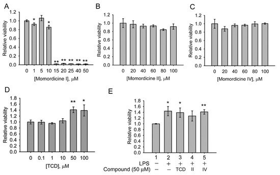
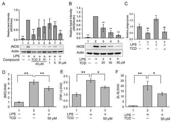
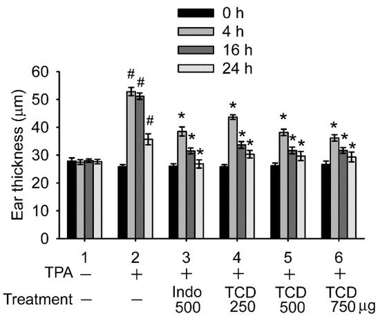
4. Anti-Inflammatory Effects of Triterpenoids

The anti-inflammatory effects of momordicine II, momordicine IV, and TCD were compared in LPS-treated RAW 264.7 cells. As presented in Figure 5A, LPS increased the expression of inducible nitric oxide synthase (iNOS) in the cells (Lane 2 vs. Lane 1), whereas the addition of 40 µM TCD (Lane 3) significantly suppressed iNOS expression compared with LPS treatment alone (Lane 2). By contrast, momordicine II (Lane 4) or momordicine IV (Lane 5) did not significantly inhibit iNOS expression. Although momordicine I is toxic to RAW 264.7 cells, we evaluated its anti-inflammatory effect using sublethal concentrations. As displayed in Figure 5A (Lanes 6–8), 1–10 µM momordicine I dose-dependently inhibited iNOS expression, implying that it has anti-inflammatory activity at sublethal concentrations.
Figure 5. TCD reduced the expression of LPS-induced inflammatory biomarkers in RAW 264.7 cells. (A,B) Western blotting analysis of inducible nitric oxide synthase (iNOS) expression: (A) cells were stimulated with 100 ng/mL LPS (Lanes 2–8) and cotreated with 40 µM TCD (Lane 3), momordicine II (Lane 4), or momordicine IV (Lane 5) for 16 h or with 1, 5, or 10 µM momordicine I (Lanes 6, 7, and 8, respectively) for 16 h; (B) cells were treated with LPS and 20, 40, or 50 µM TCD (Lanes 3, 4, and 5, respectively), or LPS alone (Lane 2) for 16 h. Relative band intensity (normalized by actin) versus Lane 2 was determined. Data are presented as the means ± standard deviations of four (A) or three (B) independent experiments. * p < 0.05 and ** p < 0.005 versus Lane 2. (C) Cells were stimulated with solvent (Group 1), 50 µM TCD (Group 2), 100 ng/mL LPS (Group 3), or cotreated with LPS and TCD (Group 4) for 24 h. This was followed by a phagocytosis assay. Phagocytic activity relative to that of Group 1 was determined. Data are presented as the means ± standard deviations of experiments performed in triplicate. * p < 0.05 versus Group 1; ## p < 0.005 versus Group 3. (DF) Cells were stimulated with the solvent (Group 1) or 100 ng/mL LPS (Group 2) or cotreated with LPS and 50 µM TCD (Group 3) for 12 h (E) or 16 h (D,F), and the medium was subjected to [NO] (D), tumor necrosis factor α ([TNF-α]) (E), or interleukin-6 ([IL-6]) (F) analysis. Data are presented as means ± standard deviations (N = 9 in (D); N = 3 in (E) and (F)). * p < 0.05, ** p < 0.005 between the indicated groups.
Overall, the results suggest that TCD and momordicine I both possess anti-inflammatory activities. However, momordicine I is cytotoxic. Therefore, we further characterized the anti-inflammatory effects of TCD and the underlying mechanisms.
As presented in Figure 5B, 20–50 µM TCD dose-dependently inhibited LPS-induced iNOS expression. Moreover, compared with the control, LPS increased the phagocytic activity of RAW 264.7 cells (Group 3), whereas TCD alone (Group 2) or with LPS (Group 4) significantly reduced the phagocytic activity (Figure 5C). Furthermore, LPS triggered NO production in RAW 264.7 cells (Figure 5D, Group 2), and TCD partially but significantly inhibited LPS activity (Figure 5D, Group 3). Similarly, LPS increased the expression of tumor necrosis factor α (TNF-α) (Figure 5E, Group 2) and interleukin-6 (IL-6) (Figure 5F, Group 2), both of which were significantly suppressed by the addition of TCD (Figure 5E,F, Group 3). These results demonstrate that TCD possesses anti-inflammatory activity.
As shown in Figure 6, 12-O-tetradecanoylphorbol-13-acetate (TPA) caused ear edema, a sign of ear inflammation, in treated mice 4, 16, and 24 h after TPA stimulation (Group 2). TCD treatment ameliorated ear edema. All tested doses (250, 500, and 750 µg/ear in Groups 4, 5, and 6, respectively) of TCD significantly reduced the degree of ear edema in mice 4, 16, and 24 h after TPA stimulation compared with the TPA control (Group 2). The effects of 500 and 750 µg/ear TCD on ear inflammation reduction were comparable to that of 500 µg/ear indomethacin, a nonsteroidal anti-inflammatory drug (Group 3). These results confirm that TCD can ameliorate inflammation in vivo.
Figure 6. TCD ameliorated 12-O-tetradecanoylphorbol-13-acetate (TPA)-induced ear edema in vivo. Mice were treated with TPA (Groups 2–6) or the solvent (Group 1) in one ear. One hour later, solvent (Groups 1 and 2), 500 μg/ear indomethacin (Group 3), or 250, 500, or 750 μg/ear TCD (Groups 4, 5, and 6, respectively) was applied on the ear. Ear thickness was measured before (0 h) and at 4, 16, and 24 h after TPA treatment. Data are presented as the means ± standard deviations of each group (N = 6). # p < 0.001 versus 0 h of Group 2; * p < 0.001 versus the same time point of Group 2.

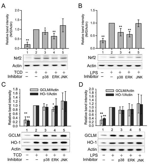
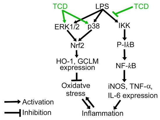
5. Anti-Inflammatory Mechanism of TCD

LPS activates the inhibitor kappa B kinase (IKK)/nuclear factor-κB (NF-κB) pathway through the activation of toll-like receptor 4 [10,11][10][11]. Thus, we determined whether TCD inhibited the IKK/NF-κB pathway. As presented in Figure 7A, LPS caused an apparent increase in phosphorylated IKK (Lane 2) in RAW 264.7 cells. The addition of 20–50 μM TCD (Lanes 3, 4, 5) dose-dependently inhibited LPS-induced IKK phosphorylation. Moreover, Figure 7B demonstrates that LPS increased the phosphorylation of the inhibitor of NF-κB (IκB) (Lane 2), a substrate of IKK, but that cotreatment of 20–50 μM TCD with LPS dose-dependently reduced the level of IκB phosphorylation (Lanes 3–5). Furthermore, the nuclear translocation of NF-κB was examined using confocal microscopy. As presented in Figure 7C, in solvent-treated control cells, the location of the NF-κB subunit p65 was perinuclear, suggesting that NF-κB is located outside the nucleus. In LPS-treated cells, the location of p65 overlapped with the 4′,6-diamidino-2-phenylindole (DAPI)-labeled nucleus, indicating that NF-κB translocated into the nucleus in these cells. Under cotreatment with LPS and TCD, p65 exhibited perinuclear distribution in most cells, suggesting that TCD inhibited the LPS-induced nuclear translocation of NF-κB. Furthermore, the morphologies of RAW 264.7 cells differed among groups. In the control and LPS + TCD groups, cell morphology was similar to that of fibroblasts or endothelial cells. By contrast, in the LPS group, the cells exhibited a macrophage-like morphology, with numerous needle-like protrusions extending from the cell membrane. This suggested that LPS induced the differentiation of RAW 264.7 cells into macrophage-like proinflammatory cells [12,13][12][13], yet TCD suppressed the differentiation. Overall, the results imply that TCD suppressed the LPS-activated IKK/NF-κB pathway.
Figure 7. TCD inhibited the inhibitor kappa B kinase (IKK)/nuclear factor-κB (NF-κB) pathway. (A,B) Western blot analysis. RAW 264.7 cells were treated with the solvent (Lane 1) or 100 ng/mL LPS (Lane 2), or co-treated with LPS and 20, 40, or 50 μM TCD (Lanes 3, 4, and 5, respectively) for 1 h. The levels of IKK phosphorylation (A) and the inhibitor of NF-κB (IκB) phosphorylation (B) were analyzed. Normalized relative band intensity versus Lane 2 was determined. Data are presented as the means ± standard deviations of four independent experiments. ** p < 0.005 versus Lane 2. (C) Confocal microscopy analysis. RAW 264.7 cells were treated with the solvent (control), 100 ng/mL LPS alone (LPS), or LPS and 50 μM TCD (LPS + TCD) for 1 h. Cells were labeled with a p65-specific antibody and an FITC-conjugated secondary antibody (green fluorescence), rhodamine phalloidin (actin labeling for locating the cell membrane; red fluorescence), and 4′,6-diamidino-2-phenylindole (DAPI; nuclear staining; blue fluorescence). The superimposed images of DAPI staining and rhodamine phalloidin staining (DAPI + Actin), the results of FITC labeling (p65), and the superimposed images of all staining (DAPI + Actin + p65) are shown. Scale bars: 5 μm.
In addition to activating IKK, TLR4 also activates mitogen-activated protein kinases (MAPKs) p38, c-Jun-N-terminal kinase (JNK), and extracellular signal-regulated kinase1/2 (ERK1/2), which activate AP-1 and promote the expression of some proinflammatory cytokines [10,11][10][11]. Therefore, the effects of LPS and TCD on MAPKs were also explored. Figure 8A–C reveals that TCD (Lane 2), LPS (Lane 3), and LPS + TCD (Lane 4) clearly activated p38 (A), ERK1/2 (B), and JNK (C) compared with the control (Lane 1). The results indicate that TCD and LPS both activated MAPKs and that TCD did not suppress LPS-induced MAPK activation.
Figure 8. TCD activated mitogen-activated protein kinases (MAPKs) and the nuclear factor erythroid 2-related factor 2 (Nrf2)/heme oxygenase-1 (HO-1) pathway. RAW 264.7 cells were treated with the solvent (Lane 1), 50 μM TCD (Lane 2), 100 ng/mL LPS (Lane 3), or LPS and TCD (Lane 4) for 1 h (AC), 24 h (D,E), 2 h (F), or 6 h (G,H). The levels of phosphorylated p38 (A), phosphorylated extracellular signal-regulated kinase1/2 (ERK1/2) (B), phosphorylated c-Jun N-terminal kinase (JNK) (C), HO-1 (D,G), glutamate–cysteine ligase modifier subunit (GLCM) (E,H), and Nrf2 (F) were analyzed using Western blotting. In (F,G,H), an inhibitor (20 μM) of p38 (Lane 5), ERK1/2 (Lane 6), or JNK (Lane 7) was also added to cells cotreated with LPS and TCD. Normalized relative band intensity versus Lane 3 was determined. Data are presented as the means ± standard deviations of four independent experiments. * p < 0.05, ** p < 0.005 versus Lane 1. In (F,G,H), statistical analysis of Lanes 5, 6, and 7 versus Lane 4 was also performed, but no significant difference was found.
Nonetheless, MAPKs also activate cytoprotective and anti-inflammatory factors. They have been shown to activate nuclear factor erythroid 2-related factor 2 (Nrf2), leading to the expression of antioxidant enzymes, including heme oxygenase-1 (HO-1) and the glutamate–cysteine ligase modifier subunit (GCLM) [14,15,16][14][15][16]. Therefore, we further determined whether TCD activated the Nrf2/HO-1 pathway. As displayed in Figure 8D,E, TCD (Lane 2), LPS (Lane 3), and LPS + TCD (Lane 4) all significantly enhanced the expression of HO-1 (D) and GCLM (E) compared with the control (Lane 1). Consistently, these treatments also elevated Nrf2 expression (Figure 8F, Lanes 2–4). These data confirm that TCD and LPS both activated the Nrf2/HO-1 pathway. To further assess the roles of MAPKs, a p38, ERK1/2, or JNK inhibitor was added to cells cotreated with LPS and TCD (Figure 8F; Lanes 5, 6, and 7, respectively). None of the inhibitors significantly blocked Nrf2 activation by LPS and TCD. Similarly, the expression of HO-1 and GCLM induced by LPS + TCD (Figure 8G,H, Lane 4) was not apparently inhibited by any single MAPK inhibitor (Figure 8G,H, Lanes 5–7). Furthermore, the respective effect of TCD or LPS on MAPKs was also examined using the MAPK inhibitors. Figure 9A shows that TCD-promoted Nrf2 expression (Lane 2) was partially yet significantly inhibited by p38 inhibitor (Lane 3) or ERK1/2 inhibitor (Lane 4), but not by JNK inhibitor (Lane 5). Similarly, LPS-elevated Nrf2 expression (Figure 9B, Lane 2) was also partially and significantly suppressed by p38 inhibitor or ERK1/2 inhibitor (Figure 9B, Lanes 3 and 4), but not by JNK inhibitor (Figure 9B, Lane 5). However, Figure 9C,D exhibit that TCD- (C, Lane 2) or LPS-induced (D, Lane 2) GCLM and HO-1 expression was repressed by p38 inhibitor (Lane 3) or ERK inhibitor (Lane 4) in a milder, less apparent extent. In agreement with the results of Figure 9A,B, JNK inhibitor did not inhibit the effect of TCD or LPS on inducing GCLM and HO-1 expression (Figure 9C,D, Lane 5). The results of Figure 9 suggest that JNK is probably not involved in activating the Nrf2/HO-1 pathway.
Figure 9. The effects of MAPK inhibitors on TCD- or LPS-activated Nrf2/HO-1 pathway. RAW 264.7 cells were treated with the solvent (Lane 1), 50 μM TCD ((A,C), Lanes 2–5), or 100 ng/mL LPS ((B,D), Lanes 2–5) for 2 h (A,B) or 6 h (C,D). An inhibitor (20 μM) of p38 (Lane 3), ERK1/2 (Lane 4), or JNK (Lane 5) was also added to cells treated with TCD or LPS. The levels of Nrf2 (A,B), GCLM (C,D), and HO-1 (C,D) were analyzed using Western blotting. Normalized relative band intensity versus Lane 2 was determined. Data are presented as the means ± standard deviations of four independent experiments. * p < 0.05, ** p < 0.005 versus Lane 2.

6. Discussion

In this study, chemical isolation from the triterpenoid-rich fractions of the extracts of the vines and leaves of M. charantia yielded predominantly four cucurbitane-type triterpenoids: momordicines I, II, and IV, and TCD. Momordicine I exerted deleterious effects on the growth of normal cell lines, momordicine II had a milder adverse effect on cells, and momordicine IV and TCD were not harmful to normal cells at 0.1–100 μM. Therefore, in view of safety, momordicine IV and TCD are more appropriate for potential development in biomedical applications than are momordicine I or II. However, these findings suggest that the extract of vines and leaves from M. charantia contains potentially toxic chemicals and thus should be used with caution.
The anti-inflammatory potential of TCD was more notable than those of momordicines II or IV; thus, we further characterized it. To reiterate, TCD was indicated to have anti-inflammatory activity both in vivo and in vitro because it inhibited LPS-induced phagocytosis and the expression of iNOS, NO, TNF-α, and IL-6 in a macrophage cell model. Furthermore, it ameliorated ear edema in an animal model. The underlying mechanism likely hinges on the suppression of the IKK/NF-κB pathway. However, LPS enhanced the expression of Nrf2, HO-1, and GCLM, which may provide protection against excessive inflammatory responses [17], whereas TCD also increased the expression of these proteins whether when used alone or together with LPS. HO-1 reduces cellular oxidative damage and mitigates inflammatory responses [18,19,20][18][19][20]. Thus, TCD likely protected cells from oxidative damage by activating the Nrf2/HO-1 pathway, which also helped reduce inflammation. However, TCD activated MAPKs and did not suppress MAPK activation by LPS. MAPKs increase the expression of some proinflammatory cytokines, which may explain why TCD only partially inhibited LPS-induced production of inflammatory cytokines, as shown in Figure 5E,F.
MAPKs are suggested to activate the Nrf2/HO-1 pathway [14[14][15][16],15,16], and ERK1/2 and p38 have been reported to activate Nrf2 [18[18][21][22],21,22], whereas the role of JNK is less clear. TCD and LPS both activated p38, ERK1/2, and JNK. Our data suggest that p38 and ERK1/2 are both involved in LPS- or TCD-induced activation of Nrf2, whereas JNK is not. TCD- or LPS-promoted Nrf2 expression was both partially yet significantly inhibited by either p38 inhibitor or ERK inhibitor, though the suppressive effects of these inhibitors declined on the Nrf2 downstream factors HO-1 and GCLM. Theoretically, if p38 and ERK1/2 are both involved in activating Nrf2, when one of these kinases is inhibited, Nrf2 can still be activated by the other one. Therefore, the observed partial decrease of Nrf2 level by either p38 or ERK1/2 inhibitor is reasonable. In TCD and LPS-cotreated cells (Figure 8F–H), LPS and TCD may synergistically activate p38 or ERK1/2 when one of these kinases is inhibited, resulting in a higher extent of Nrf2 activation. Consequently, significant Nrf2 suppression by either inhibitor could not be observed in Figure 8F.
On the basis of our findings, we propose the anti-inflammatory mechanism of TCD (Figure 10). TCD can reduce LPS-induced inflammation through inhibiting IKK activation by LPS. Moreover, TCD and LPS both activate ERK1/2 and p38. These kinases activate Nrf2 and increase the expression of the antioxidant enzymes HO-1 and GCLM. Inflammation can exacerbate oxidative stress; conversely, oxidative stress can aggravate inflammation. Antioxidant enzymes can protect cells from oxidative damage and thus prevent excessive amounts of inflammation. In other words, TCD reduces the extent of inflammation and protects cells from oxidative damage.
Figure 10. The proposed anti-inflammatory mechanism of TCD. TCD inhibits the LPS-induced activation of IKK. Moreover, TCD, together with LPS, activates ERK1/2 and p38, resulting in elevated expression of Nrf2 and the antioxidant enzymes HO-1 and GCLM to protect cells from oxidative damage. P-IκB, phosphorylated IκB.
Momordicines II and IV are the glucosides of momordicine I. In cytotoxicity assays, the GI50 of momordicine I was lower than that of momordicine II in IEC-18 cells. Moreover, momordicine I was lethal to FL83B cells and RAW 264.7 cells in concentrations higher than 10 μM, whereas 20–100 μM momordicine II did not inhibit the growth of these cells. Momordicine IV exhibited no harmful effect on any of the three tested cell lines. Thus, the order of cytotoxicity is momordicine I > momordicine II > momordicine IV. This suggests that glycosylation reduces the cytotoxicity of momordicine I, and that glycosylation on the –OH group of C-7 (momordicine IV) reduces toxicity to a greater extent than does glycosylation on the –OH group of C-23 (momordicine II).
TCD and momordicine I are isomers with similar structures, but their cytotoxic effects differ substantially. TCD exerts considerably less deleterious effects on cells than does momordicine I. These two isomers differ structurally between C-23 and C-25. The –OH group on C-25 of TCD is shifted to C-23 in momordicine I; the double bond between C-23 and C-24 in TCD is moved to between C-24 and C-25. This structural rearrangement notably affects the cytotoxicity. Future studies should investigate the cellular targets that interact with momordicine I and cause the cytotoxic effect.
Although cytotoxic, momordicine I exhibited a potential anti-inflammatory effect in that it reduced the LPS-induced iNOS expression at its sublethal concentration (10 μM). Thus, momordicine I and TCD both likely contribute to the anti-inflammatory activity of the extract of vines and leaves from M. charantia. However, momordicine I may also contribute to the adverse effects of the extract.
High lipophilicity may facilitate the permeation of a compound through biomembranes, resulting in higher bioactivity or cytotoxicity of the compound [23,24,25][23][24][25]. The lipophilicity of the compounds reported here is TCD > momordicine I > momordicine II = momordicine IV according to their logP values. This lipophilicity order may explain why TCD and momordicine I are more bioactive than momordicine II and momordicine IV, but it cannot explain why momordicine I is the most cytotoxic among them. Moreover, momordicine I exhibited a potential anti-inflammatory effect at its sublethal concentration, suggesting that momordicine I may be as effective as or even better than TCD in anti-inflammation. Therefore, in addition to lipophilicity, there are likely other factors that influence the bioactivity or cytotoxicity of these compounds. For example, MAPKs regulate mitochondrial metabolism [26], and some compounds were reported to target mitochondria for their cytotoxic or anti-inflammatory effects [24,27,28][24][27][28]. Thus, whether the triterpenoids reported in this study target mitochondria deserves to be addressed. Moreover, some lipophilic compounds including nobiletin, kaempferol, luteolin, quercetin, and tangeretin were suggested to enter cells through an energy independent carrier-mediated system [29]. It cannot be excluded that such a system also exists for the compounds reported here.
TCD was also found in the fruits of M. charantia L. [30]. This compound has been reported to reduce blood glucose levels in diabetic animal models [30] and serve as an insulin sensitizer and insulin substitute [31]. Our findings further demonstrate that it is an anti-inflammatory agent. Moreover, TCD was also isolated from the leaves of wild varieties of M. charantia previously [32] and was demonstrated to reduce periodontal pathogen- or Cutibacterium acnes-induced inflammatory responses in human monocytic THP-1 cells in vitro and ameliorate inflammation in the corresponding pathogen-stimulated mouse models in vivo [32,33][32][33]. Therefore, studies from different groups all support that TCD is an anti-inflammatory agent. In sum, TCD, which is distributed in the fruits, vines, and leaves of M. charantia L., has hypoglycemic effects and anti-inflammatory functions. It can be regarded as a potential new agent for developing therapeutic or health-care products of anti-inflammation or glycemic control, or as an index ingredient for the bioactivity of bitter melon extracts.
Our results indicate that although momordicine I is cytotoxic, TCD is relatively safe and exerts notable anti-inflammatory effects. Therefore, a protocol that can enrich the extraction of TCD from the vines and leaves of M. charantia but remove momordicine I would likely enhance the anti-inflammatory effect and improve the safety of the extract.

References

  1. Jia, S.; Shen, M.; Zhang, F.; Xie, J. Recent advances in Momordica charantia: Functional components and biological activities. Int. J. Mol. Sci. 2017, 18, 2555.
  2. Bortolotti, M.; Mercatelli, D.; Polito, L. Momordica charantia, a nutraceutical approach for inflammatory related diseases. Front. Pharmacol. 2019, 10, 486.
  3. Minihane, A.M.; Vinoy, S.; Russell, W.R.; Baka, A.; Roche, H.M.; Tuohy, K.M.; Teeling, J.L.; Blaak, E.E.; Fenech, M.; Vauzour, D.; et al. Low-grade inflammation, diet composition and health: Current research evidence and its translation. Br. J. Nutr. 2015, 114, 999–1012.
  4. Chang, C.I.; Chen, C.R.; Liao, Y.W.; Cheng, H.L.; Chen, Y.C.; Chou, C.H. Cucurbitane-type triterpenoids from Momordica charantia. J. Nat. Prod. 2006, 69, 1168–1171.
  5. Cheng, H.L.; Huang, H.K.; Chang, C.I.; Tsai, C.P.; Chou, C.H. A cell-based screening identifies compounds from the stem of Momordica charantia that overcome insulin resistance and activate AMP-activated protein kinase. J. Agric. Food Chem. 2008, 56, 6835–6843.
  6. Nagarani, G.; Abirami, A.; Siddhuraju, P. Food prospects and nutraceutical attributes of Momordica species: A potential tropical bioresources—a review. Food Sci. Hum. Well. 2014, 3, 117–126.
  7. Yasuda, M.; Iwamoto, M.; Okabe, H.; Yamauchi, T. Structures of Momordicines I, II and III, the bitter principles in the leaves and vines of Momordica charantia L. Chem. Parm. Bull. 1984, 32, 2044–2047.
  8. Fatope, M.O.; Takeda, Y.; Yamashita, H.; Okabe, H.; Yamauchi, T. New cucurbitane triterpenoids from Momordica-Charantia. J. Nat. Prod. 1990, 53, 1491–1497.
  9. Mekuria, D.B.; Kashiwagi, T.; Tebayashi, S.; Kim, C.S. Cucurbitane glucosides from Momordica charantia leaves as oviposition deterrents to the leafminer, Liriomyza trifolii. Z. Naturforsch. C 2006, 61, 81–86.
  10. Mohan, S.; Gupta, D. Crosstalk of toll-like receptors signaling and Nrf2 pathway for regulation of inflammation. Biomed. Pharmacother. 2018, 108, 1866–1878.
  11. Tajalli-Nezhad, S.; Karimian, M.; Beyer, C.; Atlasi, M.A.; Azami Tameh, A. The regulatory role of Toll-like receptors after ischemic stroke: Neurosteroids as TLR modulators with the focus on TLR2/4. Cell. Mol. Life Sci. 2019, 76, 523–537.
  12. Liu, L.; Guo, H.; Song, A.; Huang, J.; Zhang, Y.; Jin, S.; Li, S.; Zhang, L.; Yang, C.; Yang, P. Progranulin inhibits LPS-induced macrophage M1 polarization via NF-κB and MAPK pathways. BMC Immunol. 2020, 21, 32.
  13. Gong, J.; Li, J.; Dong, H.; Chen, G.; Qin, X.; Hu, M.; Yuan, F.; Fang, K.; Wang, D.; Jiang, S.; et al. Inhibitory effects of berberine on proinflammatory M1 macrophage polarization through interfering with the interaction between TLR4 and MyD88. BMC Complement. Altern. Med. 2019, 19, 314.
  14. Bryan, H.K.; Olayanju, A.; Goldring, C.E.; Park, B.K. The Nrf2 cell defence pathway: Keap1-dependent and -independent mechanisms of regulation. Biochem. Pharmacol. 2013, 85, 705–717.
  15. Niture, S.K.; Khatri, R.; Jaiswal, A.K. Regulation of Nrf2-an update. Free Radic. Biol. Med. 2014, 66, 36–44.
  16. Yu, C.; Xiao, J.H. The Keap1-Nrf2 system: A mediator between oxidative stress and aging. Oxid. Med. Cell Longev. 2021, 2021, 6635460.
  17. Rushworth, S.A.; MacEwan, D.J.; O'Connell, M.A. Lipopolysaccharide-induced expression of NAD(P)H:quinone oxidoreductase 1 and heme oxygenase-1 protects against excessive inflammatory responses in human monocytes. J. Immunol. 2008, 181, 6730–6737.
  18. Tran, P.L.; Tran, P.T.; Tran, H.N.K.; Lee, S.; Kim, O.; Min, B.S.; Lee, J.H. A prenylated flavonoid, 10-oxomornigrol F, exhibits anti-inflammatory effects by activating the Nrf2/heme oxygenase-1 pathway in macrophage cells. Int. Immunopharmacol. 2018, 55, 165–173.
  19. Paine, A.; Eiz-Vesper, B.; Blasczyk, R.; Immenschuh, S. Signaling to heme oxygenase-1 and its anti-inflammatory therapeutic potential. Biochem. Pharmacol. 2010, 80, 1895–1903.
  20. Yang, C.H.; Yen, T.L.; Hsu, C.Y.; Thomas, P.A.; Sheu, J.R.; Jayakumar, T. Multi-targeting andrographolide, a novel NF-kappaB inhibitor, as a potential therapeutic agent for stroke. Int. J. Mol. Sci. 2017, 18, 1638.
  21. Xu, Y.; Yuan, H.; Luo, Y.; Zhao, Y.J.; Xiao, J.H. Ganoderic acid D protects human amniotic mesenchymal stem cells against oxidative stress-induced senescence through the PERK/NRF2 signaling pathway. Oxid. Med. Cell. Longev. 2020, 2020, 8291413.
  22. Song, C.; Heping, H.F.; Shen, Y.S.; Jin, S.X.; Li, D.Y.; Zhang, A.G.; Ren, X.L.; Wang, K.L.; Zhang, L.; Wang, J.D.; et al. AMPK/p38/Nrf2 activation as a protective feedback to restrain oxidative stress and inflammation in microglia stimulated with sodium fluoride. Chemosphere 2020, 244, 125495.
  23. Dubinin, M.V.; Semenova, A.A.; Ilzorkina, A.I.; Markelova, N.Y.; Penkov, N.V.; Shakurova, E.R.; Belosludtsev, K.N.; Parfenova, L.V. New quaternized pyridinium derivatives of betulin: Synthesis and evaluation of membranotropic properties on liposomes, pro- and eukaryotic cells, and isolated mitochondria. Chem. Biol. Interact. 2021, 349, 109678.
  24. Dubinin, M.V.; Semenova, A.A.; Ilzorkina, A.I.; Penkov, N.V.; Nedopekina, D.A.; Sharapov, V.A.; Khoroshavina, E.I.; Davletshin, E.V.; Belosludtseva, N.V.; Spivak, A.Y.; et al. Mitochondria-targeted prooxidant effects of betulinic acid conjugated with delocalized lipophilic cation F16. Free Radic. Biol. Med. 2021, 168, 55–69.
  25. Dubinin, M.V.; Semenova, A.A.; Ilzorkina, A.I.; Mikheeva, I.B.; Yashin, V.A.; Penkov, N.V.; Vydrina, V.A.; Ishmuratov, G.Y.; Sharapov, V.A.; Khoroshavina, E.I.; et al. Effect of betulin and betulonic acid on isolated rat liver mitochondria and liposomes. Biochim. Biophys. Acta Biomembr. 2020, 1862, 183383.
  26. Javadov, S.; Jang, S.; Agostini, B. Crosstalk between mitogen-activated protein kinases and mitochondria in cardiac diseases: Therapeutic perspectives. Pharmacol. Therapeut. 2014, 144, 202–225.
  27. Belosludtsev, K.N.; Belosludtseva, N.V.; Kosareva, E.A.; Talanov, E.Y.; Gudkov, S.V.; Dubinin, M.V. Itaconic acid impairs the mitochondrial function by the inhibition of complexes II and IV and induction of the permeability transition pore opening in rat liver mitochondria. Biochimie 2020, 176, 150–157.
  28. Kodr, D.; Stankova, J.; Rumlova, M.; Dzubak, P.; Rehulka, J.; Zimmermann, T.; Krizova, I.; Gurska, S.; Hajduch, M.; Drasar, P.B.; et al. Betulinic acid decorated with polar groups and blue emitting BODIPY dye: Synthesis, cytotoxicity, cell-cycle analysis and anti-HIV profiling. Biomedicines 2021, 9, 1104.
  29. Kimura, O.; Ohta, C.; Koga, N.; Haraguchi, K.; Kato, Y.; Endo, T. Carrier-mediated uptake of nobiletin, a citrus polymethoxyflavonoid, in human intestinal Caco-2 cells. Food Chem. 2014, 154, 145–150.
  30. Harinantenaina, L.; Tanaka, M.; Takaoka, S.; Oda, M.; Mogami, O.; Uchida, M.; Asakawa, Y. Momordica charantia constituents and antidiabetic screening of the isolated major compounds. Chem. Pharm. Bull. 2006, 54, 1017–1021.
  31. Chang, C.I.; Chou, C.H.; Liao, M.H.; Chen, T.M.; Cheng, C.H.; Anggriani, R.; Tsai, C.P.; Tseng, H.I.; Cheng, H.L. Bitter melon triterpenes work as insulin sensitizers and insulin substitutes in insulin-resistant cells. J. Funct. Foods 2015, 13, 214–224.
  32. Tsai, T.H.; Huang, W.C.; Ying, H.T.; Kuo, Y.H.; Shen, C.C.; Lin, Y.K.; Tsai, P.J. Wild bitter melon leaf extract inhibits Porphyromonas gingivalis-induced inflammation: Identification of active compounds through bioassay-guided isolation. Molecules 2016, 21, 454.
  33. Chuang, L.T.; Huang, W.C.; Hou, Y.C.; Chyuan, J.H.; Chang, H.; Chang, C.I.; Tsai, T.H.; Tsai, P.J. Suppressive effect of two cucurbitane-type triterpenoids from Momordica charantia on Cutibacterium acnes-induced inflammatory responses in human THP-1 monocytic cell and mouse models. Molecules 2021, 26, 579.
More
ScholarVision Creations