Mucosal DNA Vaccines Inoculated via the Airway: Comparison
Please note this is a comparison between Version 4 by Jessie Wu and Version 3 by Jessie Wu.

The highly vascularized URT is the primary route of ingress of inhaled pathogens. A dense network of mucosal-associated lymphoid tissues (MALTs) is in the mucosal tissues to help induce pathogen-specific immune responses, reducing occurrences of infections. 

  • DNA vaccine
  • nanoparticles
  • mucosal vaccine
  • pulmonary delivery

1. Background

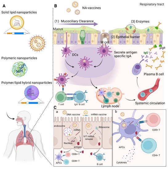
Recent advancements in the field of in vitro transcribed mRNA (IVT-mRNA) vaccination have attracted considerable attention to such vaccination as a cutting-edge technique against infectious diseases including COVID-19 caused by SARS-CoV-2. While numerous pathogens infect the host through the respiratory mucosa, conventional parenterally administered vaccines are unable to induce protective immunity at mucosal surfaces. Mucosal immunization enables the induction of both mucosal and systemic immunity, efficiently removing pathogens from the mucosa before an infection occurs. Although respiratory mucosal vaccination is highly appealing, successful nasal or pulmonary delivery of nucleic acid-based vaccines is challenging because of several physical and biological barriers at the airway mucosal site, such as a variety of protective enzymes and mucociliary clearance, which remove exogenously inhaled substances. Hence, advanced nanotechnologies enabling delivery of DNA and IVT-mRNA to the nasal and pulmonary mucosa are urgently needed.

2. DNA Vaccines

The first proof of concept for in vivo protein expression with nucleic acids was reported in 1990 by injecting DNA or RNA molecules into mouse skeletal muscle for the expression of chloramphenicol acetyltransferase, luciferase, and galactosidase [1]. It was thereafter demonstrated that the production of cytotoxic T lymphocytes for influenza could be induced by injecting plasmid DNA (pDNA) encoding influenza A nucleoproteins into the quadriceps of BALB/c mice [2]. These pioneer studies confirmed sufficient immunogenicity of DNA vaccines in animal models, providing evidence of this immunization platform’s promising ramifications. DNA vaccines are generally constructed by inserting gene fragments encoding immunogenic antigens into a bacterial plasmid vector, forming pDNA. After pDNA is delivered into the host cell nucleus, antigenic proteins are subsequently expressed. Generally, APCs are the primary targets to be transfected with the genetic material. Following effective presentation in APCs, foreign antigenic proteins initiate specific immune responses [3]. DNA vaccines offer several advantages over conventional vaccines (e.g., live-attenuated, inactivated, or subunit vaccines). Also, DNA vaccines are generally stable at room temperature (though this may vary between formulations), avoiding the need for an uninterrupted cold chain during storage and transport [4]. Large-scale manufacturing of DNA vaccines primarily involves synthesis of relevant nucleic acids followed by standard cloning into plasmid vectors, avoiding the time- and labor-intensive culturing procedures required by traditional subunit and virus-based vaccines [5]. In contrast to subunit vaccines, DNA vaccines have been demonstrated to induce more potent cytotoxic T cell responses without severe side effects [6]. Although the potential possibility of genome integration remains the primary theoretical safety concern with DNA vaccines, this scenario has not been realized across large numbers of studies and reports [6]. Numerous clinical investigations have also demonstrated DNA vaccines to be largely safe in humans [7][8][9]. All these advantages make DNA vaccines an ideal candidate for rapid responses in the event of epidemic and pandemic outbreaks. Indeed, the first DNA vaccine to be approved for human use was a COVID-19 DNA vaccine (ZyCoV-D) developed in India. It was found to be 67% protective in clinical trials [10], providing evidence that DNA vaccines can be effective in controlling the pandemic [11].
In past decades, DNA vaccines have been studied for the prevention and treatment of a variety of diseases, such as infectious diseases, cancer, autoimmune diseases, and allergies. However, the immunogenicity of DNA vaccines in humans is not as sufficient as that in mouse studies to elicit significant clinical benefits [7]. The poor transport of pDNA into the nucleus of host cells results in low antigen synthesis, limiting the protective immunological responses in the recipient. Several approaches have been explored in recent years to address this issue, including the optimization of codon sequences/transcriptional elements, the incorporation of adjuvants, and enhancing delivery technologies. In this section, we discuss several well-studied delivery vehicles used for DNA vaccines that are administered via nasal or pulmonary routes.

2.1. Delivery of DNA Vaccines via Respiratory Routes

Intranasal and pulmonary administration of DNA vaccines have attracted widespread attention recently because of various enticing properties. Early reports on inhaled DNA vaccines combined plasmids encoding ovalbumin, hepatitis B surface antigen, and HLA-A*0201-restricted T cell epitopes of Mycobacterium tuberculosis (M. tuberculosis), which resulted in enhanced immunity as indicated by antibodies and cytokine production [12][13]. These pioneering investigations suggested that mucosal immune responses can be more effectively elicited when DNA vaccines are delivered directly to mucosal sites. However, there are still many obstacles that need to be overcome to more effectively utilize mucosal DNA vaccines. Vaccines must penetrate the mucus layer, translocate into target cells, and avoid extracellular and intracellular degradation in order to be effective (Figure 1). For example, delivery via the nasal cavity exposes DNA vaccines to being trapped by the nasal mucus, resulting in enzymatic breakdown. The viscosity and pore size of the mucus layer markedly affect the effective diffusivity of particles on the airway surfaces. Mucociliary clearance from cilia cells also significantly determines the fate of entrapped DNA vaccines. It continuously pushes mucus outwards, expelling mucus from the nasal channel and limiting residence time at the mucosal surface. The dilution effect in bulk mucosal fluids can also impede successful deposition onto the epithelium.
Figure 1. Overview of nucleic acid-based (NA-) vaccines administrated via the respiratory tract using nanotechnologies. (A) Schematic view of different nanoparticles used for intranasal and pulmonary vaccinations. (B) Physical and biological barriers at the airway mucosal site and mechanism of immune responses in the respiratory tract mediated by mucosal-associated lymphoid tissues (MALTs). NA-vaccines transcytose from the mucus layer into the epithelial tissues by microfold cells (M cells) or passively diffuse through epithelial cell junctions. Other NA-vaccines are captured and internalized by APCs, such as DCs, from their extension through epithelial junctions. APCs that have been transfected with genetic antigens migrate to the nearest lymph node to activate T cells and B cells. Activated B cells proliferate in the lymph node and enter the systemic circulation to the mucosal effector sites. B cells locally differentiate into antibody-secreting plasma cells to produce IgA dimers. IgA dimers are secreted via pIgR at the mucosal surface. Antigen-specific systemic IgG is also produced. (C) NA-vaccines are taken up by epithelial cells (a), and pathogen-derived antigens are then transcribed and translated from plasmid DNA or IVT mRNA and secreted into the extracellular space, where they can be taken up by professional APCs such as DCs. (b). APCs then present antigens to naïve T cells for activation and differentiation, promoting humoral and cell-mediated immune responses against the encoded antigen.
As a result, a safe and effective DNA delivery mechanism must be designed to overcome these obstacles. A suitable delivery system should target mucosal APCs for antigens processing, resulting in selective B and T cell activation. The ultimate goals of DNA delivery systems are to promote uptake of DNA into target tissues and cells, protect DNA from enzymatic breakdown, extend residence time at the target site, boost antigen expression, and optimize immune response, all without sacrificing safety. Section 2.2 discusses in detail several DNA vaccine delivery technologies that have been evaluated for respiratory administration.

2.2. Delivery Systems for DNA Vaccines via Respiratory Routes

The most prevalent technologies in the delivery of DNA constructs, such as electroporation, particle bombardment, and jet injectors, have exhibited improved transfection efficiency and huge potential in clinical trials when compared to traditional intramuscular injection of naked pDNA [14][15]. However, these attractive methods are inapplicable for the respiratory route, necessitating the use of a potent delivery strategy to overcome the barriers in the respiratory system. Advancements in nanotechnologies and material science have proven to be beneficial in the effective delivery of pDNA by the synthesis of DNA nanoparticles with diverse structures. Nanoparticles can better pass cell membranes via endocytosis while preventing premature degradation of pDNA and subsequently promote intracellular trafficking into the nucleus following endosomal escape, increasing immunogenicity in animal models and humans. Most importantly, sustained and controlled release of pDNA from nanocarriers at the delivery site recruits APCs, resulting in an improved antigen-specific immune response. The immunogenicity of DNA by vaccines can be further enhanced employing additional adjuvants. Currently, cationic lipids and polymers are two of the most employed nanomaterials for DNA vaccines.

2.2.1. Liposomes and Niosomes

Liposomes and some other vesicular systems are widely used as delivery systems for DNA vaccines. Liposomes generally consist of aqueous cores surrounded by phospholipid bilayers. For antigen delivery, two approved liposomal vaccine formulations used to be available: Inflexal® V (influenza vaccine) and Epaxal® (hepatitis A vaccine). Both of these formulations use virosome-based technology in which viral proteins are bonded to the surface of a liposome carrier similarly to how viral particles are bound [16]. The goal is to imitate a safe viral-like particle capable of eliciting significant protective immune responses. Although both examples have been discontinued, comparable technology could be used to increase the immunogenicity of DNA vaccines. To achieve this, pDNA could be either electrostatically complexed on the surface of cationic liposomes or encapsulated in the aqueous core by a dehydration–rehydration procedure. In general, cationic liposomes have shown higher in vitro transfection efficiency, while nonionic or anionic counterparts have shown enhanced antibody responses in animal models [17][18]. Surface modifications with antigenic components or targeting ligands further enhance immune responses of liposome-based vaccines [18]. It has been shown that liposomes coated with glycol chitosan are mucus adhesive and immune system stimulating [19]. Surface-modified cationic liposomes such as phosphatidylcholine (PC), dioleoyl phosphatidylethanolamine (DOPE), and cholesterol (Chol) elicited stronger humoral, mucosal, and cell-mediated immune responses post intranasal administration in mice against hepatitis than uncoated counterparts [18]. Liposomes containing noncoding pDNA have also been shown to exhibit adjuvant-like behavior, inducing elevated antibody levels and T cell immunity in mice and nonhuman primates [20].
Liposomes have been used as DNA vaccine carriers for intranasal delivery in several investigations to induce efficient immune responses against respiratory pathogens. To induce immune protection against M. tuberculosis, D’Souza et al. adopted pDNA encoding antigen 85A formulated with a (+/−)-N-(3-aminopropyl)-N,N-dimethyl-2,3-bis (dodecyloxy)-1-propanaminium bromide (GAP-DLRIE):DOPE liposome [21]. After intranasal immunization in mice, a positive splenic Th1-type cytokine response was induced in GAP-DLRIE:DOPE formulations, but this response was still weaker than that obtained from intramuscular administration with the same dosage. However, the combination of intranasal and intramuscular injections elicited even stronger Th1 type immune responses in the lungs [21]. Another study, performed by Rosada et al., described how a single intranasal immunization with liposome-based formulations of pDNA encoding HSP65 against M. tuberculosis led to a remarkable reduction in the amount of bacilli in lungs of mice [22]. The authors employed egg phosphatidylcholine (EPC), DOPE, and 1,2-dioleoyl-3-trimethylammonium-propane (DOTAP) to formulate the delivery system. This formulation also increased the production of IFN-γ and lung parenchyma protection to a level similar to that in mice vaccinated intramuscularly four times the dosage of naked pDNA encoding HSP65 [22]. In addition, intranasal immunization with liposome-based DNA vaccine provided complete protection against influenza after a viral challenge assay [23]. Mice immunized intranasally with liposome-encapsulated pDNA encoding hemagglutinin (HA) protein, but not naked plasmid, were found to produce strong serum IgA/IgG responses and increased IgA titers in bronchoalveolar lavage fluid (BALF) [24]. T cell-proliferative responses were also successfully induced in both intranasal and intramuscular administration [24]. These studies demonstrated the ability of liposomes in the delivery of DNA vaccines inoculated via the intranasal route to confer significant immune protection against respiratory infections in animal models. However, widespread adoption of liposome-based vaccines remains stunted by their relatively lower physical and chemical stability in aqueous dispersions during long-term storage [25]. Accordingly, numerous methods to improve the stability of liposome formulations during storage have been investigated, including freeze-drying, spray-drying, supercritical fluid technology, and lyophilization [26][27][28].
Niosomes, which are nonionic surfactant-based vesicles, have been developed as alternative delivery systems to liposomes because of their advantages such as cost-effective manufacturing, large-scale producibility, and stability [29][30]. Because of their structural similarities to liposomes, niosomes were also applied as vehicles for pDNA, small interference RNAs (siRNAs), and aptamers in target cells [31]. Cationic niosomes, containing cationic lipids, made an effective vector for pDNA delivery and achieved ~95% transfection efficiency in vitro [32]. Later, the same research team reported successful transfection of human tyrosinase gene (pMEL34) and the stability of developed cationic niosomes in transdermal delivery [33]. Perrie et al. reported that niosomes carried with H3N2 influenza virus resulted in enhanced immune response after subcutaneous administration in mice [34]. Mannolysated niosomes encapsulated with pDNA encoding HBsAg were reported to provoke protective immunity against hepatitis B as both a DNA vaccine carrier and adjuvant for oral immunization [35]. However, there have been no reports utilizing niosomes as a mucosal delivery platform in the respiratory tract as far as we know. Their efficacy for the intranasal and pulmonary delivery of DNA vaccine needs further investigation.

2.2.2. Polymers

One of the most appealing characteristics of polymer-based DNA delivery technologies is their flexibility in structure design and modification. Electrostatic interactions allow cationic polymers to form complexes (polyplexes) with DNA vaccines. Polymer synthesis is also relatively inexpensive and simple to scale up. To maximize cellular uptake and transfection effectiveness, the size and surface characteristics of polymeric particles can be adjusted by employing different polymers and methods of preparation [36][37]. It has been found that alveolar macrophages are particularly effective in absorbing particles with diameters ranging from 300 to 600 nm, so the particle size should be less than 3 μm (preferably under 500 nm) for DC-targeted absorption in the respiratory tract [38]. Aside from particle size, particle charge also influences cellular absorption in APCs in the respiratory tract. Where both DCs and macrophages are substantially present, preferential uptake of DNA vaccines into DCs is desirable. DCs can produce large quantities of peptide–MHC II complexes, which are then presented on the cellular surface to initiate T cell activation and differentiation [39]. It has been found that macrophages have higher phagocytic activity than DCs, but also that absorption in DCs can be increased by imparting positive charge to the particle [40]. Polymeric particles can also be modified with functional groups or ligands to improve the cellular uptake of DCs. DCs can preferentially uptake ligand-modified nanoparticles via receptor-mediated endocytosis using C-type lectin receptors or mannose receptors [41].

Polyethylenimine

Polyethylenimine (PEI) is one of the most well-studied polymers with high transfection efficiency and has been extensively applied for mediating in vitro and in vivo transfection of DNA molecules [42]. Compared to lipid-based formulations, DNA complexed with PEI has shown improved stability and higher levels of pulmonary transfection, even after nebulization [43][44]. For intranasal immunization, PEI/pDNA complexes encoding SARS-CoV spike proteins induced higher antigen-specific Th2 dominant IgG and IgA antibodies in BALF than naked plasmid counterparts [45]. Cellular immune responses were also detected in a PEI/pDNA treated group, with increased B cells and higher numbers of IFN-γ-, TNF-α-, and IL-2-producing T cells in the lungs [45]. A H5N1 intranasal vaccine with DNA encoding HA formulated with PEI induced potent mucosal and systemic immune responses and elicited both full protection against the parental strain and partial cross-protection against a distinct highly pathogenic strain [46]. Pulmonary immunization of DNA vaccines formulated with PEI also induced robust systemic and CD8+ T-cell responses in the gut and vaginal mucosa [47]. Furthermore, mice inoculated via the intratracheal (i.t.) route elicited higher levels of interleukin-2 than those inoculated by intramuscular immunization in lung-associated antigen-specific CD4+ T cells [47]. These robust T cell responses, which were induced by i.t. but not intramuscular administration, protected mice from a lethal recombinant vaccinia virus challenge [47]. A similar study also reported that robust pulmonary CD8+ T cell populations effectively mediated protective immunity against influenza respiratory challenges after pulmonary immunization with PEI/pDNA [48].
Although PEI appears to be a promising delivery vector for airway inoculated DNA vaccines, one remaining major limitation is its toxicity due to its highly positively-charged and nondegradable nature. Immunization with PEI vaccine was found to provoke the activation of genes with apoptosis, stress responses, and oncogenesis [49]. As a result, biodegradable PEI derivatives with low-toxic profiles have been developed for DNA delivery. A less toxic form of PEI called deacylated PEI (dPEI) with potent transfection efficiency was applied in delivering pDNA encoding HA [50]. Essentially, dPEI is a completely hydrolyzed linear PEI with 11% more free protonatable nitrogen atoms than conventional PEI. Following intranasal administration, dPEI-complexed pDNA vaccine formulations were capable of generating strong systemic and mucosal humoral responses, activating cellular responses, and mediating a higher degree of protection in a challenge study against influenza [50]. Other strategies using covalent modification or electrostatic neutralization of PEI’s cationic group to reduce zeta potential have also been investigated. Poly-lactic-co-glycolic acid (PLGA), a synthetic biodegradable copolymer, has been approved by the Food and Drug Administration (FDA) for human use in delivering therapeutic agents such as proteins and nucleic acids [51]. The negative charge and hydrophobic nature of PLGA could be used to neutralize the positive charge of PEI for safe nucleic acid delivery. Bivas-Benita et al. developed PLGA nanoparticles bearing PEI on their surfaces. Internalization of the DNA-loaded PLGA–PEI nanoparticles was also studied in the human airway submucosal epithelial cell line, Calu-3 [52]. The results suggested that DNA could be detected in the endolysosomal compartment after 6 h incubation with Calu-3 cells and that and the optimal cell viability was achieved when the weight ratio of PEI to DNA was between 1:1 and 0.5:1 [52]. A similar study reported the formulation of PLGA–PEI microparticles, in which 10% PEI (w/w) efficiently adsorbed DNA and protected DNA from enzymatic degradation [53]. Intramuscular immunization of mice with such PLGA–PEI formulations loaded with pDNA encoding immunodominant antigens of Listeria monocytogenes demonstrated that the formulation had an adjuvant effect [53]. These studies indicated that PEI has a favorable profile to be a nonviral gene carrier for DNA vaccines delivered through the respiratory tract, but also that further optimization is still necessary to realize their full potential.

Chitosan

Chitosan is a biodegradable and biocompatible polysaccharide derived from chitin and has been frequently employed as a DNA delivery vector because of its biodegradability and biocompatibility [54]. Furthermore, chitosan and its derivatives possess substantial mucoadhesive properties, making them ideal for intranasal administration [55][56]. Chitosan has also been reported to be immune stimulating by enhancing macrophage accumulation and activation, increasing cytokines’ resilience against infections, and promoting cytotoxic T cell response [57][58]. To assess its potential in mediating mucosal immunization, chitosan was used to complex with pDNAs encoding nine different antigens (NS1, NS2, M, SH, F, M2, N, G, and P) from respiratory syncytial virus (RSV) [58]. A single intranasal administration of chitosan–pDNA resulted in a significant reduction of viral titers and viral antigen load in the lungs after an acute RSV infection [58]. In addition, significantly elevated levels of serum RSV-specific IgG antibodies, nasal IgA antibodies, cytotoxic T lymphocytes, and IFN-γ production in the lung and splenocytes were detected in comparison with controls [58]. However, when pDNA encoding the M2 proteins of RSV antigens was formulated with chitosan, virus-specific CTL responses in BALB/c mice were induced only at a level that was comparable to those induced via intradermal immunization [59]. Nonetheless, aerosolized pDNA–chitosan nanoparticles induced higher levels of IFN-γ through pulmonary administration than counterparts immunized by intratracheal and intramuscular administration [13].
In order to achieve targeted delivery of antigen to DCs, biotinylated chitosan nanoparticles loaded with pDNA encoding the nucleocapsid (N) protein of SARS-CoV were developed by Raghuwanshi et al. [60]. Chitosan was modified with bifunctional fusion protein (bfFp) consisting of truncated core-streptavidin fused with anti-DEC-205 single chain antibody (scFv) [60]. The core-streptavid in the arm of bfFp bonded with biotinylated nanoparticles, while anti-DEC-205 scFv imparted targeting specificity to the DCs’ DEC-205 receptors. Intranasal administration of such targeted formulations led to the detection of an enhanced number of N protein-specific systemic IgG and nasal IgA antibodies [60]. In another study, mannosylated chitosan (MCS) formulated with DNA vaccine encoding a multi-T-epitope was employed to facilitate airway delivery and antigen targeting to the APCs in the alveoli [61]. Following intranasal immunization, HSP65-specific sIgA in the BALF was significantly elevated. A modest antigen-specific Th1 (IFN-γ, TNF-α, and IL-2) response and a potent polyfunctional CD4+ T response were induced for enhancing mucosal immune protection against M. tuberculosis in the spleen and lung, respectively [61].
Thiolated chitosan derivatives have been found to improve the transfection efficiency of chitosan for intranasal delivery. In a study performed by Bernkop-Schnürch et al., thiol-bearing moieties were introduced on the polymeric backbone of chitosan in order to prepare thiolated chitosan that could interact with mucus glycoproteins via the formation of disulfide bonds [62]. The results indicated that thiolated chitosan improved mucosa adhesiveness to the mucus layer 6- to 100-fold compared to the unmodified counterpart, resulting in enhanced mucus permeation. Simultaneously, increased penetration at the mucosal surface was also observed in chitosan-coated PLGA nanoparticles encapsulating macromolecules [63][64]. Based on this fact, an emulsion–diffusion–evaporation technique was employed to prepare cationic nanospheres composed of biodegradable and biocompatible copolyester PLGA, with a PVA–chitosan blend stabilizing the PLGA nanospheres [65]. Despite the charge on the nanospheres being sufficient to bind the negatively charged DNA, the immunity of DNA vaccines complexed by this formulation remains to be illuminated. In one study, chitosan-coated PLGA was employed to deliver pDNA encoding foot-and-mouth disease (FMDV) capsid protein and bovine IL-6 to protect mice against FMDV infections [66]. This chitosan/PLGA/pDNA vaccine formulation provided enhanced protective immunity against FMDV post-intranasal immunization [66].

References

  1. Wolff, J.A.; Malone, R.W.; Williams, P.; Chong, W.; Acsadi, G.; Jani, A.; Felgner, P.L. Direct gene transfer into mouse muscle in vivo. Science 1990, 247, 1465–1468.
  2. Ulmer, J.B.; Donnelly, J.J.; Parker, S.E.; Rhodes, G.H.; Felgner, P.L.; Dwarki, V.J.; Gromkowski, S.H.; Deck, R.R.; DeWitt, C.M.; Friedman, A.; et al. Heterologous protection against influenza by injection of DNA encoding a viral protein. Science 1993, 259, 1745–1749.
  3. Silveira, M.M.; Moreira, G.M.S.G.; Mendonça, M. DNA vaccines against COVID-19: Perspectives and challenges. Life Sci. 2021, 267, 118919.
  4. Ingolotti, M.; Kawalekar, O.; Shedlock, D.J.; Muthumani, K.; Weiner, D.B. DNA vaccines for targeting bacterial infections. Expert Rev. Vaccines 2010, 9, 747–763.
  5. Xu, Y.; Yuen, P.-W.; Lam, J.K.-W. Intranasal DNA Vaccine for Protection against Respiratory Infectious Diseases: The Delivery Perspectives. Pharmaceutics 2014, 6, 378–415.
  6. Mallapaty, S.; Callaway, E. What scientists do and don’t know about the Oxford-AstraZeneca COVID vaccine. Nature 2021, 592, 15–17.
  7. Kutzler, M.A.; Weiner, D.B. DNA vaccines: Ready for prime time? Nat. Rev. Genet. 2008, 9, 776–788.
  8. Tebas, P.; Yang, S.; Boyer, J.D.; Reuschel, E.L.; Patel, A.; Christensen-Quick, A.; Andrade, V.M.; Morrow, M.P.; Kraynyak, K.; Agnes, J.; et al. Safety and immunogenicity of INO-4800 DNA vaccine against SARS-CoV-2: A preliminary report of an open-label, Phase 1 clinical trial. EClinicalMedicine 2020, 31, 100689.
  9. Modjarrad, K.; Roberts, C.C.; Mills, K.T.; Castellano, A.R.; Paolino, K.; Muthumani, K.; Reuschel, E.L.; Robb, M.L.; Racine, T.; Oh, M.-D.; et al. Safety and immunogenicity of an anti-Middle East respiratory syndrome coronavirus DNA vaccine: A phase 1, open-label, single-arm, dose-escalation trial. Lancet. Infect. Dis. 2019, 19, 1013–1022.
  10. Sheridan, C. First COVID-19 DNA vaccine approved, others in hot pursuit. Nat. Biotechnol. 2021, 39, 1479–1482.
  11. Mallapaty, S. India’s DNA COVID vaccine is a world first—More are coming. Nature 2021, 597, 161–162.
  12. Lombry, C.; Marteleur, A.; Arras, M.; Lison, D.; Louahed, J.; Renauld, J.-C.; Préat, V.; Vanbever, R. Local and systemic immune responses to intratracheal instillation of antigen and DNA vaccines in mice. Pharm. Res. 2004, 21, 127–135.
  13. Bivas-Benita, M.; van Meijgaarden, K.E.; Franken, K.L.M.C.; Junginger, H.E.; Borchard, G.; Ottenhoff, T.H.M.; Geluk, A. Pulmonary delivery of chitosan-DNA nanoparticles enhances the immunogenicity of a DNA vaccine encoding HLA-A*0201-restricted T-cell epitopes of Mycobacterium tuberculosis. Vaccine 2004, 22, 1609–1615.
  14. Low, L.; Mander, A.; McCann, K.; Dearnaley, D.; Tjelle, T.; Mathiesen, I.; Stevenson, F.; Ottensmeier, C.H. DNA vaccination with electroporation induces increased antibody responses in patients with prostate cancer. Hum. Gene Ther. 2009, 20, 1269–1278.
  15. Rottinghaus, S.T.; Poland, G.A.; Jacobson, R.M.; Barr, L.J.; Roy, M.J. Hepatitis B DNA vaccine induces protective antibody responses in human non-responders to conventional vaccination. Vaccine 2003, 21, 4604–4608.
  16. Brito, L.A.; Malyala, P.; O’Hagan, D.T. Vaccine adjuvant formulations: A pharmaceutical perspective. Semin. Immunol. 2013, 25, 130–145.
  17. Legendre, J.Y.; Szoka, F.C.J. Delivery of plasmid DNA into mammalian cell lines using pH-sensitive liposomes: Comparison with cationic liposomes. Pharm. Res. 1992, 9, 1235–1242.
  18. Khatri, K.; Goyal, A.K.; Gupta, P.N.; Mishra, N.; Mehta, A.; Vyas, S.P. Surface modified liposomes for nasal delivery of DNA vaccine. Vaccine 2008, 26, 2225–2233.
  19. Gogev, S.; de Fays, K.; Versali, M.-F.; Gautier, S.; Thiry, E. Glycol chitosan improves the efficacy of intranasally administrated replication defective human adenovirus type 5 expressing glycoprotein D of bovine herpesvirus 1. Vaccine 2004, 22, 1946–1953.
  20. Lay, M.; Callejo, B.; Chang, S.; Hong, D.K.; Lewis, D.B.; Carroll, T.D.; Matzinger, S.; Fritts, L.; Miller, C.J.; Warner, J.F.; et al. Cationic lipid/DNA complexes (JVRS-100) combined with influenza vaccine (Fluzone) increases antibody response, cellular immunity, and antigenically drifted protection. Vaccine 2009, 27, 3811–3820.
  21. D’Souza, S.; Rosseels, V.; Denis, O.; Tanghe, A.; De Smet, N.; Jurion, F.; Palfliet, K.; Castiglioni, N.; Vanonckelen, A.; Wheeler, C.; et al. Improved tuberculosis DNA vaccines by formulation in cationic lipids. Infect. Immun. 2002, 70, 3681–3688.
  22. Rosada, R.S.; de la Torre, L.G.; Frantz, F.G.; Trombone, A.P.F.; Zárate-Bladés, C.R.; Fonseca, D.M.; Souza, P.R.M.; Brandão, I.T.; Masson, A.P.; Soares, E.G.; et al. Protection against tuberculosis by a single intranasal administration of DNA-hsp65 vaccine complexed with cationic liposomes. BMC Immunol. 2008, 9, 38.
  23. Wong, J.P.; Zabielski, M.A.; Schmaltz, F.L.; Brownlee, G.G.; Bussey, L.A.; Marshall, K.; Borralho, T.; Nagata, L.P. DNA vaccination against respiratory influenza virus infection. Vaccine 2001, 19, 2461–2467.
  24. Wang, D.; Christopher, M.E.; Nagata, L.P.; Zabielski, M.A.; Li, H.; Wong, J.P.; Samuel, J. Intranasal immunization with liposome-encapsulated plasmid DNA encoding influenza virus hemagglutinin elicits mucosal, cellular and humoral immune responses. J. Clin. Virol. Off. Publ. Pan Am. Soc. Clin. Virol. 2004, 31 (Suppl. 1), 99–106.
  25. Chen, C.; Han, D.; Cai, C.; Tang, X. An overview of liposome lyophilization and its future potential. J. Control. Release 2010, 142, 299–311.
  26. Mishima, K. Biodegradable particle formation for drug and gene delivery using supercritical fluid and dense gas. Adv. Drug Deliv. Rev. 2008, 60, 411–432.
  27. Lo, Y.; Tsai, J.; Kuo, J. Liposomes and disaccharides as carriers in spray-dried powder formulations of superoxide dismutase. J. Control. Release 2004, 94, 259–272.
  28. Kadimi, U.S.; Balasubramanian, D.R.; Ganni, U.R.; Balaraman, M.; Govindarajulu, V. In vitro studies on liposomal amphotericin B obtained by supercritical carbon dioxide-mediated process. Nanomedicine 2007, 3, 273–280.
  29. Baldino, L.; Reverchon, E. Niosomes formation using a continuous supercritical CO2 assisted process. J. CO2 Util. 2021, 52, 101669.
  30. Durak, S.; Esmaeili Rad, M.; Alp Yetisgin, A.; Eda Sutova, H.; Kutlu, O.; Cetinel, S.; Zarrabi, A. Niosomal Drug Delivery Systems for Ocular Disease-Recent Advances and Future Prospects. Nanomaterials 2020, 10, 1191.
  31. Grijalvo, S.; Puras, G.; Zárate, J.; Sainz-Ramos, M.; Qtaish, N.A.L.; López, T.; Mashal, M.; Attia, N.; Díaz, D.; Pons, R.; et al. Cationic Niosomes as Non-Viral Vehicles for Nucleic Acids: Challenges and Opportunities in Gene Delivery. Pharmaceutics 2019, 11, 50.
  32. Rose, J.K.; Buonocore, L.; Whitt, M.A. A new cationic liposome reagent mediating nearly quantitative transfection of animal cells. Biotechniques 1991, 10, 520–525.
  33. Manosroi, J.; Khositsuntiwong, N.; Manosroi, W.; Götz, F.; Werner, R.G.; Manosroi, A. Enhancement of transdermal absorption, gene expression and stability of tyrosinase plasmid (pMEL34)-loaded elastic cationic niosomes: Potential application in vitiligo treatment. J. Pharm. Sci. 2010, 99, 3533–3541.
  34. Perrie, Y.; Barralet, J.E.; McNeil, S.; Vangala, A. Surfactant vesicle-mediated delivery of DNA vaccines via the subcutaneous route. Int. J. Pharm. 2004, 284, 31–41.
  35. Jain, S.; Singh, P.; Mishra, V.; Vyas, S.P. Mannosylated niosomes as adjuvant-carrier system for oral genetic immunization against hepatitis B. Immunol. Lett. 2005, 101, 41–49.
  36. Singh, M.; Chakrapani, A.; O’Hagan, D. Nanoparticles and microparticles as vaccine-delivery systems. Expert Rev. Vaccines 2007, 6, 797–808.
  37. Fifis, T.; Gamvrellis, A.; Crimeen-Irwin, B.; Pietersz, G.A.; Li, J.; Mottram, P.L.; McKenzie, I.F.C.; Plebanski, M. Size-dependent immunogenicity: Therapeutic and protective properties of nano-vaccines against tumors. J. Immunol. 2004, 173, 3148–3154.
  38. Gratton, S.E.A.; Ropp, P.A.; Pohlhaus, P.D.; Luft, J.C.; Madden, V.J.; Napier, M.E.; DeSimone, J.M. The effect of particle design on cellular internalization pathways. Proc. Natl. Acad. Sci. USA 2008, 105, 11613–11618.
  39. Blank, F.; Wehrli, M.; Lehmann, A.; Baum, O.; Gehr, P.; von Garnier, C.; Rothen-Rutishauser, B.M. Macrophages and dendritic cells express tight junction proteins and exchange particles in an in vitro model of the human airway wall. Immunobiology 2011, 216, 86–95.
  40. Thiele, L.; Rothen-Rutishauser, B.; Jilek, S.; Wunderli-Allenspach, H.; Merkle, H.P.; Walter, E. Evaluation of particle uptake in human blood monocyte-derived cells in vitro. Does phagocytosis activity of dendritic cells measure up with macrophages? J. Control. Release 2001, 76, 59–71.
  41. De Temmerman, M.-L.; Rejman, J.; Demeester, J.; Irvine, D.J.; Gander, B.; De Smedt, S.C. Particulate vaccines: On the quest for optimal delivery and immune response. Drug Discov. Today 2011, 16, 569–582.
  42. Boussif, O.; LezoualC’H, F.; Zanta, M.A.; Mergny, M.D.; Scherman, D.; Demeneix, B.; Behr, J.P. A versatile vector for gene and oligonucleotide transfer into cells in culture and in vivo: Polyethylenimine. Proc. Natl. Acad. Sci. USA 1995, 92, 7297–7301.
  43. Densmore, C.L.; Orson, F.M.; Xu, B.; Kinsey, B.M.; Waldrep, J.C.; Hua, P.; Bhogal, B.; Knight, V. Aerosol Delivery of Robust Polyethyleneimine-DNA Complexes for Gene Therapy and Genetic Immunization. Mol. Ther. 2000, 1, 180–188.
  44. Gautam, A.; Densmore, C.L.; Xu, B.; Waldrep, J.C. Enhanced gene expression in mouse lung after PEI-DNA aerosol delivery. Mol. Ther. 2000, 2, 63–70.
  45. Shim, B.-S.; Park, S.-M.; Quan, J.-S.; Jere, D.; Chu, H.; Song, M.K.; Kim, D.W.; Jang, Y.-S.; Yang, M.-S.; Han, S.H.; et al. Intranasal immunization with plasmid DNA encoding spike protein of SARS-coronavirus/polyethylenimine nanoparticles elicits antigen-specific humoral and cellular immune responses. BMC Immunol. 2010, 11, 65.
  46. Torrieri-Dramard, L.; Lambrecht, B.; Ferreira, H.L.; Van den Berg, T.; Klatzmann, D.; Bellier, B. Intranasal DNA vaccination induces potent mucosal and systemic immune responses and cross-protective immunity against influenza viruses. Mol. Ther. 2011, 19, 602–611.
  47. Bivas-Benita, M.; Bar, L.; Gillard, G.O.; Kaufman, D.R.; Simmons, N.L.; Hovav, A.-H.; Letvin, N.L. Efficient Generation of Mucosal and Systemic Antigen-Specific CD8+ T-Cell Responses following Pulmonary DNA Immunization. J. Virol. 2010, 84, 5764–5774.
  48. Bivas-Benita, M.; Gillard, G.O.; Bar, L.; White, K.A.; Webby, R.J.; Hovav, A.H.; Letvin, N.L. Airway CD8+ T cells induced by pulmonary DNA immunization mediate protective anti-viral immunity. Mucosal Immunol. 2013, 6, 156–166.
  49. Regnström, K.; Ragnarsson, E.G.E.; Köping-Höggård, M.; Torstensson, E.; Nyblom, H.; Artursson, P. PEI—A potent, but not harmless, mucosal immuno-stimulator of mixed T-helper cell response and FasL-mediated cell death in mice. Gene Ther. 2003, 10, 1575–1583.
  50. Mann, J.F.S.; McKay, P.F.; Arokiasamy, S.; Patel, R.K.; Klein, K.; Shattock, R.J. Pulmonary delivery of DNA vaccine constructs using deacylated PEI elicits immune responses and protects against viral challenge infection. J. Control. Release 2013, 170, 452–459.
  51. Panyam, J.; Labhasetwar, V. Biodegradable nanoparticles for drug and gene delivery to cells and tissue. Adv. Drug Deliv. Rev. 2003, 55, 329–347.
  52. Bivas-Benita, M.; Romeijn, S.; Junginger, H.E.; Borchard, G. PLGA-PEI nanoparticles for gene delivery to pulmonary epithelium. Eur. J. Pharm. Biopharm. Off. J. Arb. fur Pharm. Verfahr. e.V 2004, 58, 1–6.
  53. Oster, C.G.; Kim, N.; Grode, L.; Barbu-Tudoran, L.; Schaper, A.K.; Kaufmann, S.H.E.; Kissel, T. Cationic microparticles consisting of poly(lactide-co-glycolide) and polyethylenimine as carriers systems for parental DNA vaccination. J. Control. Release 2005, 104, 359–377.
  54. Kang, M.L.; Cho, C.S.; Yoo, H.S. Application of chitosan microspheres for nasal delivery of vaccines. Biotechnol. Adv. 2009, 27, 857–865.
  55. Casettari, L.; Vllasaliu, D.; Lam, J.K.W.; Soliman, M.; Illum, L. Biomedical applications of amino acid-modified chitosans: A review. Biomaterials 2012, 33, 7565–7583.
  56. Issa, M.M.; Köping-Höggård, M.; Artursson, P. Chitosan and the mucosal delivery of biotechnology drugs. Drug Discov. Today. Technol. 2005, 2, 1–6.
  57. Seferian, P.G.; Martinez, M.L. Immune stimulating activity of two new chitosan containing adjuvant formulations. Vaccine 2000, 19, 661–668.
  58. Kumar, M.; Behera, A.K.; Lockey, R.F.; Zhang, J.; Bhullar, G.; De La Cruz, C.P.; Chen, L.-C.; Leong, K.W.; Huang, S.-K.; Mohapatra, S.S. Intranasal gene transfer by chitosan-DNA nanospheres protects BALB/c mice against acute respiratory syncytial virus infection. Hum. Gene Ther. 2002, 13, 1415–1425.
  59. Iqbal, M.; Lin, W.; Jabbal-Gill, I.; Davis, S.S.; Steward, M.W.; Illum, L. Nasal delivery of chitosan-DNA plasmid expressing epitopes of respiratory syncytial virus (RSV) induces protective CTL responses in BALB/c mice. Vaccine 2003, 21, 1478–1485.
  60. Raghuwanshi, D.; Mishra, V.; Das, D.; Kaur, K.; Suresh, M.R. Dendritic cell targeted chitosan nanoparticles for nasal DNA immunization against SARS-CoV nucleocapsid protein. Mol. Pharm. 2012, 9, 946–956.
  61. Wu, M.; Zhao, H.; Li, M.; Yue, Y.; Xiong, S.; Xu, W. Intranasal vaccination with mannosylated chitosan formulated DNA vaccine enables robust IgA and cellular response induction in the lungs of mice and improves protection against pulmonary mycobacterial challenge. Front. Cell. Infect. Microbiol. 2017, 7, 445.
  62. Bernkop-Schnürch, A.; Hornof, M.; Guggi, D. Thiolated chitosans. Eur. J. Pharm. Biopharm. Off. J. Arb. fur Pharm. Verfahrenstechnik e.V 2004, 57, 9–17.
  63. Nafee, N.; Taetz, S.; Schneider, M.; Schaefer, U.F.; Lehr, C.-M. Chitosan-coated PLGA nanoparticles for DNA/RNA delivery: Effect of the formulation parameters on complexation and transfection of antisense oligonucleotides. Nanomedicine 2007, 3, 173–183.
  64. Janes, K.A.; Calvo, P.; Alonso, M.J. Polysaccharide colloidal particles as delivery systems for macromolecules. Adv. Drug Deliv. Rev. 2001, 47, 83–97.
  65. Ravi Kumar, M.N.V.; Bakowsky, U.; Lehr, C.M. Preparation and characterization of cationic PLGA nanospheres as DNA carriers. Biomaterials 2004, 25, 1771–1777.
  66. Wang, G.; Pan, L.; Zhang, Y.; Wang, Y.; Zhang, Z.; Lü, J.; Zhou, P.; Fang, Y.; Jiang, S. Intranasal delivery of cationic PLGA nano/microparticles-loaded FMDV DNA vaccine encoding IL-6 elicited protective immunity against FMDV challenge. PLoS ONE 2011, 6, e27605.
More
Video Production Service