Your browser does not fully support modern features. Please upgrade for a smoother experience.
Please note this is a comparison between Version 1 by Atanu Jana and Version 2 by Camila Xu.
Luminescent micelles are extensively studied molecular scaffolds used in applied supramolecular chemistry.
- luminescent micelle
- functional nano-materials
- explosive sensor
1. Introduction
In the last decades, significant advancement has been made to construct the luminescent micellar systems for sensing target analytes, ion transport, and bioimaging. These luminophoric materials are used to demonstrate the rudimentary models for various CT/ET processes in an aqueous solution in the presence of various chemical entities [1][2][3][4][5][6][7][1,2,3,4,5,6,7]. Based on the fundamental idea of CT/ET transition, a considerable number of elegant luminous micellar systems are developed for sensing metal ions [8][9][10][11][12][8,9,10,11,12] and anions [13][14][15][13,14,15], biomolecules [16][17][18][19][20][16,17,18,19,20], detection of explosive [21][22][23][24][25][21,22,23,24,25], etc. to name a few. Moreover, these micellar systems can be typically considered as the simplest biological mimic of cellular membranes having both hydrophobic and hydrophilic parts in a single molecular platform. Therefore, they might be useful materials considering the artificial synthetic models compatible for biosensing. In addition to that, augmenting particular luminophoric moieties to the micellar systems may provide specific hybrid nano-composites selective for target analytes. It is noteworthy that compared to the conventional luminescent/optical sensors, these luminescent micellar sensors are a new category that require more exposure to unfurl their potential applications in sensing activity.
The ever-increasing use of explosive chemicals for homeland security, armed military forces, mining, road construction, demolition of old buildings and civil structures, and more importantly by separatist groups, terrorist organizations, and criminals has raised the necessity of their trace level detection by various sensitive techniques. Depending on the chemical structures, these explosives can be categorized as nitroaromatics, nitrate esters, nitramines, and peroxides. Among them, nitroaromatic and nitramine explosives are the most commonly used high energy explosives (secondary explosives), widely used by armed forces and also by separatist and terrorist organizations. Nitroaromatic explosives are aromatic compounds containing single or multiple electron poor nitro (–NO2) groups. The most common examples of nitroaromatic compounds are DNT, TNT, TNP (or PA), etc. On the other hand, the representative examples of chemical explosives fall under the category of nitramine, which includes RDX, HMX, and Tetryl, etc. Sensing of these explosive analytes for forensic applications by using various “chemical noses” in different medium, is therefore, an important topic in applied supramolecular chemistry research, which is growing rapidly in the present era. In current literature, explosive sensing and their materials applications by various purely organic or composite materials have been described in a few review articles [26][27][26,27]. However, to the best of our knowledge, a detailed review article based on supramolecular micellar systems exclusively meant for forensic applications has yet to appear in the current literature. Considering the applied supramolecular chemistry, materials chemistry research, sensor development, and timeliness of this topic, we therefore, strongly believe that a comprehensive review article is urgently necessary to summarize the recent development of the luminescent micellar sensors. Particularly, in this review article we will highlight the progress on various luminescent micellar systems which are effective in an aqueous medium. The efforts have also been made on the working principles of the sensing behavior of each micellar–analyte supramolecular system. We also discuss the role of explosive analytes to control the CT phenomena, which has tremendous influence on both ground and excited state dynamics. In general, the luminescence quenching processes are involved (with few exceptions of enhanced emission) in the sensing of explosive analytes depending on their acceptor strengths and geometries.
2. Luminescent Micellar Systems
The chemistry of luminescent micellar systems originally developed decades ago, however, a short discussion might be relevant here for neophytes. Figure 1a shows a typical model structure of a micelle that is structurally different from reverse micelle liposome and lipid bilayer (Figure 1b–d). Typically, a micellar system consists of a hydrophobic core resulted from self-assembled organic synthons having a hydrophilic head and hydrophobic tail groups (cf. Figure 1, Inset) [28][29][30][28,29,30]. To construct a luminescent micellar system, either polyaromatic hydrocarbon (PAH)-based fluorophore(s) or a luminescent metal complex tag should be attached or grafted to the skeleton. While the hydrophobic and hydrophilic part self-assembled to form a micellar structure, the luminophores too stacked either on the surface or within the core. The specific arrangement of luminophores in the core/surface/intermediate layer of micelle results in monomer/excimer emission with much enhanced quantum yield compared to that in organic solvents under identical conditions. In the presence of explosive materials (which are generally electron poor in nature due to the presence of multiple NO2 groups present in a single unit), a D–A type of host–guest complex can be produced with micellar systems where the explosive molecules are intercalated within the lyophilic part of the micelles. Normally, the sensing behavior resulted from either by quenching of initial luminescence of the existing luminophore or quenching of the excimer emission by intermolecular D–A charge transfer transitions. Few cases are also reported where the luminescence sensing was achieved by the fluorescence “Turn ON” mechanism. The efficacy of such a micellar system as a sensor can be easily determined by the Stern–Volmer constant (KSV) using the Stern–Volmer equation given below:
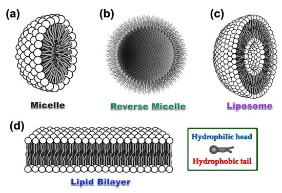
Figure 1. Graphical representation of a standard micelle (a), reverse micelle (b), liposome (c), and lipid bilayer (d), respectively. Inset: A single unit consists of hydrophobic aliphatic tail and hydrophilic head, which takes part to construct all the above-mentioned self-assembled structures with various geometries under different environments.

Figure 2. The proposed mechanism involved in the “Turn OFF” luminescent sensing of explosive analytes. (a) Luminescence quenching of excimer-based emission in the presence of explosive analytes. (b) Luminescence quenching of a surface-modified micellar system by supramolecular D–A CT transition within the cyclometallated luminophore (D) and explosive analytes (A). The models are redrawn based on the concept used in references [22][23][22,23].
2.1. Luminescent Micellar Systems for Metal Ion Sensing
A wide variety of luminescent micellar systems have been reported so far in the current literature demonstrating metal ion sensing [8][9][10][11][12][31][32][33][34][35][36][37][38][39][40][41][42][43][44][45][46][47][48][49][50][8,9,10,11,12,31,32,33,34,35,36,37,38,39,40,41,42,43,44,45,46,47,48,49,50]. However, as our main focus is on explosive sensing in a micellar medium, we will cover the metal ion sensing in this section in brief. Most well-known cases that have fused heterocyclic organic fluorophores or traditional polyaromatic hydrocarbons (PAH) have been utilized (see Chart 1 for details) to construct luminescent micellar sensors. The major advantages of these micellar organic luminophores include: (i) Enhanced emission intensity of the monomer or excimer through vibrational rigidity, (ii) selective site binding with metal ions as compared to bulk phase, (iii) mimicking biological membrane-like systems and in vivo sensing feasibility, (iv) selective and sensitive sensing of metal ions through enhanced emission of organic luminophore via controlled binding, and (v) the use of lipophilic fluorophores in aqueous medium solubilized by micelles, etc. to name a few.
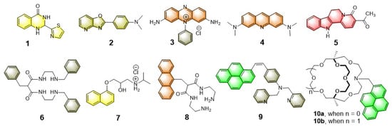
Chart 1. Chemical structures of few representative fluorophores and receptors (both chelated and macrobicyclic) used for metal ion sensing in an aqueous micellar medium.
In addition to that, depending on the charge state of micellar systems (cationic, anionic, or neutral) and the nature of coordinating atoms, the sensing efficacy is directly related to the pH of the medium. Based on these ideas, various luminescent probes to detect transition metal ions (viz. Cu2+, Fe3+, Hg2+, Cd2+, etc.) have been successfully demonstrated, which are able to work in an aqueous medium. For example, the fluorescent “Turn ON” metal ion sensors 10a,b are reported by Nakatsuji and Akashi et al. which are constructed by using laterally unsymmetric bicyclic oxa-aza cryptands [34]. These photosensitive monoazacryptand derivatives are used to sense the alkali and alkaline earth metal ions in the presence of various surfactants, depending on the size of the inner core. Typically, the sensing mechanism involves the deactivation pathway of the PET process between cryptands and fluorophore (pyrene), which results in the “Turn ON” emission switching upon metal binding. It was noticed that the 15-crown-5 -based fluorophoric system (10a) is selective for K+, however, the corresponding larger analogue 10b (18-crown-6 -based system) is selective for Ba2+ in an aqueous medium at ambient condition. It is speculated that a bigger core diameter of oxa-aza cryptand 10b provides a suitable platform to encapsulate Ba2+ that make the receptor 10b selective for Ba2+ ion.
Taking advantage of the aggregated pyrene-based excimer emission, Ding and co-workers used a series of synthetic luminescent molecular probes for constructing the surfactant ensembled metal ion sensors, which are nicely summarized in a recent article [12]. Among the series, the neutral bispyrene derivative shown in Figure 3 inset provides substantial sensing behavior to various di- and tri-valent metal ions. Interestingly, when the bispyrene/SDS assemblies are titrated against Fe3+, Cu2+, Hg2+, emission quenching was observed (cf. Figure 3a,b in case of Fe3+, Cu2+ respectively), however, incremental addition of Mg2+, Ca2+, Co2+, Ni2+, Zn2+, Cd2+, and Pb2+ exhibit ratiometric response (cf. Figure 3c,d in case of Co2+ and Zn2+ as representative examples). A prominent optical change in the presence of these metal ions under exposure of UV light reflects their potential applications in metal ion sensing. The suggested mechanism involving the sensing behavior of bispyrene/SDS adduct against these di- and tri-valent metal ions by emission quenching and ratiometric pathway is shown in Figure 3e. The flexibility of the long aliphatic bridge of bispyrene derivative plays an important role during the sensing process.
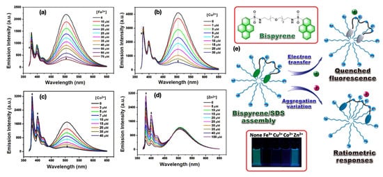
Figure 3. (a–c) Emission quenching of bispyrene/SDS assemblies with the incremental addition of Fe3+, Cu2+, and Co2+ respectively, in an aqueous medium at ambient condition. (d) The ratiometric change of bispyrene/SDS assemblies when titration was performed against Zn2+ ion. (e) The schematic representation of a bispyrene/SDS self-assembled sensor towards various metal ions. Inset: Top, the Chemdraw representation of bispyrene derivative; bottom, the photograph of bispyrene/SDS sensor in the presence of Fe3+, Cu2+, Co2+, and Zn2+ respectively, under exposure of UV light. This figure is reproduced with permission from reference [12], Copyright © 2022, American Chemical Society.
Apart from these organic fluorophores, a DANSYL appended macromolecular system is reported by Zhao et al. to detect Hg2+ ion selectively in an aqueous medium. This lyophilic organic molecular probe is solubilized in an aqueous medium by the help of surfactant-based micellar systems. A more detailed account on luminescent micellar systems for metal ion sensing can be found in the following recent review article [33].
2.2. Luminescent Micellar Systems for Anion Sensing
Among various anionic species, sensing of halides (e.g., F−, Cl−, Br−, and I−) and cyanide (CN−) by luminescent micellar probes have been extensively studied [13][14][15][51][52][53][54][55][13,14,15,51,52,53,54,55]. Although a large number of model sensors are reported in current literature, however, as mentioned earlier, our primary focus in this current review is to highlight the most compelling applications of micellar systems for explosive sensing. Therefore, exclusive discussion on anion sensing with micellar systems in this part is intentionally precluded to avoid repetition. Nevertheless, emphasis is placed on some quintessential micellar systems as the representative examples which can be treated as model micellar systems for anion sensing in an aqueous medium. The sensing strategies mostly relied on using the receptors containing the metal complexes which selectively binds specific anions to give a substantial luminescence signal. In addition to that, another interesting class of anion sensor is also observed in current literature where the anion reacts with the receptor/fluorophore to produce a substituted/addition product with changed luminescence output. Few representative examples of such fluorophoric systems reported so far in the current literature for anion sensing in aqueous micellar medium are shown in Chart 2. Considering CN− sensing in water, the bis-indolyl fluorophores, 11–13, undergo Michael addition reaction with CN− which give rise to the new luminophores. Nevertheless, the probes exhibit very weak detection limit due to poor solubility as a result of aggregation. However, in micellar media, the probes behave effectively with a much lower detection limit (8 ppb) through improved solubilization of the probes. The other fluorophores used for CN− sensing includes 2,4,6- triarylthiopyrylium (14) encapsulated with a neutral surfactant (Triton X-100)-based micelle reported by Manez et al. [13]. In a similar fashion, the probes 15–20 has been used for sensing the halides (Cl−, F−) and cyanide (CN−) ions after being solubilized through micellar media.
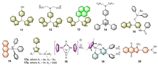
Chart 2. Chemical structures of various luminophores used for sensing specific anions in the aqueous micellar media.
To demonstrate Cl− sensing by metal complexes, the work of Severin and coworkers is noteworthy [15]. Their aqueous buffered solution was loaded with a Rh(II)-complex (19) as a coordinating site and hydroxypyrenesulfonate as fluorophore, which is mixed with a supplementary surfactant. The resulting adduct remarkably increases Cl− affinity. It was described that Cl− binds to the metal center, which in turn changes the phase behavior of the whole system. The metal complex now in turn becomes amphiphilic in nature as compared to its initial ionic state, which can easily move towards the lyophilic core of the micelle. The resulting species causes significant modulation of the luminescence property. A fluorescence quenching was observed, which is directly related to the added Cl− concentration (Figure 4a,b). This in situ generated anion sensor showcased a model system which exhibits high sensitivity and selectivity, by signal transduction mechanism, which can effectively work in a neutral aqueous solution (i.e., at pH = 7).
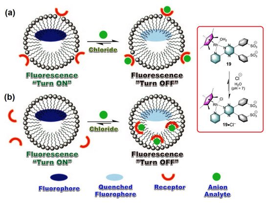
Figure 4. (a,b) The proposed mechanism involved in “Turn OFF” luminescent sensing of Cl− anion as analyte by using a Rh(II)-metal complex. Inset: The working principle of activation of metal complex by chloride anion complexation. This graphic is reproduced with permission from reference [15], Copyright © 2022 WILEY-VCH Verlag GmbH & Co. KGaA, Weinheim, Germany.
Bhattacharya et al. reported a “Turn-ON” luminescence sensor composed by the surface modification of cationic micellar system (CTAB) with naphthalimide derivative [53]. Due to the partial CT from Br− to the naphthalimide moieties, the CTAB micellar adduct is non-emissive in nature. However, upon addition of strong anions (e.g., PO43−, SO42−, MoO42−, WO42−, H2PO4− etc.) this adduct exhibits significant enhancement of emission under identical experimental condition. It was speculated that these strong anions can effectively replace the bromide counter anions from the periphery of CTAB. Therefore, partial CT transition operating within the Br− anions to the naphthalimide moieties is restricted, that results a substantial fluorescence enhancement of the adduct. Moreover, these strong anions rearrange the initial spherical micellar structure to an elongated core-modified geometry as shown in Figure 5.
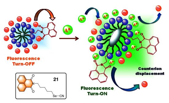
Figure 5. The proposed mechanism involved in “Turn ON” luminescent sensing of anions as analytes. Inset: The chemical structure of the naphthalimide-based fluorophoric unit 21. This figure is reproduced with permission from reference [53], Copyright © 2022 Elsevier B.V.
Sessler and Khashab et al. reported an interesting thermo-responsive amphiphilic polymer, P1 {poly(N-isopropylacrylamide)-b-poly(calix[4]pyrrole-co-methyl methacrylate} [56] where hydrophobic calix[4]pyrrole (C4P) are attached as the anion receptor (Figure 6, Inset). This polymer was designed to capture the contaminated anions within pendent C4Ps in its micellar form which effectively works under an extraction-free condition. This is a typical demonstration of a water purification process by a synthetic C4P-based polymeric system where the target anions can be effectively “captured” and “removed” from contaminated water under easy experimental conditions. Owing to the thermo-responsive characteristics of the hydrophilic block chain, the anion incorporated micelles that can be precipitated out from the aqueous phase upon heating the aqueous solution at 50 °C. Then, a simple filtration technique can be employed to remove the generated precipitate from the anion contaminated aqueous solution. Finally, the pristine polymer (P1) can be easily recovered by treating the anion-trapped micelles with a dilute nitric acid (0.2 M) solution at room temperature. The whole reversible process of anion removal and regeneration of the polymer (P1) is provided in Figure 6.
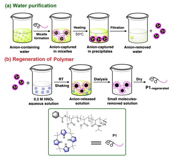
Figure 6. (a) The proposed mechanism involved in targeted anion “capture” and “remove” by the use of polymer P1 with pendent C4P receptors. (b) The sequence used to regenerate the pristine polymer P1 under ambient condition. Inset: The chemical structure and cartoon representation of P1. Reproduced with permission from reference [56], 2018 Wiley-VCH Verlag GmbH & Co. KGaA, Weinheim, Germany.
2.3. Luminescent Micellar Systems for Bio Sensing
Biosensors are the integrated receptor-transducer devices that emerge as a core research topic for many researchers in recent time. This is mainly due to their wide range of applications in the area of health care and diagnosis, environmental monitoring, etc. to name a few [57][58][57,58]. Among them, much attention has been devoted in designing the luminescent biosensors as they offer outstanding advantages, such as high sensitivity, low background noise, and facile sample preparation [59][60][61][59,60,61]. Considering the advancement of nanotechnology and nanoscience, nanostructured micellar probes have been considered as a promising candidate for bio-sensing owing to their structural flexibility and versatility [62]. However, only a handful of luminescent biosensor probes based on micellar systems appeared in the recent literature [16][17][18][19][20][63][64][65][66][67][68][69][70][71][72][73][74][16,17,18,19,20,63,64,65,66,67,68,69,70,71,72,73,74]. We will provide a very brief glimpse on these luminescent biosensors, which are used to create self-assembled nanostructure materials showing promise for use as next generation potential as “functional materials”.
Pioneering work by Li and Yang et al. on poly β-Cyclodextrin/TPdye nanomicelle-based two-photon nanoprobe is reported describing two-photon-induced fluorescent microscopic detection of enzymatic activities in living cells and tissues [63]. In this nanoprobe, a two-photon active (TPA) donor molecule trans-4-[p-(N,N-diethylamino)styryl]-N-methylpyridinium iodide (DEASPI) was used as a complementary combination with β-CDP nanomicelle. This combined adduct serves as a potential TPA fluorophore and also behaves like a carrier vehicle to deliver a specific peptide sequence to the living cell through fast endocytosis. For example, an adamantine GRRRDEVDK-BHQ2 peptide (Ad-DEVD-BHQ2) can be easily mounted to construct a robust inclusion complex, DEASPI/βCDP@Ad-DEVD-BHQ2 onto the nanomicellar surface by various noncovalent interactions (cf. Figure 7). The adduct DEASPI/βCDP@Ad-DEVD-BHQ2 was then used to demonstrate both in vitro and in vivo enzymatic activities assay of caspase-3 in the complex biological environment. From their experiments, it is evident that this “Turn-ON” fluorescent biosensor offers a new platform for high-contrast imaging of enzymatic activities within live cells and tissues. (Figure 7, Inset) Thus, DEASPI/βCDP@Ad-DEVD-BHQ2 nanoconjugate also offers the opportunity to screen enzyme inhibitors, which are also able to evaluate the apoptosis-associated disease progression.
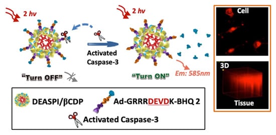
Figure 7. Schematic illustration of the DEASPI/βCDP@Ad-DEVD-BHQ2 nanoconjugate for caspase-3 activity assay. Orange Inset: The TPM image of HeLa cells treated with 4 μM STS for 3 h after incubating with DEASPI/βCDP@Ad-DEVD-BHQ2 nanoconjugate (top); and TPM image of the 1.0 mm thick cervical tumor tissue slice pretreated with doxorubicin after incubating with DEASPI/βCDP@Ad-DEVD-BHQ2 nanoconjugate (0.15 mg/mL, concentration of the nanoconjugate refers to the concentration of βCDP). Black Inset: Cartoon representations of few components. This figure is reproduced with permission from reference [63], Copyright © 2022, American Chemical Society.
Another fluorescent micellar nanoprobe (NanoDPA-NMP-tyr) is reported describing sensing of tyrosinase (TYR) in a living B16 cellular medium [16]. NanoDPA-NMP-tyr is constructed with a hydrophobic interior of the amphiphilic copolymer mPEG-DSPE that shows a very fast response towards TYR with high sensitivity and selectivity with a detection limit of 0.057 U/mL. The sensing behavior involved in this nanoprobe is thought to be due to the Förster Resonance Energy Transfer (FRET) from 9,10-diphenylanthracene (DPA) (λem = ∼445 nm) donor to the naphthalimide-tyrosene (NMP-tyr) (λabs = ∼450 nm) acceptor.
Recently Lee et al. reported an interesting model sensor [17] to sense heparin (HP) with amplified fluorescence response resulted from inter-fluorophore excitation migration within a self-assembled micellar medium. For this purpose, a coumarin-based amphiphilic fluorescent molecule (see Figure 8, Inset) is designed which forms a micellar structure consisting of a hydrophobic core and hydrophilic surface, where the aggregated coumarins promote the energy and electron transport under bioanalytical conditions. The sensing response of micellized 22 (5.0 × 10⁻⁶ M) via ionic (cationic–anionic) interactions is also highly selective for HP among the other tested analytes e.g., dextrose, sucrose, glucose, mannitol, ATP, sodium citrate, Na2SO4, Na3PO4, sodium hyaluronate (HA), and chondroitin sulfate sodium salt (ChS) in a 10-mM HEPES buffer solution at pH 7.4 at ambient condition (Figure 8a). High selectivity of the micellized 22 (5.0 × 10⁻⁶ M) for HP via fluorescence suppression, reaches a quenching maximum of ~78.4% with an incubation of HP of concentration 2.0 × 10⁻⁶ M (Figure 8b). The sensing mechanism for the detection of heparin by the conjugated micellar system of coumarin derivative 22 is shown in Figure 8c.
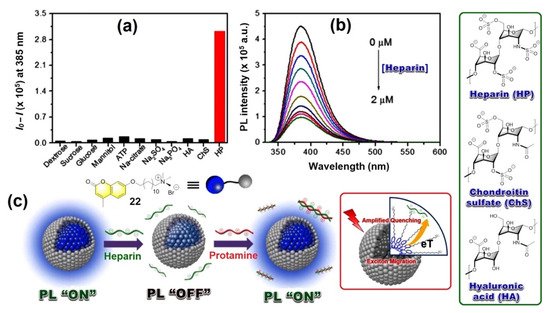
Figure 8. (a) Fluorescence response of micellized 22 (5.0 × 10⁻⁶ M) towards various analytes (1.7 × 10⁻⁶ M), and (b) Fluorescence intensity changes of micellized 22 upon gradual addition of HP (0 ~ 2.0 × 10⁻⁶ M) in 10 mM HEPES buffer solution at pH 7.4. λexc = 320 nm and fluorescence intensity were monitored at a 385 nm wavelength. (c) The proposed mechanism involving “ON”/”OFF” fluorescence switching by heparin. Green Inset: Structure of various biomolecules. Red Inset: The schematic diagram of amplified quenching mechanism involved with micellized 22.
Fan et al. demonstrated discriminative luminescence sensors [18] for metal and nonmetal proteins by employing a pyrene/bispyrene-based fluorophoric system which are encapsulated in a cationic micellar structure. In case of a metal-free protein system, comparative luminescence intensity of monomer and excimer emission of pyrenes displayed quenching of an excimer emission whereas the monomer emission remain unchanged. On the other hand, metal containing proteins showed substantial quenching of both monomer and excimer emission under identical experimental conditions.
Selective detection of ATP is another important case of bio-sensing in recent times. Pang et al. have demonstrated a cationic squaraine dye encapsulated by CTAB based micelles, which can selectively detect ATP [19]. It is speculated that the ionic interactions between cationic CTAB head groups and anionic ATP along with the π−π staking of ATP and the dye molecules enormously contributed towards sensing behavior.
Likewise, Jiang et al. recently reported a luminescent micellar system [20] for ATP, comprised of a cationic surfactant, dodecyltrimethylammonium bromide (DTAB), and a near infrared (NIR) emissive cyanine dye. As mentioned above, a similar π−π staking within ATP and dye molecule triggered an effective energy transfer (ET) from the dye to ATP that resulted in significant quenching of emission intensity. A more detailed account on luminescent micellar sensors for sensing bioanalytes can be found in reference [4][5][4,5].