Extremophilic Fungi from Marine Environments: Comparison
Please note this is a comparison between Version 2 by Vivi Li and Version 1 by Lesley-Ann Giddings.

Marine environments are underexplored terrains containing fungi that produce a diversity of natural products given unique environmental pressures and nutrients. While bacteria are commonly the most studied microorganism for natural products in the marine world, marine fungi are also abundant but remain an untapped source of bioactive metabolites. Given that their terrestrial counterparts have been a source of many blockbuster antitumor agents and anti-infectives, including camptothecin, the penicillins, and cyclosporin A, marine fungi also have the potential to produce new chemical scaffolds as leads to potential drugs. Fungi are more phylogenetically diverse than bacteria and have larger genomes that contain many silent biosynthetic gene clusters involved in making bioactive compounds. However, less than 5% of all known fungi have been cultivated under standard laboratory conditions. While the number of reported natural products from marine fungi is steadily increasing, their number is still significantly lower compared to those reported from their bacterial counterparts.

  • extreme environments
  • marine fungi
  • bioactive compounds
  • marine natural products
  • fungal cultivation strategies

1. Introduction

Natural products have been a rich source of therapeutics since the beginning of civilization. To date, ~60% of all small-molecule drugs approved by the US Food and Drug Administration (FDA), or their equivalent in other countries, have been inspired by these secondary metabolites [1]. Many are anti-infectives and antitumor agents that have been isolated from microbes, including those with symbiotic associations. While microbes are a prolific source of bioactive molecules used to treat cancer and infectious diseases, chemists have been repetitively rediscovering natural products, thus leading to the requirement for a system for the rapid dereplication of known compounds and/or their close relatives. Microbes usually express fewer biosynthetic gene clusters (BGCs) when grown under standard laboratory conditions, even though they have the genetic potential to express more secondary metabolic BGCs. To circumvent these limitations, scientists are either cultivating microbes under nominally non-standard laboratory conditions to activate the expression of cryptic BGCs [2], using genetic engineering and ‘omics methods to identify and directly express BGCs [3], or they are isolating microbes from understudied environments [4].
Extreme ecosystems represent new frontiers for drug discovery, as these locations contain unique environmental variables that are not found in mesophilic locales and influence microbial metabolism. Extreme environments have been underexplored due to the challenges in accessing these locales. However, more sampling tools and techniques have been developed to study marine environments, presenting new opportunities for exploration. The marine world is vast, covering ~70% of Earth, including some of the most extreme ecosystems on the planet. Extreme marine environments are characterized as having high pressures, and temperatures that can range from close to freezing to over 250 °C at the “black smokers”, severe ultraviolet radiation, high salt or metal concentrations, symbiosis, or more than one combination of these variables. Microbes constitute much of the biomass in the sea and are prominent producers of secondary metabolites, with a wide range of potent bioactivities against tumor cells and other pathogens, plus potential in other diseases. Thus, bioprospecting these environments for new drugs is an attractive prospect, especially as marine natural products have high hit rates in a broad range of bioassays, with many in clinical and preclinical trials. For a current assessment, the web site, www.marinepharmacology.org (accessed on 12 December 2021), is kept up to date, covering approved drugs from marine sources and compounds that are in clinical trials from Phase I to Phase III plus selected preclinical candidates. Most of the information is from the NIH trials database (www.clinicaltrials.gov accessed on 12 December 2021), but it also includes data from comparable databases from other countries.
Most microbe-derived marine natural products tend to be isolated from bacteria, which make up much of the biomass in seawater (i.e., >105 cells per milliliter) and are extremely well represented in deep oceanic “muds”. However, fungi are also abundant, as 103 to 104 fungal cells are contained in a milliliter of seawater. Approximately 38% of the reported ~23,000 bioactive microbial metabolites are of fungal origin and only 5% of the microbial taxa had been identified as of 2008, 10% of which are fungi [5]. Fungi have larger genomes with more BGCs, resulting in greater chemodiversity [6,7][6][7]. Furthermore, they have produced numerous clinically approved anti-infectives and antitumor drugs, as mentioned earlier, but remain understudied.
Why are there disparities in the isolation of marine natural products from fungi? While genetic engineering and ‘omics methods are being used to isolate an increasing amount of bacterial natural products, these methods are still in their infancy insofar as fungi are concerned. Cultivation which is still one of the main ways to discover bioactive metabolites continues to be a challenge when working with marine fungi. Scientists are still using antiquated methods that promote the growth of generalist genera, such as Penicillium and Aspergillus to culture marine fungi [8]. In 2019, we published a compendium of bioactive compounds isolated from marine fungi [9]. Herein, we update that list, focusing on anti-infective and antitumor agents as well as other fungal natural products with potential from marine environments. This review aims to highlight the bioactive metabolites found from culturable fungi isolated from marine environments and cultivation methods that have been found to increase the production of diverse bioactive metabolites.
Although we are listing significant numbers of compounds (approximately 200) in this review, we will only show the structures of compounds for which biological activities have been reported and will give the names of the others with suitable references, so that readers may consult the original literature for related compounds without a biological activity reported at that time.

1.1. Antitumor Agents from Deep-Sea Sediments

It should be mentioned at this point that it was well known in the early days of antimicrobial assays, though never formally reported from the pharmaceutical industry, that compounds with antitumor activities in vitro also inhibited Gram-positive bacteria at comparable concentrations in disc diffusion assays (DJN Personal Observations). The following examples will be arranged as best we can, by the area(s) from which the sediments were collected for subsequent isolation of fungi.

1.2. Indian Ocean

Numerous potential antitumor agents (some also with antibacterial activity) continue to be isolated and reported from deep-sea-derived fungi collected from the Indian Ocean. The new diterpenoids, longidiacids A (1) and B, polyketides, and the cytochalasin analogs, longichalasins A and B (2), were isolated from the fungus Diaporthe longicolla FS429 [10] obtained from deep-sea sediment collected at a depth of 3000 m. Longidiacid A (1) and longichalasin B (2) inhibited tyrosine phosphatase B in Mycobacterium tuberculosis cells by 35.4% and 53.5%, respectively, and longichalasin B (2) exhibited antiproliferative activity against glioblastoma cells (SF-268) with an IC50 value of 16.44 μM.
From sediment collected at a depth of 3471 m, the fungus Cladosporium cladosporiodes HDN14-342 yielded the polyketides, clindanones A and B as well as cladosporols F and G (34) following cultivation [11]. Cladosporols F and G exhibited cytotoxic activities against the human cervical (HeLa), leukemia (K562), and colon (HCT-116) cancer cells with IC50 values ranging from 3.9 to 23.0 μM depending upon the particular cell line. In 2017, Li et al. published revisions of the initial structures of these two metabolites [12].
Phomopsis lithocarpus FS508 was isolated from a sediment sample collected at 3606 m and fermented, producing several benzophenone aldehydes, including tenellone H (5). This benzophenone exhibited cytotoxic activity against liver (HepG-2; IC50 16 μM) and lung (A549; IC50 17.6 μM) cancer cell lines [13]. At the even deeper depth of 5752 m, the trimeric peniphenylanes A and B and dimeric peniphenylanes C–G were reported from Penicillium fellutanum HDN14-323 isolated from that deep-sea sediment. Of these seven compounds, only peniphenylane D (6) exhibited reasonable cytotoxic activity against the cervical cancer cell (HeLa; IC50 = 9.3 μM) [14].

1.3. Seas near China

Moving further East, several cytotoxic agents have been reported from fungi from deep-sea sediment collected in the South China Sea. For example, the cytotoxic agent acaromycin A (7) along with acaromyester A were isolated from Acaromyces ingoilii FS121 collected from deep-sea sediment at a depth of 3415 m. The naptha-[2,3-b] pyrandione analog acaromycin A (7) exhibited cytotoxic activities against human breast (MCF-7), brain (SF-268), liver (HepG-2), and lung (NCI-H460) cancer cells with IC50 values less than 10 μM [15].
At a deeper depth of 3739 m, engyodontiumones A–J and 2-methoxyl-cordyol C were isolated from Engyodontium album DFFSCS02. Engyodontiumone H (8) exhibited cytotoxic activity against human histiocytic lymphoma U937 cells (IC50 = 4.9 μM) together with antibacterial activity against Escherichia coli and Bacillus subtilis at a concentration of 25 μg/disc (which is low activity for a disc diffusion assay). Engyodontiumin A (9), isolated from the same fungus, exhibited antimicrobial activities against Aspergillus niger, multidrug-resistant Staphylococcus aureusVibrio vulnificusV. rotiferianus, and V. campbellii [16].

1.4. Eastern Pacific Ocean

Several new breviane spiroditerpenoids, breviones F–I, were isolated from a deep ocean sediment-derived Penicillium sp. (MCCC 3A00005) collected in the East Pacific Ocean at a depth of 5115 m [17]. Brevione F (10) inhibited HIV-1 replication in C8166 cells with an EC50 value of 14.7 μM. Breviones F–H (1012) exhibited minimal cytotoxic activity against HeLa cells (25 to 50% growth inhibition at 10 μg/mL). Finally, from these agents, brevione I (13) was cytotoxic against the breast cancer line MCF-7 (IC50 = 7.44 μM) but barely active against the lung cancer A549 cell line (IC50 = 32.5 μM) [18].
A series of sorbicillinoid derivatives, trisorbicillinone A–D, oxosorbiquinol, dihydrooxosorbiquinol, dihydrodemethylsorbicillin, dihydrotrichodermolide, and phialofurone, were isolated from the deep ocean fungus Phialocephala sp. Fl30 r collected from sediment at 5059 m in the East Pacific Ocean [19,20,21][19][20][21]. Of these agents, only trisorbicillinone A (14) exhibited any significant cytotoxic activity against the HL60 tumor cell line (IC50 = 3.14 μM) [19]. Oxosorbiquinol (15) and its dihydro derivative (16) were marginally to effectively inactive as cytotoxins against the following tumor cell lines; leukemia (P388, HL60, and K562) and the hepatocellular BEL7402 cell lines with IC50 values from 8.9 to 68.2 μM, respectively [19]. By contrast, dihydrodemethylsorbicillin (17), dihydrotrichodermolide (18), and phialofurone (19Figure 1) exhibited cytotoxic activity against the leukemia cell lines (P388 and K562) with IC50 values ranging from 0.1 to 22.9 μM [21].
Figure 1. Antitumor Active Agents {Multiple Ocean Areas} (Structures 1 to 19).

2. Anti-Infective Agents from Deep-Sea Fungi

2.1. South Atlantic Ocean

Marine fungi are also abundant sources of anti-infectives, including potential antifungal, antibacterial, antiprotozoal, and antiviral agents. For example, 19 thiodiketopiperazine alkaloids, eutypellazines A–S, were isolated from the marine-derived fungus Eutypella sp. MCCC 3A00281 collected at a depth of 5610 m in the South Atlantic Ocean [22,23][22][23]. Eutypellazines A–L (2031) exhibited anti-HIV activity against pNL4.3Env-Luc co-transfected 293T cells with IC50 values ranging from 3.2 to 18.2 μM. Eutypellazines P–S (3235Figure 2) inhibited the growth of S. aureus ATCC 25923 and vancomycin-resistant Enterococci with MIC values ranging from 16 to 32 μM [22,23][22][23].
Figure 2. Anti-HIV and Antibacterial Active Agents {South Atlantic Ocean, South China Sea, Indian and West Pacific Oceans} (Structures 20 to 35).

2.2. South China Sea

The alkaloids arthpyrones D–K were isolated from Arthrinium sp. UJNMF0008 collected from the South China Sea at a depth of 3858 m. Of these eight, four, arthpyrones F–I (3639), exhibited antibacterial activity against Mycobacterium smegmatis and S. aureus with IC50 values ranging from 1.66 to 42.8 μM [24]. From the deep-sea fungus, P. brevicompactum DFFSCS025, isolated from sediment collected at 3928 m depth in the South China Sea, two alkaloids, breviamides X and Y, and two mycochromenic acid derivatives, 6-(methyl 3-methylbutanoate)-7-hydroxy-5-methoxy-4-methylphthalan-1-one (40) and (3′S)-(E)-7-hydroxy-5-methoxy-4-methyl-6-(2-(2-methyl-5-oxotetrahydrofuran-2-yl)vinyl) isobenzofuran-1(3H)-one, were purified. Of these four, only compound 40 exhibited significant antifouling activity against the bryozoan Bugula neritina with an EC50 value of 13.7 μM and an LC50/EC50 value > 100 [25].
A small series of compounds were isolated from fermentation of the fungus Emericella sp. SCSIO 05240, collected from sediment at a depth of 3258 m in the South China sea. These were four new prenylxanthones, named as emerixanthones A–D. These four agents structurally differ by chlorination, methylation, and hydroxylation, and of the four congeners, two, emerixanthones A (41) and C (42), exhibited weak antibacterial activity against several bacterial pathogens, including E. coliKlebsiella pneumoniaS. aureusE. faecalisAcinetobacter baumanni, and Aeromonas hydrophilia. Interestingly, one of the remaining two compounds, emerixanthone D (43), exhibited mild antifungal activity against several agricultural pathogens, including Fusarium sp., Penicillium sp., A. nigerRhizoctonia solaniFusarium oxysporium f. sp. niveum, and F. oxysporium f. sp. cucumeris [26].

2.3. Indian Ocean

From sediment collected in the Indian Ocean at a depth of 3972 m, the versicoloids A and B as well as two 4-aryl-quinolin-2-one alkaloids and prenylated xanthones, versicones A–D, were isolated from culturing the fungus A. versicolor SCSIO 05879. Of these four compounds, only versicoloids A and B (4445) demonstrated activity against the plant pathogen Colletotrichum acutatum (MIC 1.6 μg/mL) [27].

2.4. West Pacific Ocean

From sediment collected at a depth of 2869 m in the West Pacific Ocean, the fungus A. versicolor was isolated and upon fermentation produced the anthraquinone antimicrobial agent 2-(dimethoxymethyl)-1-hydroxyanthracene-9,10-dione (46Figure 3). This compound exhibited antibacterial activity against multidrug-resistant strains of S. aureus ATCC 43300 (MIC 3.9 μg/mL) and CGMCC 1.12409 (MIC 7.8 μg/mL). In addition, it was also weakly active against strains of Vibrio (MICs of 15.6–62.5 μg/mL). Molecular docking studies with topoisomerase IV and AmpC β-lactamase further supported its antibacterial properties [28].
Figure 3. Biologically Active Agents, Indian and West Pacific Ocean (Structures 36 to 46).

3. Bioactive Compounds from Fungal Endophytes of Mangroves

Marine fungi are often involved in mutualistic interactions, as their survival may well require relationships including symbiosis with other organisms. Many endophytic fungi have been isolated from mangroves, which are intertidal wetland environments comprising closely associated plants, animals, and microbes from many genera. These environments line (sub)tropical habitats, comprising one-quarter of the tropical coastline in the world (over 15.5 million hectares) [31][29].
Mangroves are frontiers between land and sea, a mixture of freshwater and saltwater, exporting plant detritus and faunal biomass to support life offshore. Currently, the Bandaranayake paper [31][29] has been cited over 350 times, demonstrating the “ecological value” of these marine/freshwater environments. These environments are complex and rich in fungal species diversity, largely based on the availability of colonizable substrata. Within these environments, fungi play an important role in the nutrient cycles, transforming polymeric substances into smaller and simpler organic matter that can be used by other organisms [32][30]. These secondary metabolic products help organisms cope with biotic stress factors, such as varying water and salt levels. There is also intense competition for space; thus, secondary metabolites are used as a form of chemical defense and/or offense to combat pathogens, herbivores, and other organisms.
Two recent reviews on anti-infectives from mangrove endophytic fungi were published by Deshmukh et al. in 2020 [33][31] and Cadamuro et al. in 2021 [34][32]. Many of these fungi have been reported from China, primarily due to their research groups now studying mangrove endophytes instead of their medicinal plant hosts [35][33]. These fungi typically belong to the genera AspergillusPhomopsisPestalotiopsis, and Penicillium, which is not surprising due to the cosmopolitan nature of these genera. The Chinese have a longstanding history of using traditional herbal mixtures to treat disease and have known about the toxic properties of marine natural products for centuries. However, many of these newly isolated compounds have been published in local journals or those dedicated to plant research, such as Phytochemistry, Planta Medica, Phytomedicine, or Phytochemical letters, and thus were not frequently read/cited by scientists involved in marine chemistry.
The She group at Sun Yat-Sen University has focused on identifying bioactive metabolites from mangrove-derived endophytic fungi from the South China Sea. Many of these metabolites have been published in Phytochemical Letters, Planta Medica, as well as Marine Drugs. For example, in 2014, Liu and coworkers published a paper in Planta Medica on three new vermistatin derivatives produced by a mangrove endophytic Penicillium sp. HN29-3B1 isolated from the sea mango Cerbera manghas in the South China Sea. With the exception of 5′-hydroxypenisimplicissin, both 6-demethylpenisimplicissin (50) and 2′-epihydroxydihydro-vermistatin (51) exhibited α-glucosidase inhibitory activity with IC50 values of 9.5 and 8.0 μM, respectively [36][34]. A year later, this group reported the production of pinazaphilones A and B, 4′-(S)-(3,5-dihydroxyphenyl)-4′-hydroxy-6′-methylcyclopent-1′-en-5′-one, 6′-methyl-[1,1′-biphenyl]-3,3′,4′,5-tetraol, and penicidone D from the same fungal isolate. Pinazaphilone B (52) and 6′-methyl-[1,1′-biphenyl]-3,3′,4′,5-tetraol (53) inhibited α-glucosidase with IC50 values of 28.0 and 2.2 μM, respectively, demonstrating their potential as antidiabetic agents [37][35].
The She group has reported several metabolites produced by endophytic Talaromyces. In 2011, they reported cytotoxic norsesquiterpene peroxides produced by an endophytic T. flavus isolated from the leaves of the mangrove plant Sonneratia apetala collected along the saltmarsh of the South China Sea [38][36]. Talaperoxides A to D (5457), constituted two isomeric pairs exhibiting toxicity to brine shrimp at median lethal doses (LD50) of less than 10 ppm. These compounds were also evaluated for cytotoxicity against human breast (MCF-7 and MDA-MB-435), hepatoma (HepG2), cervical (HeLa), and prostate (PC-3) cancer cell lines. Talaperoxides B (55) and D (57) were cytotoxic against these cell lines with IC50 values ranging from 0.89 to 1.92 μg/mL. The group later reported two new benzophenone derivatives, peniphenone and methylpeniphenone, from the mycelia of an endophytic Penicillium sp. ZJ-SY2 isolated from the same plant [39][37]. Peniphenone (58) exhibited immunosuppressive activity against T-cell (concanavalin A-induced) and B-cell (liposaccharide-induced) proliferation in mouse splenic lymphocytes with IC50 values of 8.1 and 9.3 μg/mL, respectively. The methyl ester methylpeniphenone (59) exhibited weaker activity against T cells and B cells with IC50 values of 17.5 and 23.7 μg/mL, respectively. More recently, the group reported two new depsidones, talaromyones A and B, produced by cultures of Talaromyces stipitatus SK-4 isolated from the leaves of the mangrove plant Acanthus ilicifolius from the Shankou Mangrove Nature Reserve in China. The acetylated depsidone talaromyone B (60) exhibited antibacterial activity against B. subtilis (MIC = 12.5 μg/mL) as well as α-glucosidase activity (IC50 = 48.4 μM) [40][38].
Over the last decade, reports on the number of mangrove-derived strains of Talaromyces that produce bioactive metabolites have increased, especially as taxonomic revisions have been made to species with symmetrical biverticillate conidiophores. For more information on secondary metabolites from mangrove-associated strains of Talaromyces, the 2018 review by Nicoletti et al. in Marine Drugs is an excellent source of information [41][39].
Other research groups have reported bioactive compounds from fungal endophytes isolated from mangrove-associated medicinal plants. For example, Lin and coworkers published a paper in Phytochemistry describing four new polyketides produced by endophytic Penicillium sp. JP-1 isolated from the inner bark of Aegiceras corniculatum collected in Fujian, China [42][40]A. corniculatum is a shrub known for its analgesic, cytotoxic, and antidiabetic properties [43,44][41][42]. Leptosphaerone C, penicillenone, arugosin I, and 9-demethyl FR-901235 isolated from fungal endophytes of this plant were evaluated for cytotoxic activity against human lung cancer (A-549) and murine lymphocytic leukemia (P388) cell lines. Leptosphaerone C (61) exhibited cytotoxic activity against human lung cancer cells (A-549; IC50 1.45 μM) while penicillenone (62) was cytotoxic against murine lymphocytic leukemia (P388; IC50 = 1.38 μM).
In 2012, the Wang group published a communication reporting a new cytotoxic and antifungal metabolite, chaetoglobosin X (63) from an endophytic fungus isolated from the leaves of Curcuma wenyujin Y.H. Chen et C. Ling, which belongs to the ginger family, collected in Zhejiang Province, Wenzhou, China [45][43]. Chaetoglobosin X (63) exhibited antifungal activity against several plant pathogens, including Exerohilum turcicumF. oxysporum f. sp. CucumerinimCurvularia lunata (MIC; 3.125 μg/mL), as well as F. graminearum and F. moniliforme (MIC; 6.25 μg/mL). Other chaetoglobosins have been reported from the mangrove endophytic fungus P. chrysogenum V11 isolated from the vein of the medicinal plant Myoporum bonitioides A. Gray in the Leizhou Penninsula in China [46][44]. These included penochalasin I (64), containing an unprecedented six-membered fused ring system, and penochalasin J (65). Penochalasin I (65) exhibited cytotoxic activity against gastric (SGC-7901) and breast cancer cell lines (MDA-MB-435) with IC50 values below 10 μM. Penochalasin J (65) exhibited antifungal activity against the plant pathogen Colletotrichum gloeosporioides with an MIC of 25.08 μM, which was more active than the known antifungal agent carbendazim. Other antifungal agents have been reported from endophytic fungi from this plant. These agents can be found in the following reports by Li et al. [47,48,49][45][46][47].

4. Bioactive Compounds from Fungi Isolated from Marine Vertebrates and Selected Invertebrates

In addition to mangrove plants, marine fungi have mutualistic interactions with other organisms, including marine vertebrates, animals with backbones including fish, amphibians, mammals, reptiles, and birds. These animals have commensal, competitive, and predatory interactions with each other. For example, mammals find food by following birds and birds take advantage of the herding efforts of predatory fishes and mammals for food. Furthermore, these animals are active and mobile, covering wide distances to avoid competitors, reducing the rate of evolutionary divergence. Yet, their mobility also provides opportunities to obtain nutrients from different locations, increasing the diversity of their microbiomes. From initial sequencing studies, the microbiomes of some mammals and marine vertebrates were found, as expected, to have gut microbiota diversity across species [59,60,61,62][48][49][50][51]. Several fish microbiomes have been sequenced and found to be unique environmental niches for the microbial production of new bioactive molecules [63][52]. While fish are the most dominant marine vertebrates, only a small number of natural products have so far been reported from these organisms. Fumiquinazolines A–G (6975) were produced by the fungus A. fumigatus isolated from the gastrointestinal tract of the Japanese saltwater fish Pseudolabrus japonicas [64,65][53][54]. These peptidyl alkaloids have variable degrees of oxygenation, methylation, and substitution on their indole moieties and all exhibited cytotoxic activity against the murine lymphocytic leukemia (P388) cells with ED50 values of 6.1, 16.0, 14.6, 17.7, 52.0, 13.5 and 13.8 μg/mL, respectively. This next section might seem out of place, but we have put it here due to the similarity in the structures that were isolated, as this demonstrates that the “nominal host” is perhaps not the controlling factor. The structurally related fumiquinazolines H–L were isolated from fermentation of an Acremonium sp. obtained from the non-vertebrate marine tunicate Ecteinascidia turbinata [66][55], as well as other marine-derived A. fumigatus and endophytic Scopulariopsis sp. isolated from non-vertebrate gorgonians [67][56]. Some of these fumiquinazolines inhibited the proliferation of mouse CDC2-mutant (tsFT210) cells, including compounds 6971, and 74 from the fish-related fungus and fumiquinazoline J (78Figure 6) from the tunicate, while fumiquinazolines H–I (7677) exhibited antifungal activity. The 2019 review by Resende et al. [68][57] is an excellent discussion on the fumiquinazolines, covering their source(s), chemical and biological activities. This review along with the 2020 article in Marine Drugs by Han et al. [69][58] should be consulted for current information on this class of fungal metabolites and relatives, including genomic aspects underlying their production.
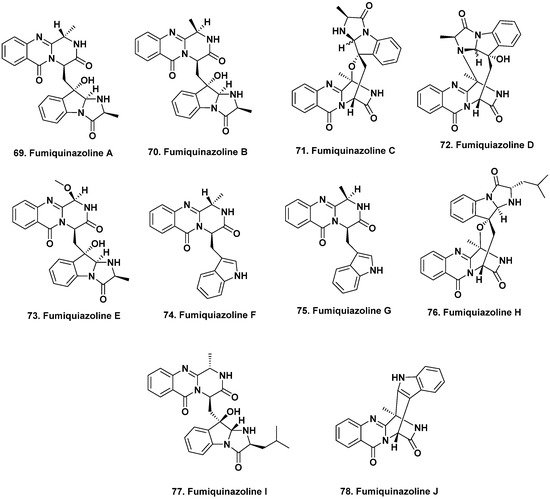
Figure 6. Bioactive Fumiquinazolines from Vertebrates and Invertebrates (Structures 69 to 78).
Due to the duplication of structures and names related to fumiquinazolines published from different sources, with approximately 80 mentioned in the Resende et al. review [68][57], we have used the structures for fumiquinazoline A to J from that source, rather than those first reported in 1992 [64][53].

References

  1. Newman, D.J.; Cragg, G.M. Natural products as sources of new drugs over the nearly four decades from 01/1981 to 09/2019. J. Nat. Prod. 2020, 83, 770–803.
  2. Bode, H.B.; Bethe, B.; Höfs, R.; Zeeck, A. Big effects from small changes: Possible ways to explore nature’s chemical diversity. ChemBioChem 2002, 3, 619–627.
  3. Harvey, A.L.; Edrada-Ebel, R.; Quinn, R.J. The re-emergence of natural products for drug discovery in the genomics era. Nat. Rev. Drug Discov. 2015, 14, 111–129.
  4. Giddings, L.-A.; Newman, D.J. Bioactive compounds from marine extremophiles. In Extremophilic Bacteria, Springer Briefs in Microbiology; Tiquia-Arashiro, S.M., Mormile, M., Eds.; Springer: Cham, Switzerland, 2015; pp. 1–150.
  5. Schulz, B.; Draeger, S.; de la Cruz, T.E.; Rheinheimer, J.; Siems, K.; Loesgen, S.; Bitzer, J.; Schloerke, O.; Zeeck, A.; Kock, I.; et al. Screening strategies for obtaining novel, biologically active, fungal secondary metabolites from marine habitats. Bot. Mar. 2008, 51, 219–234.
  6. Keller, N.P.; Turner, G.; Bennett, J.W. Fungal secondary metabolism-from biochemistry to genomics. Nat. Rev. Micro. 2005, 3, 937–947.
  7. Rokas, A.; Mead, M.E.; Steenwyk, J.L.; Raja, H.A.; Oberlies, N.H. Biosynthetic gene clusters and the evolution of fungal chemodiversity. Nat. Prod. Rep. 2020, 37, 868–878.
  8. Overy, D.P.; Rama, T.; Oosterhuis, R.; Walker, A.K.; Panf, K.-L. The neglected marine fungi, sensu stricto, and their isolation for natural products’ discovery. Mar. Drugs 2019, 17, 42.
  9. Giddings, L.-A.; Newman, D.J. Bioactive compounds from extremophilic marine fungi. In Fungi in Extreme Environments: Ecological Role and Biotechnological Significance; Tiquia-Arashiro, S.M., Grube, M., Eds.; Springer: Cham, Switzerland, 2019; pp. 349–382.
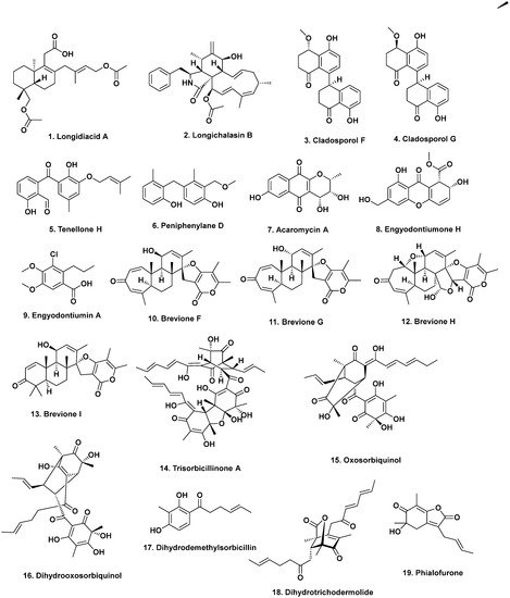
  10. Liu, Z.; Chen, Y.; Li, S.; Wang, Q.; Hu, C.; Liu, H.; Zhang, W. Bioactive metabolites from the deep-sea-derived fungus Diaporthe longicolla fs429. Mar. Drugs 2020, 18, 381.
  11. Zhang, Z.; He, X.; Liu, C.; Che, Q.; Zhu, T.; Gu, Q.; Li, D. Clindanones A and B and cladosporols F and G, polyketides from the deep-sea derived fungus Cladosporium cladosporioides HDN14-342. RSC Adv. 2016, 6, 76498–76504.
  12. Li, H.-L.; Li, X.-M.; Mándi, A.; Antus, S.; Li, X.; Zhang, P.; Liu, Y.; Kurtán, T.; Wang, B.-G. Characterization of cladosporols from the marine algal-derived endophytic fungus Cladosporium cladosporioides EN-399 and configurational revision of the previously reported cladosporol derivatives. J. Org. Chem. 2017, 82, 9946–9954.
  13. Xu, J.-L.; Liu, H.-X.; Chen, Y.-C.; Tan, H.-B.; Guo, H.; Xu, L.-Q.; Li, S.-N.; Huang, Z.-L.; Li, H.-H.; Gao, X.-X.; et al. Highly substituted benzophenone aldehydes and eremophilane derivatives from the deep-sea derived fungus Phomopsis lithocarpus FS508. Mar. Drugs 2018, 16, 329.
  14. Zhang, Z.; Guo, W.; He, X.; Che, Q.; Zhu, T.; Gu, Q.; Li, D. Peniphenylanes A–G from the deep-sea-derived fungus Penicillium fellutanum HDN14-323. Planta Med. 2016, 82, 872–876.
  15. Gao, X.-W.; Liu, H.-X.; Sun, Z.-H.; Chen, Y.-C.; Tan, Y.-Z.; Zhang, W.-M. Secondary metabolites from the deep-sea derived fungus Acaromyces ingoldii FS121. Molecules 2016, 21, 371.
  16. Wang, W.; Li, S.; Chen, Z.; Li, Z.; Liao, Y.; Chen, J. Secondary metabolites produced by the deep-sea-derived fungus Engyodontium album. Chem. Nat. Compds. 2017, 53, 224–226.
  17. Li, Y.; Ye, D.; Chen, X.; Lu, X.; Shao, Z.; Zhang, H.; Che, Y. Breviane spiroditerpenoids from an extreme-tolerant Penicillium sp. isolated from a deep sea sediment sample. J. Nat. Prod. 2009, 72, 912–926.
  18. Li, Y.; Ye, D.; Shao, Z.; Cui, C.; Che, Y. A sterol and spiroditerpenoids from a Penicillium sp. isolated from a deep sea sediment sample. Mar. Drugs 2012, 10, 497–508.
  19. Li, D.; Wang, F.; Cai, S.; Zeng, X.; Xiao, X.; Gu, Q.; Zhu, W. Two new bisorbicillinoids isolated from a deep-sea fungus, Phialocephala sp. Fl30r. J. Antibiot. 2007, 60, 317–320.
  20. Li, D.; Cai, S.; Zhu, T.; Wang, F.; Xiao, X.; Gu, Q. Three new sorbicillin trimers, trisorbicillinones B, C and D, from a deep ocean sediment derived fungus, Phialocephala sp. Fl30r. Tetrahedron 2010, 66, 5101–5106.
  21. Li, D.-H.; Cai, S.-X.; Zhu, T.-J.; Wang, F.-P.; Xiao, X.; Gu, Q.-Q. New cytotoxic metabolites from a deep-sea-derived fungus, Phialocephala sp., strain FL30r. Chem. Biodiv. 2011, 8, 895–901.
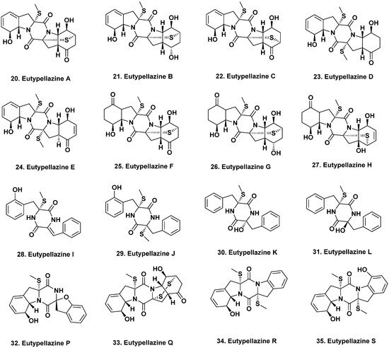
  22. Niu, S.; Liu, D.; Shao, Z.; Proksch, P.; Lin, W. Eutypellazines A–M, thiodiketopiperazine -type alkaloids from deep sea derived fungus Eutypella sp. MCCC 3A00281. RSC Adv. 2017, 7, 33580–33590.
  23. Niu, S.; Liu, D.; Shao, Z.; Proksch, P.; Lin, W. Eutypellazines N–S, new thiodiketo-piperazines from a deep sea sediment derived fungus Eutypella sp. With anti-VRE activities. Tetrahedron Lett. 2017, 58, 3695–3699.
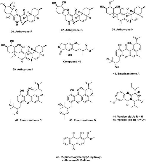
  24. Bao, J.; Zhai, H.; Zhu, K.; Yu, J.-H.; Zhang, Y.; Wang, Y.; Jiang, C.-S.; Zhang, X.; Zhang, Y.; Zhang, H. Bioactive pyridone alkaloids from a deep-sea-derived fungus Arthrinium sp. UJNMF0008. Mar. Drugs 2018, 16, 174.
  25. Xu, X.; Zhang, X.; Nong, X.; Wang, J.; Qi, S. Brevianamides and mycophenolic acid derivatives from the deep-sea-derived fungus Penicillium brevicompactum DFFSCS025. Mar. Drugs 2017, 15, 43.
  26. Fredimoses, M.; Zhou, X.; Lin, X.; Tian, X.; Ai, W.; Wang, J.; Liao, S.; Liu, J.; Yang, B.; Yang, X.; et al. New prenylxanthones from the deep-sea derived fungus Emericella sp. SCSIO 05240. Mar. Drugs 2014, 12, 3190–3202.
  27. Wang, J.; He, W.; Huang, X.; Tian, X.; Liao, S.; Yang, B.; Wang, F.; Zhou, X.; Liu, Y. Antifungal new oxepine-containing alkaloids and xanthones from the deep-sea-derived fungus Aspergillus versicolor SCSIO 05879. J. Agric. Food Chem. 2016, 64, 2910–2916.
  28. Wang, W.; Chen, R.; Luo, Z.; Wang, W.; Chen, J. Antimicrobial activity and molecular docking studies of a novel anthraquinone from a marine-derived fungus Aspergillus versicolor. Nat. Prod. Res. 2017, 32, 558–563.
  29. Bandaranayake, W.M. Traditional and medicinal uses of mangroves. Wetl. Ecol. Manag. Mangroves Salt Marshes 1998, 2, 133–148.
  30. Thatoi, H.; Behera, B.C.; Mishra, R.R. Ecological role and biotechnological potential of mangrove fungi: A review. Mycology 2013, 4, 54–71.
  31. Deshmukh, S.K.; Agrawal, S.; Prakash, V.; Gupta, M.K.; Reddy, M.S. Anti-infectives from mangrove endophytic fungi. S. A. J. Bot. 2020, 134, 237–263.
  32. Cadamuro, R.D.; da Silveira Bastos, I.M.A.; Silva, I.T.; Cabral da Cruz, A.C.; Robl, D.; Sandjo, L.P.; Alves, S., Jr.; Lorenzo, J.M.; Rodríguez-Lázaro, D.; Treichel, H.; et al. Bioactive compounds from mangrove endophytic fungus and their uses for microorganism control. J. Fungi 2021, 7, 455.
  33. Sun, W.; Wu, W.; Liu, X.; Zaleta-Pinet, D.A.; Clark, B.R. Bioactive compounds isolated from marine-derived microbes in China: 2009–2018. Mar. Drugs 2019, 17, 339.
  34. Liu, Y.; Xia, G.; Li, H.; Ma, L.; Ding, B.; Lu, Y.; He, L.; Xia, X.; She, Z. Vermistatin derivatives with α-glucosidase inhibitory activity from the mangrove endophytic fungus Penicillium sp. HN29-3B1. Planta Med. 2014, 80, 912–917.
  35. Liu, Y.; Yang, Q.; Xia, G.; Huang, H.; Li, H.; Ma, L.; Lu, Y.; He, L.; Xia, X.; She, Z. Polyketides with α-glucosidase inhibitory activity from a mangrove endophytic fungus, Penicillium sp. HN29-3B1. J. Nat. Prod. 2015, 78, 1816–1822.
  36. Li, H.; Huang, H.; Shao, C.; Huang, H.; Jiang, J.; Zhu, X.; Liu, Y.; Liu, L.; Lu, Y.; Li, M.; et al. Cytotoxic norsesquiterpene peroxides from the endophytic fungus Talaromyces flavus isolated from the mangrove plant Sonneratia apetala. J. Nat. Prod. 2011, 74, 1230–1235.
  37. Liu, H.; Chen, S.; Liu, W.; Liu, Y.; Huang, X.; She, Z. Polyketides with immunosuppressive activities from mangrove endophytic fungus Penicillium sp. ZJ-SY2. Mar. Drugs 2016, 14, 217.
  38. Cai, R.; Chen, S.; Long, Y.; Li, C.; Huang, X.; She, Z. Depsidones from Talaromyces stipitatus SK-4, an endophytic fungus of the mangrove plant Acanthus ilicifolius. Phytochem. Lett. 2017, 20, 196–199.
  39. Nicoletti, R.; Salvatore, M.M.; Andolfi, A. Secondary metabolites of mangrove-associated strains of Talaromyces. Mar. Drugs 2018, 16, 12.
  40. Lin, Z.J.; Zhu, T.J.; Fang, Y.C.; Gu, Q.Q.; Zhu, W.M. Polyketides from Penicillium sp JP-1, an endophytic fungus associated with the mangrove plant Aegiceras corniculatum. Phytochemistry 2008, 69, 1273–1278.
  41. Xu, M.; Deng, Z.; Li, M.; Li, J.; Fu, H.; Proksch, P.; Lin, W. Chemical constituents from the mangrove plant, Aegiceras corniculatum. J. Nat. Prod. 2004, 67, 762–766.
  42. Bandaranayake, W.M. Bioactivities, bioactive compounds and chemical constituents of mangrove plants. Wetlands Ecol. Managmt. 2002, 10, 421–452.
  43. Wang, Y.; Xu, L.; Ren, W.; Zhao, D.; Zhu, Y.; Wu, X. Bioactive metabolites from Chaetomium globosum l18, an endophytic fungus in the medicinal plant Curcuma wenyujin. Phytomedicine 2012, 19, 364–368.
  44. Huang, S.; Chen, H.; Li, W.; Zhu, X.; Ding, W.; Li, C. Bioactive chaetoglobosins from the mangrove endophytic fungus Penicillium chrysogenum. Mar. Drugs 2016, 14, 172.
  45. Li, C.; Gong, B.; Cox, D.G.; Li, C.; Wang, J.; Ding, W. Dichlorodiaportinol a—A new chlorine-containing isocoumarin from an endophytic fungus Trichoderma sp. 09 from Myoporum bontioides a. Gray and its cytotoxic activity. Pharmacogn. Mag. 2014, 10, 153–158.
  46. Li, W.; Xu, J.; Li, F.; Xu, L.; Li, C. A new antifungal isocoumarin from the endophytic fungus Trichoderma sp. 09 of Myoporum bontioides a. Gray. Pharmacogn. Mag. 2016, 12, 259–261.
  47. Li, W.; Xiong, P.; Zheng, W.; Zhu, X.; She, Z.; Ding, W.; Li, C. Identification and antifungal activity of compounds from the mangrove endophytic fungus Aspergillus clavatus R7. Mar. Drugs 2017, 15, 259.
  48. Kim, D.-H.; Brunt, J.; Austin, B. Microbial diversity of intestinal contents and mucus in rainbow trout (Oncorhynchus mykiss). J. Appl. Microbiol. 2006, 102, 1654–1664.
  49. Ward, N.L.; Steven, B.; Penn, K.; Methe, B.A.; Detrich III, W.H. Characterization of the intestinal microbiota of two antarctic notothenioid fish species. Extremophiles 2009, 13, 679–685.
  50. Smriga, S.; Sandin, S.A.; Azam, F. Abundance, diversity, and activity of microbial assemblages associated with coral reef fish guts and feces. FEMS Microbiol. Ecol. 2010, 73, 31–42.
  51. Roeselers, G.; Mittge, E.K.; Stephens, W.Z.; Parichy, D.M.; Cavanaugh, C.M.; Guillemin, K.; Rawls, J.F. Evidence for a core gut microbiota in the zebrafish. ISME J. 2011, 5, 1595–1608.
  52. Sanchez, L.M.; Wong, W.R.; Riener, R.M.; Schulze, C.J.; Linington, R.G. Examining the fish microbiome: Vertebrate-derived bacteria as an environmental niche for the discovery of unique marine natural products. PLoS ONE 2012, 7, e35398.
  53. Numata, A.; Takahashi, C.; Matsushita, T.; Miyamoto, T.; Kawai, K.; Usami, Y.; Matsumura, E.; Inoue, M.; Ohishi, H.; Shingu, T. Fumiquinazolines, novel metabolites of a fungus isolated from a saltfish. Tetrahedron Lett. 1992, 33, 1621–1624.
  54. Takahashi, C.; Matsushita, T.; Doi, M.; Minoura, K.; Shingu, T.; Kumeda, Y.; Numata, A. Fumiquinazolines A-G, novel metabolites of a fungus separated from a Pseudolabrus marine fish. J. Chem. Soc. Perkin Trans. 1995, 1, 2345–2353.
  55. Belofsky, G.N.; Anguera, M.; Jensen, P.R.; Fenical, W.; Kock, M. Oxepinamides A-C and fumiquinazolines H-I: Bioactive metabolites from a marine isolate of a fungus of the genus Acremonium. Chem.-Eur. J. 2000, 6, 1355–1360.
  56. Shao, C.L.; Xu, R.F.; Wei, M.Y.; She, Z.G.; Wang, C.Y. Structure and absolute configuration of fumiquinazoline l, an alkaloid from a gorgonian-derived Scopulariopsis sp. fungus. J. Nat. Prod. 2013, 76, 779–782.
  57. Resende, D.I.S.P.; Boonpothong, P.; Sousa, E.; Kijjoa, A.; Pinto, M.M.M. Chemistry of the fumiquinazolines and structurally related alkaloids. Nat. Prod. Rep. 2019, 36, 7–34.
  58. Han, J.; Liu, M.; Jenkins, I.D.; Liu, X.; Zhang, L.; Quinn, R.J.; Feng, Y. Genome-inspired chemical exploration of marine fungus Aspergillus fumigatus MF071. Mar. Drugs 2020, 18, 352.
More
ScholarVision Creations