Perilymph Sampling Advances Inner Ear Diagnostics: Comparison
Please note this is a comparison between Version 2 by Beatrix Zheng and Version 1 by Madeleine St. Peter.

In the clinical setting, the pathophysiology of sensorineural hearing loss is poorly defined and there are currently no diagnostic tests available to differentiate between subtypes. This often leaves patients with generalized treatment options such as steroids, hearing aids, or cochlear implantation. The gold standard for localizing disease is direct biopsy or imaging of the affected tissue; however, the inaccessibility and fragility of the cochlea make these techniques difficult. Thus, the establishment of an indirect biopsy, a sampling of inner fluids, is needed to advance inner ear diagnostics and allow for the development of novel therapeutics for inner ear disease. A promising source is perilymph, an inner ear liquid that bathes multiple structures critical to sound transduction. Intraoperative perilymph sampling via the round window membrane of the cochlea has been successfully used to profile the proteome, metabolome, and transcriptome of the inner ear and is a potential source of biomarker discovery. Here, we discuss the various applications of human perilymph sampling and propose a design for a sampling needle.

  • perilymph
  • round window
  • stapedectomy
  • cochlear implantation
  • sensorineural hearing loss

1. Applications of Human Perilymph Sampling

Opening the inner ear was historically considered impossible due to the risk of hearing loss; however, recent experience with hearing preservation cochlear implantation and past sampling studies on stapedectomy patients indicates that it is possible to manipulate the inner ear with no or minimal loss of residual hearing in most patients[1]. We propose that perilymph can be sampled as a stand-alone procedure. Several different existing ear surgeries can be used to model the development of a perilymph sampling procedure including cochlear implantation, stapedectomy, and cochleosacculotomy. Initial applications of perilymph sampling will likely be in enhancing CI and evaluating progressive hearing loss. As more targeted therapeutics for hearing loss emerge, further applications will develop[2].

1.1. Cochlear Implantation

A cochlear implant is a prosthetic device inserted into the inner ear of patients with severe SNHL and poor speech perception who have minimal improvement with the use of hearing aids. Criteria for undergoing implantation have been recently expanded from including only patients with profound hearing loss to those patients with significant residual hearing but poor speech understanding. The surgery is performed by drilling a mastoidectomy and then entering the middle ear space through a posterior tympanotomy (the space between the incus, chorda tympani, and facial nerves)[3]. The bony overhang of the round window is then drilled down to visualize the RWM. An incision is made in the RWM, allowing for the sampling of a small amount of perilymph. The electrode can then be advanced through the window.
CI can improve the speech perception ability in 82.0% of adults with post lingual hearing loss and 53.4% of adults with prelingual hearing loss and can markedly improve quality of life in the responders[4]. However, there is a proportion of patients who do not benefit, and most continue to lose hearing that was present before the operation[2]. Unfortunately, clinicians currently have no way to predict which patients will respond to CI. Although duration of hearing loss and pre-implantation speech perception are thought to be correlated to outcomes, studies have shown mixed results in small sample sizes, and there is still no consensus regarding which patient factors predict functional hearing over time[5]. Characteristics such as sex and age also have mixed results regarding correlation to residual hearing, and do not account for the large variability in patient response to CI[2]. In animal models, some groups have shown that trauma during surgery can induce inflammation and affect post-op hearing[6]; however, human studies have shown that hearing continues to decline long after post-operative inflammation has resolved[2]. Taken together, this has led to the hypothesis that etiology of SNHL rather than patient profile or surgical factors may have the most influence on CI outcomes, further demonstrating the need for subclassification of SNHL.
Most of the current perilymph sampling studies have focused on perilymph sampled from cochlear implant patients and information derived from these studies may yield information on optimal pharmacologic intervention to protect hearing during the implantation process. Sampling could also help predict who would be a candidate for supplementation with neurotrophins or who could benefit from drug eluting cochlear implant electrodes[7]. Since patients with significant residual hearing are being successfully implanted, perilymph sampling at the time of implantation can give us initial safety data on the procedure when it is performed at the same time as cochlear implantation. Hannover Medical School has been routinely sampling perilymph on all implant patients and has not seen a decline in their hearing preservation rates[8][9]. However, safety data should not be gleaned solely from CI procedures. Both the perilymph sampling procedure and CI electrode insertion require puncture of the RWM, which causes perilymph egress. Therefore, if these are done simultaneously, it will be difficult to draw conclusions regarding the safety of perilymph sampling specifically.

1.2. Stapedectomy and Cochleosacculotomy

Several other operations access the inner ear fluid spaces. Stapedectomy is commonly performed for patients with otosclerosis, a cause of CHL. Stapedectomy is well tolerated and significantly improves hearing, with some studies showing up to 70% of patients achieving an air–bone gap of 20 dB or better[10][11].
Although opening the stapes footplate accesses the perilymphatic spaces of the inner ear, this approach would probably not be applicable in routine perilymph sampling in patients without a fixed stapes footplate. Additionally, in patients undergoing workup for SNHL, the stapes supra-structure would impede access to the inner ear. Access to the middle ear, which contains the stapes bone, is gained by making an incision in the auditory canal and lifting the tympanic membrane. The bony scutum is then shaved down, allowing for the visualization of multiple middle and inner ear structures, including the round window niche.
Stapedectomy is generally not indicated for SNHL and is therefore not useful for directly profiling patients with SNHL. However, it is commonly performed for CHL, which provides an opportunity for a control group. Although sampling during stapedectomy is a debated topic among some clinicians, multiple groups have reported safety using this methodology, and stapedectomy has yielded valuable information on the pathogenesis of CHL[12][13][14][15][16][17][18][19][20][21][22][23][24][25][26]. In this technique, perilymph is not collected from the stapedotomy opening, but from the surrounding footplate where perilymph has already egressed out of the vestibule. Therefore, it is unlikely that collection of perilymph is significantly altering post-operative outcomes.
The RWM itself is accessed during another open ear surgery called cochleosacculotomy, indicated for patients with refractory MD (SNHL) who have minimal or no residual hearing in the affected ear[27]. In this procedure, the middle ear is opened and the bony overhang of the round window niche is removed. A 4 mm right-angle pick is then used to obliterate the inner ear. A similar approach to the round window could be used to develop a non-destructive sampling of inner ear fluid.

1.3. Proposed Method of RWM “Tap”

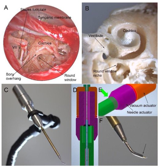
For sampling perilymph, a standard transcanal approach would be used. After making a cut in the ear canal skin, the tympanic membrane is elevated, revealing the structures of the middle ear (Figure 1A). The round window niche is identified, and the bony overhang is removed. Next, the stapes is gently palpated to look for a round window reflex to ensure the correct anatomical plane. The window is then punctured for a sample. Optimally, this should be in the inferior portion of the round window to avoid the basilar membrane. The RWM is at a median angle of 113 degrees to the ear canal; thus, a curved sampling device would be needed. As can be seen in the temporal bone specimen, this approach allows access to the basal turn of the cochlea (Figure 1B)[28]. At the conclusion of sampling, a small tissue patch can be applied. The eardrum is then moved back into position.
Figure 1. Endoscopic view of the right human middle ear (A). After lifting the tympanic membrane, the round window can be seen but is partially obscured by a bony overhang. Only a small area of the stapes footplate is visible next to the facial nerve (VII), making it difficult to access. The anatomy of the cochlea can be seen in a human temporal bone in which the cochlea has been opened (B). The round window niche allows access to the basal turn of the cochlea. A prototype sampling device features a 56 mm long shaft that can be passed down the ear canal to reach the round window (C). The device has two internal actuators, one advancing a needle and one allowing a plunger to be withdrawn from the needle/internal reservoir (D,E) through threading built into the device (green arrow, (E)). This allows advancement of a needle from the curved tip of the device in submillimeter increments and withdrawal of up to 10 µL of fluid. The tip of the device is shown in (F) and measures 0.86 mm at the tip (arrow) from which the needle is deployed.

1.4. Progress in the Design of Sampling Devices

Successful perilymph sampling depends on developing a device that safely accesses the scala tympani and atraumatically withdraws a small sample. In humans, sterile glass capillary tubes are commonly used for intraoperative sampling via the RWM with preservation of residual hearing[8][29][30][31][32]. When placed in perilymph, the capillary tube forms a meniscus. This creates a pressure gradient, causing the fluid to move into the tube. The amount of fluid drawn up depends on the radius of the tube, density of the liquid, and surface tension. The angle of approach is also an important factor, as this determines the curvature of the meniscus and thus affects the size of the pressure gradient. Benefits of using the glass capillary tube include a simple methodology and low cost. However, volume aspirated into the capillary can be non-uniform due to variable tube diameter and user technique. In addition, puncture of the RWM with the glass capillary tube can cause CSF outflow into the scala tympani and contamination of the sample[33]. A specific capillary tube has not been validated for intraoperative use in humans; however, multiple research groups have used various sterile glass capillary tubes for perilymph extraction without complication[8][29][31][30][32].
Microneedles can also be used for sampling. There are multiple types, most of which have been developed and optimized in animal models. To collect perilymph, the needle is advanced into the RWM and perilymph is drawn up using a syringe. Multiple studies have shown that perforation of the RWM with microneedles does not affect hearing threshold in animals and is generally atraumatic[34][35][36][37]. Microneedles have not yet been tested in humans intraoperatively; however Early et al. recently tested a novel microneedle in fresh frozen human temporal bones. They found that the microneedle with syringe could reliably withdraw 5 µL of perilymph from the scala tympani with minimal contamination and little trauma to the RWM[38]. Although microneedles have a more complex design than glass capillary tubes, they allow for controlled aspiration of perilymph. This may result in more consistent volumes sampled and may decrease the likelihood of CSF contamination. Using the approach to the round window outlined above and a curved sampling device, articulated instruments that allow incremental advancement of a needle through the round window and subsequent microfluidic withdrawal of 10 µL of perilymph could also be designed (Figure 1C–F).

1.5. Safety and Limitations

Although sampling has been conducted for many years across multiple institutions, there are very few studies directly examining the effects of intraoperative perilymph sampling on post-operative outcomes. Schmitt et al. is the only group to specifically address potential effects on post-CI residual hearing. They compared pre- and post-operative hearing thresholds between patients who underwent CI plus perilymph sampling and randomly selected patients that underwent only CI. No significant differences in residual hearing or speech perception were found between the groups[8].
There is a long history of sampling perilymph in stapedectomy patients. This technique has been used previously for profiling perilymph with no apparent complications but, as noted above, is probably not applicable to routine perilymph sampling for sensorineural hearing loss. Some additional insight can be gained from the surgical procedures in which the inner ear is opened. Stapedectomy is considered a safe procedure having only a minimal incidence of SNHL[39]. However, sampling through the stapes footplate would require manipulation of a mobile stapes. Hearing preservation CI in which the ear is not only opened but an implant placed has shown complete hearing preservation rates of 45%, and partial hearing preservation rates of 100%[40][41]. Analysis of cochlear microphonics during implantation suggests that if hearing loss occurs, it is not related to opening the RWM but occurs fairly late in the implantation process[41]. Therefore, a controlled puncture with a sampling of 5–10 μL is unlikely to cause any hearing loss.
There are also some technical limitations of the sampling procedure. Studies in guinea pigs show that perforation of the RWM induces perilymph outflow driven by CSF pressure, leading to possible CSF contamination of the sample, and that samples greater than 10 μL can be significantly contaminated with CSF[42]. There can also be interparticipant variations in perilymph volume, and samples can contain differing amounts of CSF and blood contamination. However, these limitations can be overcome through proper training, sample quality checks, and further optimization of instrumentation such as the microneedles used for extraction. Individual anatomic differences in the cochlear aqueduct must also be considered in sampling methodology. If the cochlear aqueduct is widely patent, there may be excessive perilymph and CSF outflow when the RWM is punctured during CI prior to electrode placement[43]. Only the fluid that first flows out of the RWM is pure perilymph. Therefore, if there is a large volume outflow, the fluid is likely to be contaminated with CSF that has entered the inner ear via the cochlear aqueduct. The likelihood of a CSF gusher is not entirely predictable but has been associated with malformation of inner ear structures, which may be detected on CT[44]. Finally, to move forward with developing this technique, large animal models such as pigs will be needed to test novel devices[45].

References

  1. Harold C. Pillsbury; Margaret T. Dillon; Craig A. Buchman; Hinrich Staecker; Sandra M. Prentiss; Michael J. Ruckenstein; Douglas C. Bigelow; Fred F. Telischi; Diane M. Martinez; Christina Runge; et al.David R. FriedlandNikolas H. BlevinsJannine B. LarkyGeorge AlexiadesDavid M. KayliePeter S. RolandRichard T. MiyamotoDouglas D. BackousFrank M. WarrenHussam K. El-KashlanHeidi K. SlagerCarisa ReyesAllison I. RaceyOliver F. Adunka Multicenter US Clinical Trial With an Electric-Acoustic Stimulation (EAS) System in Adults: Final Outcomes. Otology & Neurotology 2018, 39, 299-305, 10.1097/mao.0000000000001691.
  2. Anne G.M. Schilder; Matthew P. Su; Rishi Mandavia; Caroline R. Anderson; Evie Landry; Tanjinah Ferdous; Helen Blackshaw; Early phase trials of novel hearing therapeutics: Avenues and opportunities. Hearing Research 2019, 380, 175-186, 10.1016/j.heares.2019.07.003.
  3. Chantal Snels; Joanna IntHout; Emmanuel Mylanus; Wendy Huinck; Ingeborg Dhooge; Hearing Preservation in Cochlear Implant Surgery: A Meta-Analysis. Otology & Neurotology 2019, 40, 145-153, 10.1097/mao.0000000000002083.
  4. Yvette E. Smulders; Thomas Hendriks; Robert Eikelboom; Inge Stegeman; Peter L. Santa Maria; Marcus D. Atlas; Peter L. Friedland; Predicting Sequential Cochlear Implantation Performance: A Systematic Review. Audiology and Neurotology 2016, 22, 356-363, 10.1159/000488386.
  5. Hideaki Moteki; Shin-Ya Nishio; Maiko Miyagawa; Keita Tsukada; Satoshi Iwasaki; Shin-Ichi Usami; Long-term results of hearing preservation cochlear implant surgery in patients with residual low frequency hearing. Acta Oto-Laryngologica 2016, 137, 516-521, 10.1080/00016489.2016.1252061.
  6. L. Astolfi; E. Simoni; N. Giarbini; P. Giordano; M. Pannella; S. Hatzopoulos; A. Martini; Cochlear implant and inflammation reaction: Safety study of a new steroid-eluting electrode. Hearing Research 2016, 336, 44-52, 10.1016/j.heares.2016.04.005.
  7. Bryan E. Pfingst; Deborah J. Colesa; Donald L. Swiderski; Aaron P. Hughes; Stefan B. Strahl; Moaz Sinan; Yehoash Raphael; Neurotrophin Gene Therapy in Deafened Ears with Cochlear Implants: Long-term Effects on Nerve Survival and Functional Measures. Journal of the Association for Research in Otolaryngology 2017, 18, 731-750, 10.1007/s10162-017-0633-9.
  8. Heike A. Schmitt; Andreas Pich; Anke Schröder; Verena Scheper; Giorgio Lilli; Günter Reuter; Thomas Lenarz; Proteome Analysis of Human Perilymph Using an Intraoperative Sampling Method. Journal of Proteome Research 2017, 16, 1911-1923, 10.1021/acs.jproteome.6b00986.
  9. Gayane Sargsyan; Natalie Kanaan; Thomas Lenarz; Anke Lesinski-Schiedat; Comparison of speech recognition in cochlear implant patients with and without residual hearing: A review of indications. Cochlear Implants International 2021, 1, 1-8, 10.1080/14670100.2021.1898111.
  10. Farid Alzhrani; Mohammad M. Mokhatrish; Murad O. Al-Momani; Hassan AlShehri; Abdulrahman Hagr; Soha N. Garadat; Effectiveness of stapedotomy in improving hearing sensitivity for 53 otosclerotic patients: retrospective review. Annals of Saudi Medicine 2016, 37, 49-55, 10.5144/0256-4947.2017.49.
  11. Pekka Persson; Henrik Harder And; Bengt Magnuson; Hearing Results in Otosclerosis Surgery after Partial Stapedectomy, Total Stapedectomy and Stapedotomy. Acta Oto-Laryngologica 1996, 117, 94-99, 10.3109/00016489709117998.
  12. K. Schindler; E. A. Schnieder; H. L. Wullstein; Vergleichende Bestimmung Einiger Elektrolyte und Organischer Substanzen in Der Perilymphe Otosklerosekranker Patienten. Acta Oto-Laryngologica 1964, 59, 309-319, 10.3109/00016486509124564.
  13. Hans P. Niedermeyer; Georg Zahneisen; Peter Luppa; Raymonde Busch; Wolfgang Arnold; Cortisol Levels in the Human Perilymph after Intravenous Administration of Prednisolone. Audiology and Neurotology 2003, 8, 316-321, 10.1159/000073516.
  14. John K. Niparko; Spoken Language Development in Children Following Cochlear Implantation. JAMA 2010, 303, 1498-1506, 10.1001/jama.2010.451.
  15. Mark J. Levenson; Rosemary B. Desloge; Simon C. Parisier; Beta-2 Transferrin. The Laryngoscope 1996, 106, 159-161, 10.1097/00005537-199602000-00010.
  16. Steven D. Rauch; Transferrin Microheterogeneity in Human Perilymph. The Laryngoscope 2000, 110, 545-552, 10.1097/00005537-200004000-00006.
  17. Giuseppe Attanasio; Marika Viccaro; Edoardo Covelli; Elio De Seta; Antonio Minni; Federica Pizzoli; Roberto Filipo; Cyclo-oxygenase enzyme in the perilymph of human inner ear. Acta Oto-Laryngologica 2010, 131, 242-246, 10.3109/00016489.2010.522593.
  18. Ottó Ribári; István Sziklai; Cathepsin D Activity in Otosclerotic Bone and Perilymph. Acta Oto-Laryngologica 1987, 105, 549-552, 10.3109/00016488809119518.
  19. Causse, J.R.; Uriel, J.; Berges, J.; Shambaugh, G.E.; Bretlau, P.; Causse, J.B; The enzymatic mechanism of the . Am. J. Otol 1982, 3, 297-314.
  20. J. H. Fritsch; C. R. Jolliff; XC Protein Components of Human Perilymph. Annals of Otology, Rhinology & Laryngology 1966, 75, 1070-1076, 10.1177/000348946607500416.
  21. R. Hladk; Z. Brada; A. Kočent; Versuch Einer Biochemischen Biopsie Der Perilymphe Bei Operierten Kranken. Acta Oto-Laryngologica 1959, 51, 424-428, 10.3109/00016486009124515.
  22. Jacob, M.; Causse, J.; Gaudy, D.; Duru, C.; Causse, J.B.; Puech, A.; Antibacterial therapy in surgery of the inner and middle ear. A study of co-trimoxazole penetration into the perilymph . Nouv. Presse Med 1982, 11, 2205-2209.
  23. L. Rüedi; M. C. Sanz; U. Fisch; Untersuchung Der Perilymphe Nach Stapedktomie in Otosklerosefàallen. Acta Oto-Laryngologica 1964, 59, 289-308, 10.3109/00016486509124563.
  24. Franz Altmann; Mario Kornfeld; John J. Shea; I Inner Ear Changes in Otosclerosis. Annals of Otology, Rhinology & Laryngology 1966, 75, 5-32, 10.1177/000348946607500101.
  25. L.-G. Chevance; J. R. Causse; 1-Antitrypsin Activity of Perilymph: Occurrence During Progression of Otospongiosis. Archives of Otolaryngology - Head and Neck Surgery 1976, 102, 363-364, 10.1001/archotol.1976.00780110075008.
  26. Matthew Shew; Athanasia Warnecke; Thomas Lenarz; Heike Schmitt; Sumedha Gunewardena; Hinrich Staecker; Feasibility of microRNA profiling in human inner ear perilymph. NeuroReport 2018, 29, 894-901, 10.1097/wnr.0000000000001049.
  27. William C. Kinney; Nancy Nalepa; Gordon B. Hughes; Sam E. Kinney; Cochleosacculotomy for the treatment of meniere's disease in the elderly patient. The Laryngoscope 1995, 105, 934-937, 10.1288/00005537-199509000-00012.
  28. Takeshi Fujita; Jung Eun Shin; Marybeth Cunnane; Kyoko Fujita; Simon Henein; Demetri Psaltis; Konstantina M. Stankovic; Surgical Anatomy of the Human Round Window Region. Otology & Neurotology 2016, 37, 1189-1194, 10.1097/mao.0000000000001074.
  29. Hsiao-Chun Lin; Yin Ren; Andrew C. Lysaght; Shyan-Yuan Kao; Konstantina M. Stankovic; Proteome of normal human perilymph and perilymph from people with disabling vertigo. PLOS ONE 2019, 14, e0218292, 10.1371/journal.pone.0218292.
  30. Andrew C. Lysaght; Shyan-Yuan Kao; Joao A. Paulo; Saumil N. Merchant; Hanno Steen; Konstantina M. Stankovic; Proteome of Human Perilymph. Journal of Proteome Research 2011, 10, 3845-3851, 10.1021/pr200346q.
  31. Kyu Yup Lee; Takayuki Nakagawa; Takayuki Okano; Ryusuke Hori; Kazuya Ono; Yasuhiko Tabata; Sang Heun Lee; Juichi Ito; Novel Therapy for Hearing Loss. Otology & Neurotology 2007, 28, 976-981, 10.1097/mao.0b013e31811f40db.
  32. Matthew Shew; Helena Wichova; Andres Bur; Devin C. Koestler; Madeleine St Peter; Athanasia Warnecke; Hinrich Staecker; MicroRNA Profiling as a Methodology to Diagnose Ménière’s Disease: Potential Application of Machine Learning. Otolaryngology–Head and Neck Surgery 2020, 164, 399-406, 10.1177/0194599820940649.
  33. Stefan K. Plontke; Jared J. Hartsock; Ruth M. Gill; Alec N. Salt; Intracochlear Drug Injections through the Round Window Membrane: Measures to Improve Drug Retention.. Audiology and Neurotology 2016, 21, 72-9, 10.1159/000442514.
  34. Aykut Aksit; Daniel N. Arteaga; Miguel Arriaga; Xun Wang; Hirobumi Watanabe; Karen Kasza; Anil K. Lalwani; Jeffrey W. Kysar; In-vitro perforation of the round window membrane via direct 3-D printed microneedles. Biomedical Microdevices 2018, 20, 1-12, 10.1007/s10544-018-0287-3.
  35. Harry Chiang; Michelle Yu; Aykut Aksit; Wenbin Wang; Sagit Stern-Shavit; Jeffrey W. Kysar; Anil K. Lalwani; 3D-Printed Microneedles Create Precise Perforations in Human Round Window Membrane in Situ. Otology & Neurotology 2020, 41, 277-284, 10.1097/mao.0000000000002480.
  36. Hirobumi Watanabe; Luis Cardoso; Anil K. Lalwani; Jeffrey W. Kysar; A dual wedge microneedle for sampling of perilymph solution via round window membrane. Biomedical Microdevices 2016, 18, 1-8, 10.1007/s10544-016-0046-2.
  37. Betsy Szeto; Aykut Aksit; Chris Valentini; Michelle Yu; Emily G. Werth; Shahar Goeta; Chuanning Tang; Lewis M. Brown; Elizabeth S. Olson; Jeffrey W. Kysar; et al.Anil K. Lalwani Novel 3D-printed hollow microneedles facilitate safe, reliable, and informative sampling of perilymph from guinea pigs. Hearing Research 2020, 400, 108141, 10.1016/j.heares.2020.108141.
  38. Samuel Early; In Seok Moon; Krishna Bommakanti; Ian Hunter; Konstantina M. Stankovic; A novel microneedle device for controlled and reliable liquid biopsy of the human inner ear. Hearing Research 2019, 381, 107761, 10.1016/j.heares.2019.06.004.
  39. William H. Lippy; Leonard P. Berenholz; Revision Stapedectomy. Ear, Nose & Throat Journal 2009, 88, 1260-1260, 10.1177/014556130908801207.
  40. Wolfgang Gstoettner; Silke Helbig; Claudia Settevendemie; Uwe Baumann; Jens Wagenblast; Christoph Arnoldner; A new electrode for residual hearing preservation in cochlear implantation: first clinical results. Acta Oto-Laryngologica 2008, 129, 372-379, 10.1080/00016480802552568.
  41. Oliver F. Adunka; Stefan Mlot; Thomas A. Suberman; Adam P. Campbell; Joshua Surowitz; Craig A. Buchman; Douglas C. Fitzpatrick; Intracochlear Recordings of Electrophysiological Parameters Indicating Cochlear Damage. Otology & Neurotology 2010, 31, 1233-1241, 10.1097/mao.0b013e3181f1ffdf.
  42. Alec N. Salt; Christian Kellner; Shane Hale; Contamination of perilymph sampled from the basal cochlear turn with cerebrospinal fluid. Hearing Research 2003, 182, 24-33, 10.1016/s0378-5955(03)00137-0.
  43. Giovanni Bianchin; Valeria Polizzi; Patrizia Formigoni; Carmela Russo; Lorenzo Tribi; Cerebrospinal Fluid Leak in Cochlear Implantation: Enlarged Cochlear versus Enlarged Vestibular Aqueduct (Common Cavity Excluded). International Journal of Otolaryngology 2016, 2016, 1-9, 10.1155/2016/6591684.
  44. Liu Hongjian; Wang Guangke; Ma Song; Ding Xiaoli; Zhang Daoxing; The prediction of CSF gusher in cochlear implants with inner ear abnormality. Acta Oto-Laryngologica 2012, 132, 1271-1274, 10.3109/00016489.2012.701328.
  45. H. J. Yi; Wei Guo; N. Wu; J. N. Li; H. Z. Liu; L. L. Ren; P. N. Liu; S. M. Yang; The temporal bone microdissection of miniature pigs as a useful large animal model for otologic research. Acta Oto-Laryngologica 2013, 134, 26-33, 10.3109/00016489.2013.835866.
More
ScholarVision Creations