Your browser does not fully support modern features. Please upgrade for a smoother experience.
Please note this is a comparison between Version 1 by Michelle M. Collins and Version 2 by Yvaine Wei.
Cardiac arrhythmia, or irregular heart rhythm, is associated with morbidity and mortality and is described as one of the most important future public health challenges. In the last few decades, the zebrafish has emerged as an attractive model to reproduce in vivo human cardiac pathologies, including arrhythmias. As genetic tools in zebrafish continue to bloom, this model will be crucial for functional genomics studies and to develop personalized anti-arrhythmic therapies.
- cardiac arrhythmia
- heart development
- zebrafish
- atrial fibrillation
- cardiomyopathy
- imaging
- cardiac rhythm phenotyping
1. Introduction
The healthy human heart beats with a coordinated rhythm. Abnormal heart rhythm, or arrhythmia, refers to conditions in which heart rate or rhythmicity are altered or chaotic. The main inherited cardiac arrhythmias are long QT syndrome (LQTS), short QT syndrome (SQTS), catecholaminergic polymorphic ventricular tachycardia (CPVT), and Brugada syndrome (BrS). These diseases often result from mutations in genes encoding ion channels, leading to altered ionic currents that influence the cardiac action potential [1][2][1,2]. The most common cardiac arrhythmia is atrial fibrillation (AF), which is frequently associated with aging, inflammation, or following surgery [3][4][5][3,4,5]. A portion of AF cases arise in the absence of predisposing factors, often with a younger age of onset and with significant heritability [6]. Arrhythmias also occur in conjunction with inherited cardiomyopathies, including hypertrophic cardiomyopathy (HCM), dilated cardiomyopathy (DCM), arrhythmogenic cardiomyopathy (ACM), and left ventricular non-compaction cardiomyopathy (LVNC). These cardiomyopathies are frequently associated with mutations in genes encoding sarcomeric, desmosomal, or cytoskeletal proteins.
While often considered purely electrical diseases, primary cardiac arrhythmias have variable aetiologies. Genome-wide association studies (GWAS) and whole exome/genome sequencing techniques have implicated diverse pathways in the pathogenesis of cardiac arrhythmia, including developmental [7] and structural genes [8][9][10][8,9,10]. Many of the loci identified in GWAS are found in non-coding regions, suggesting that these variants alter gene expression which confers disease susceptibility [11][12][11,12].
The zebrafish has emerged as an exceptionally powerful model to study cardiac development and disease. From a practical perspective, zebrafish are optically transparent and develop externally, enabling observation during development. They have high fecundity such that large numbers of embryos are easily acquired. Manipulating the zebrafish genome is relatively straightforward, and numerous reporter lines allow for visualization of cellular and organ-level morphology and physiology. Notably, the small size of zebrafish enables them to survive without a functional cardiovascular system early in development, as their oxygen and nutritional needs can be met by diffusion into the embryo. This advantage permits analyses of mutants with little to no cardiac function [13][15], whereas orthologous mutants in other vertebrate models would not survive long enough to be observed.
2. Heart Development in Zebrafish
Heart development in zebrafish involves several distinct steps. This section will briefly outline the major processes that occur during each stage of heart development (Figure 1).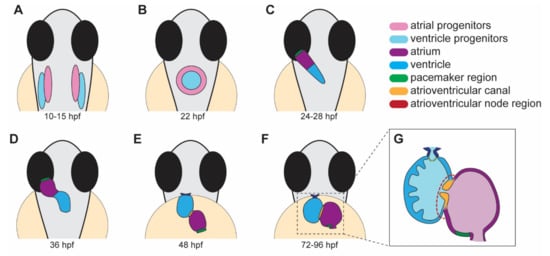
Figure 1. Stages of heart development in zebrafish from 10 to 96 h post-fertilization (hpf). (A) Atrial and ventricular cardiac progenitors are located in the anterior lateral plate mesoderm by ~15 hpf. (B) The cardiac disc is visible by 22 hpf as cardiac progenitors surround endocardial cells at the midline. (C) At 24 hpf, the linear heart tube forms and jogs to the left in preparation for heart looping. (D) At 36 hpf, the heart tube undergoes rightward looping. The AV canal (orange) begins to develop between the cardiac chambers. (E) At 48 hpf, the cardiac chambers begin to balloon and expand outwards. The bulbus arteriosus (dark blue) and AV canal (orange) continue to develop and mature. (F) From 72 to 96 hpf, the cardiac chambers expand and align beside each other. (G) A cross-section of a 96 hpf heart showing the trabeculae, the finger-like muscular projections on the inner wall of the ventricle, and endocardial leaflets (orange) of the AV canal. (A–D) dorsal views; (E–G) ventral views.
3. The Zebrafish Conduction System
The CCS is composed of pacemaker cells in the SAN, the atrioventricular node (AVN), and the fast ventricular conduction system. Although the zebrafish heart is formed by only a single atrium and single ventricle, similarities between its CCS with the human CCS supports the use of zebrafish as a model to study CCS development and function/dysfunction. Pacemaker cells have been identified in the sinus venosus and atrium junction [14][67], and slow-conducting AVC cardiomyocytes in the AVN region have been mapped in zebrafish [15][66]. While the mammalian His-Purkinje system is absent in zebrafish, the ventricular trabecular myocardium has been postulated to serve as a functional equivalent [16][68].
The molecular profiles of conducting tissues are highly conserved in zebrafish. Tomo-seq, a technique to spatially resolve genome-wide transcriptomics data, was used to profile the 2 days post-fertilization (dpf) zebrafish heart. These data identified a sub-compartment that highly expresses pacemaker development genes, including isl1 and shox2 [17][83]. Recent transcriptome profiling of the sinoatrial ring [18][84] and AVC cells [19][85] from the developing zebrafish heart confirms conserved gene expression signatures found in the mammalian SAN and AVN, respectively. Many of the core mammalian SAN/AVN genes are expressed in the developing zebrafish, including tbx18, hcn4, bmp4, cacna1ab in the sinoatrial ring [18][84], and an abundance of genes encoding connexins, T-type Ca2+ channel (cacna1g), and the pacemaker hyperpolarization channel (hcn4) in the AVN [19][85].
Cardiac physiology in zebrafish aligns closely with mammalian models. The average resting heart rate of humans is 60–90 beats per minute (bpm), while the average heart rate of zebrafish is 120–180 bpm [20][86]. This characteristic is a considerable advantage over the widely used rodent models like mouse, which have average heart rates of 300–600 bpm. Adult zebrafish basal electrocardiogram (ECG) characteristics are similar to humans, with a distinct P wave, QRS complex, and T-wave [21][22][87,88]. As in humans and large animal models, zebrafish have chamber-specific differences in action potential (AP) shape and duration (for examples, please see [21][23][24][25][87,89,90,91]). In atrial and ventricular cardiomyocytes of zebrafish, the resting membrane potential and AP amplitude are comparable with those observed in humans. Like humans, a clear plateau phase is established in ventricular APs, although a fast phase-1 repolarization is not present. Due to the elevated heart rate in zebrafish, the AP duration in the atrial and ventricular cardiomyocytes is shortened when compared to humans [25][91].
4. Zebrafish Models of Cardiac Arrhythmia
Cardiac arrhythmias have multifactorial aetiologies. Several zebrafish models with cardiac rhythm phenotypes have been reported (Table 1). Notably, these models provide unique insight toward disease initiation and mechanisms of pathogenesis.Table 1.
Zebrafish models of cardiac arrhythmia.
| Model/ Gene |
Allele | Cardiac Defect | Clinical Arrhythmia |
Human Ortholog | Ref. |
|---|---|---|---|---|---|
| atp1a1a.1 | hiphop (tx218) | 3:1 ratio of atrial contraction to ventricular contraction, bradycardia, and AV-block. |
LQTS | ATP1A1 | [26][27][102,103] |
| cacna1c | island beat (m379, m458, m231) |
Silent ventricle, uncoordinated contraction of the atrium. |
AF | CACNA1C | [13][28][15,104] |
| cmlc1 myl4 |
s977 bw24 |
Bradycardia, slow conduction in enlarged atrium, sarcomere disorganization. |
AF | MYL4 | [29][30][105,106] |
| cx43 (gja1b) | Morpholino | Bradycardia, AV-block, and fibrillation. |
AF | GJA1 | [31][107] |
| foxn4 | slipjig s644) | Peristaltic contraction with no AV delay. |
FOXN4 | [32][33][64,108] | |
| gja3/cx46 | dococ (s215, s226) | Uncoordinated conduction and contraction within the ventricle. |
CX46 | [34][109] | |
| hcn4 | Morpholino | Bradycardia and prolonged cardiac pauses. |
SSS | HCN4 | [35][110] |
| isl1 (K88X mutant) | sa0029 | 2 dpf: bradycardia due to impaired SA node function. 3–4 dpf: sinus block. |
SSS | ISL1 | [14][36][67,111] |
| kcnh6a (zerg) | breakdance (tb218) | 2:1 ratio of atrial to ventricular contraction, bradycardia, reduced cardiac output, and AV-block due to impairment of IKr channel. |
LQTS | KCNH6 (hERG) | [26][37][102,112] |
| kcnh6a (zerg) | reggae | Intermittent atrial fibrillation and acceleration of cardiomyocyte repolarization. |
SQTS | KCNH6 (hERG) | [38][113] |
| kcnma1b | Morpholino | Decreased contraction of heart chambers, sinus bradycardia. |
AF | KCNMA1 | [39][114] |
| mcu | la2446 | Cardiomyopathy. Thin, dilated atrium, small ventricle with restricted blood flow, swollen mitochondria. Heart rate variability. | SSS | MCU | [40][115] |
| nkx2.5 | vu176, vu413 | Reduced heart rate variation, increased heart rate. | CHD | NKX2-5 | [41][116] |
| pitx2c | ups6 | Embryonic: arrhythmia, sarcomere disorganization, increased ROS. Adult: extended P-wave and PR-interval, fibrosis, sarcomere disorganization. |
AF | PITX2 | [42][117] |
| pln | hu10742 | Adult: structural remodeling, immune cell infiltration, contractile defects, AP alternans, altered Ca2+ handling | ACM | PLN | [43][44][118,119] |
| scn5a | human variant |
Bradycardia, sinus pauses, AV-block. | LQTS | SCN5A | [45][120] |
| slc8a1a (ncx1) |
tremblor (tc318d, te381b, m116, m139, m158, m276, m736) | Fibrillation from onset of contraction (more prominent in the atrium than the ventricle). Absent circulation. | SLC8A1 (NCX1) |
[13][26][46][15,102,121] | |
| tbx5a | heartstrings (m21) | Slight bradycardia evident during initial heart tube stage. Heart fails to loop, contractility declines, and pericardial edema develops. |
Holt–Oram syndrome | TBX5 | [47][122] |
| tcf2 | hobgoblin (s634) | AV block at 48 hpf, silent ventricle at 96 hpf. |
TCF2 | [32][64] | |
| tmem161b | grime (uq4ks) | Bradycardia, skipped ventricular beats, increased heart rate variability | LQTS | TMEM161B | [48][123] |
| ttn.2 | sfc9 | Atrial fibrosis, compromised sarcomere assembly in atrium and ventricle, lengthened PR interval. |
AF | TTN | [49][124] |
| mobitz (s466) | AV block, sinus pause at 120 hpf. | [32][64] | |||
| elektra (s587) | AV block. | [32][64] | |||
| daredevil (s275, s563) | AV block, silent ventricle at 120 hpf. | [32][64] | |||
| bullseye (s885) | No heartbeat at 24 hpf, AV block at 36–48 hpf. |
[32][64] | |||
| kingpin (s886) | Atrial and ventricular fibrillation | [32][64] |
5. Techniques for Assessing Cardiac Rhythm and Function in Embryonic and Adult Zebrafish
5.1. Tools to Study Cardiac Rhythm at Embryonic Stages
Due to its amenability to live imaging and genetic manipulation, the zebrafish model provides a great opportunity for understanding the genetic and molecular mechanisms underlying cardiac arrhythmia.
The zebrafish embryo is easy to image due to its transparency, and light microscopy is suitable to detect early arrhythmia. High-speed acquisition of the beating heart is key to the identification of arrhythmia. One of these techniques, spinning disk microscopy, provides many attractive advantages for imaging heart contractions in vivo during development due to its speed with higher frame rates. The high speed allows the imaging of multiple samples in a short amount of time, making it an excellent instrument for high-throughput chemical screening assay in zebrafish embryos [50][169]. After acquiring heartbeat movies, a kymograph, a plot representing spatial position over time, can be generated to quantify heart rate, heart rate variability, and cardiac output in larvae [14][42][48][51][67,117,123,170].
Light-sheet microscopy is another fluorescence microscopy technique suitable to detect arrhythmia. It uses a plane of light to optically section and views tissues at a cellular resolution. Light-sheet microscopy presents the advantage of deep imaging with a thin plane of light, limiting phototoxicity and photobleaching [52][174]. By combining this approach with optogenetics, a technique in which channels can be controlled with light, Arrenberg et al. created an optically controlled pacemaker by expressing halorhodopsin and channelrhodopsin in zebrafish cardiomyocytes [15][66]. Using these tools, the cardiac pacemaker was mapped using a patterned illumination to localize the areas sensitive to hyperpolarization at the inflow tract and AV canal during early development.