In clinical applications for implantable biomedical instruments, the adhesion of harmful biomedical materials to the surface must be avoided. Thus, if the implanted system requires prevention of inflammation or infection in cell/tissue culture systems, an antibacterial surface property is highly desirable. Different methods for the development of antibacterial surfaces have been reported
[32][33][34][35][36][37][38][39][40][41][145,146,147,148,149,150,151,152,153,154]. A superhydrophobic surface inspired by biological structures has become a favorable antibacterial surface due to the anti-fouling capability that could circumvent surface bacteria from adhering
[42][43][44][45][46][155,156,157,158,159]. Privett et al.
[42][155] also produced xerogels which contained silica colloids, fluoroalkoxysilane, and silane backbone. The superhydrophobic characteristics of the coated surface of the xerogel were facilitated by fluorinated silica nanoparticles for low surface energy and a hierarchical structure. The antibacterial properties of the xerogel were described by a conventional cell flow assay using Gram-negative
Pseudomonas aeruginasa (
P. aeruginasa) and Gram-positive
Staphylococcus aureus (
S. aureus). Microbial adhesion to the superhydrophobic xerogel surface was decreased by 98% and 99% relative to the blank area. The antibacterial properties of surfaces with Gram-negative bacteria were reported by Feschauf et al.
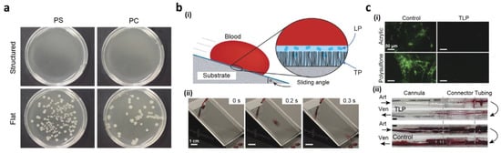
[47][29], using superhydrophobic polycarbonate (PC) and polyethylene (PE) and PS substrates. Using a basic casting method, the superhydrophobic PC, PE, and PS surfaces were acquired from a micro/nanostructured PDMS casting. A total of 10 μL of
E. coli was cultured to test standardized PS, PC, and PE surface anti-bacterial properties. Then, a bacterial
E. coli solution was cultivated for 24 h (
Figure 410). Fewer than 100 colony-forming units (CFUs) were observed on PS- and PE-based superhydrophobic surfaces after 24 h, whereas no bacterial growth was observed on the PC substrate. Moreover, 100 CFUs were observed each for flat PS and PC, while 25,800 were observed for PE. These findings suggest that, relative to flat surfaces, superhydrophobic surfaces efficiently reduce adhesion of bacteria to <0.1%. Because of the wide variety of anti-fouling properties, liquid slippery surfaces often have tremendous potential for various medical applications in comparison to different liquids and environmental pressures. Over a wide range of temperatures, heats, surface tensions, and multiple conditions they retain repellency
[46][159]. Epstein’s research team
[48][160] showed the importance of a slippery surface to avert binding biofilm. On a smooth, slippery surface, the bacteria were presented, which could not be anchored on the mobile interface compared to solid interface. Irrespective of the principal solid, porous structure, separate biofilm accumulations were stopped by a slippery surface over more than a week. In contrast to common, polyethylene glycol, anti-fouling surfaces, it also decreased bacterial attachment by 96–96.6%. To minimize the morbidity and mortality caused by thrombosis, Leslie et al.
[49][30] used slippery properties on tubing and catheters of indwelling medical devices. They developed a tethered perfluorocarbon coating (TP) at the top of the tube followed by its coating with a liquid perfluorodecalin (LP) surface to attain anti-thrombogenic and non-adhesive surfaces. The thin, moving liquid layer permitted the tethered-liquid perfluorocarbon (TLP) surface to resist fluids effectively also after the surface contacted an immiscible liquid, such as blood (
Figure 410b(i)). The surface was cleaned almost immediately from a new, whole human droplet of blood (
Figure 410b(ii)). Compared to the uncoated surfaces of acrylic and polysulfone, they showed (
Figure 410c(i)) a decrease in the adhesion and polymerization of the TLP surface. The slithery condition also decreased the adhesion of platelets in contrast to uncoated surfaces. These findings demonstrated that the smooth surface decreased fibrin polymerization and inhibited adherence, as well as plates activation. To realize an arteriogenic shunt for in vivo analysis of the anti-thrombogenic effect of the slippery surface, polycarbonate connectors, TLP-treated polyurethane cannulae, and medicinal, polyvinyl chloride (PVC)-based heart pulmonary perfusion tubes were studied. Compared with control tubes,
Figure 410c(ii) displays polymer TLP-treated tubes that minimize occlusive thrombosis post flow for 8 h. Such noteworthy, anti-fouling properties can, therefore, be used in several additional applications.
Figure 410. (
a) The growth of bacteria on structured and smooth PS and PC substrates; (
b) (
i) the smooth surfaces after coating with tethered-liquid perfluorocarbon (TLP) showing blood repellency; (
ii) the images of the slippery surface showing slipping of a blood droplet; (
c) (
i) fluorescence images of fibrinogen on polysulfone or acrylic surfaces in the presence and absence of TLP coating; (
ii) the images of polycarbonate connectors, polyurethane cannulae, and PVC tubing in the presence and absence of TLP coating. Reproduced from
[47][29] under CC BY license. Reprinted with permission from
[49][30]. Copyright 2014 Nature Publishing Group.
1.4. Lab-on-a-Chip Based on Open Channel Droplets
In terms of fast penetration into the sample fluid, energy consumption and low samples, quick chemical and biological reactions
[50][51][52][53][54][55][56][57][58][59][60][161,162,163,164,165,166,167,168,169,170,171], such as microfluidic droplet systems, have many advantages compared to traditional closed-channel, microfluidic applications. A gout-based microfluidic system
[52][163] was introduced on a superhydrophobic, porous oxide membrane by You et al. To direct the water droplets, they inserted hydrophilic PDAs on superhydrophobic surfaces. A square pDA for more complex droplet handling was also planned in the middle of the pDA micro-line. A fluid droplet traveled along the micro-lines by momentum, stopping at the square, which showed adequate surface energy to catch the droplet. Such an immobilized droplet was pushed downwards by inserting a second droplet. With this droplet capacity to combine, preparation of monodispersed gold nanoparticles and rapid, structural protein changes were introduced. For chemical and biological organic solvent-based reactions and experiments, however, superhydrophobic hydrophilic patterned surfaces could not be employed. For compatibility with different solvents, a pDA, micropatterned slippery surface was introduced by You et al.
[61][172]. To attain a fluid-guiding feature, PDA micro-lines were engineered on a nanostructured surface before lubricant infiltration. Multiple solvents, for example, gasoline, dimethyl sulfoxide, water, dimethylformamide, 1,2-dichloroethane,
n-hexane, toluene, acetone, and diesel oils, were compatible with the produced slippery device. Any solvent with a surface tension larger than that of the lubricant could be depleted by gravitational force along the micro-lines by the infused lubricant located on top of the pDA micro-line. They implemented the square shape pattern for the gout mixture in the pDA micro-line intersection with the same aforementioned process (
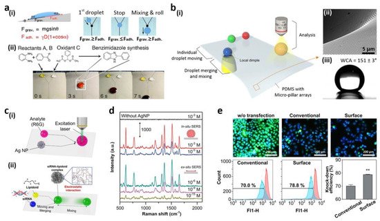
Figure 511a(i))
[52][163]. The chemical reactions using an organic solvent were carried out on the slippery surface of the pDA micro-model due to the broad compatibility of solvents. The organic reaction was performed between benzaldehyde and ortho-phenylenediamine to create 2-arylbenzimidazole. The droplet of THF solution composed of two reactants, as well as a THF, was dropped on the oily surface, as shown in
Figure 511a(ii). By fast and homogeneous mixing, the reaction rate (70.3%) improved relative to the standard bulk reaction using a vial (52.1%). Furthermore, the surface was cleared and used again after ultrasonication.
Figure 511. (
a) (
i) The movement of the droplet and its mixing on a slippery system fabricated with pDA (
ii) THF-based chemical reaction on the slithery surface; (
b) (
i) regulation of motions of water (movement, mixing, and examination) on PDMS substrate appended with micropillar arrays; (
ii) the dimple structure’s SEM picture; (
iii) water droplet image on the substrate of PDMS; (
c) (
i) the measurement system for SERS; (
ii) formation of minor, interfering lipidoid-RNA complex; (
d) SERS measurement spectra (in situ/ex situ) for R6G/Ag NP droplet mixture and an evaporated R6G/Ag NP droplet; (
e) fluorescence imaging and flow cytometry studies of transfected GFP-Hela cells. Scale bar-200 µm (n = 3, **
p < 0.01, compared to the conventional group). Reprinted with permission from
[61][62][172,173]. Copyright 2014 American Chemical Society (ACS) and copyright 2015 Nature Publishing Group.
Decorated droplet-handling systems only had minimal control over fluid processes, for instance, start and stop motion. Newly developed by Seo et al., the superhydrophobic PDMS substrate contained micropillar arrays for programmed water droplet manipulation (
Figure 511b(i))
[62][173]. When applying the substrate, the vacuum pressure on the suspended PDMS substratum was expanded (
Figure 511b(ii)) to form a local, pale structure. Decreased performance and superhydrophobicity of the micropillar array was observed (
Figure 511). In the interface region between the substrate and water droplets, the dimple arrangement could be used to monitor the motions of water droplets individually. The curvature was positive at the boundary of the pillars adjacent to the PDMS, and the width was greater than the flat substratum distance. The lower number of micropillars decreased the adhesiveness of the water and allowed isolation from the droplet substratum. The flow of water drops on the substratum also could not be conveniently regulated through the vacuum dimple structure; however, the moving direction could also be readily built without any additional pattern. Surface-enhanced Raman spectroscopy (SERS) analyzed the substrate’s analytical efficiency (
Figure 511c(i)). Rhodamine 6G (R6G) in situ/ex situ SERS tests were carried out at different concentrations (
Figure 511d). In situ, individual water droplets consisting of Ag nanoparticles and R6G molecules were determined by the SERS and combined at the Raman detection point before the collection of data. The lower limit of detection for R6G/Ag NP was calculated to be 10
−5 M, owing to disseminated R6G and Ag molecules in a mixed droplet. The mixture was evaporated and its components condensed to resolve this detection cap within an area of a certain length. The R6G molecules were also observed at 10
−15 M because of the aggregation of Ag NPs and R6G for ex situ SERS calculation. To manufacture regular, intracellular, gene transfer nanoparticles, they used the platform. In the lipid (ND98) complexes with green fluorescent protein (GFP) siRNA, a basic method of mixing and blending outlets was used (siGFP). On the platform, homogenous mixing of droplets and the efficiency of transfections to GFP-HeLa cells from the platform’s lipidoid-siRNA complexes (78.8% ± 0.5%), in contrast to classical manual pipetting mixing (70.0%), could be achieved, as shown in
Figure 511e.
1.5. Fluorescent Intelligence Sensing Based on Bioinspired Super-Wettable Patterns
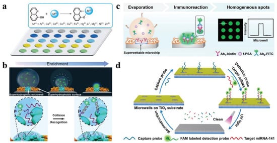
In analytical chemistry, fluorescence is a popular detection method. In a light-absorption process, fluorescence is a luminescence process induced by an appropriate molecule under light excitation. Fluorescence intensity is measurable at specific excitation/emission wavelengths. Under dilute concentrations, the amplitude of fluorescence would be equal to the fluorophore concentration. Several groups recently focused their efforts on the fabrication of bioinspired super-wettable microchips for profound fluorescence-based detection. These examples will be highlighted in the following text: mainly their features such as anchoring and enriching capabilities for biosensing. The analytes were stored in microdroplets that were enclosed inside the superhydrophilic microwells with the superhydrophobic substrate serving as a wall to keep the microdroplets from spreading. Huang and co-workers recorded sensitive detection of metal ions by integrating the fluorescence technique, as seen in
Figure 612a, by using the anchoring capacities of bioinspired micro-patterns. A hydrophilic, photonic-crystal, colloidal microchip was created by assembling hydrophilic, photonic-crystal, colloidal particles on a hydrophilic–hydrophobic patterned substrate. The developed microchip improved fluorescence detection in multiple channels selectively, as well as performed highly effective testing under discriminative conditions using twelve metal ions
[63][31]. Such a well-performing, patterned microchip demonstrates a modern use of bioinspired microchips in detection and their crucial role in the development of sophisticated fluorescent devices and advanced discriminative analysis.
Figure 612. Super-wettable fluorescence finding. (
a) High-performance metal-ion recognition using a bioinspired photonic-crystal microchip; (
b) ultra-trace DNA identification using super-wettable microchips; (
c) example of free prostate-specific antigen (f-PSA) detection on homogeneous super-wettable microchips in a schematic diagram; (
d) a green super-wettable miRNA biochip built on TiO
2 substrate. Reprinted with permission from
[63][64][65][66][31,33,37,39]. Copyright 2013, 2015 Wiley-VCH and copyright 2018 Elsevier B.V.
The “analyte solution concentration in the hydrophilic region increases as the microdroplet evaporates, which is ideal for capturing the analyte for ultra-trace detection. In
Figure 612b
[64][33], ultra-trace fluorescent DNA detection is demonstrated by using the super-wettable microchips’ enrichment ability. UV-based etching of the octadecyl trichlorosilane (OTS), functionalized, superhydrophobic, nanodendritic Si coating through a photomask and deposition of candle soot were employed to create this super-wettable microchip. The analyte was captured from an extremely diluted solution by constant evaporation and emission signals were eventually intensified to attain the recognition of DNA with a low detection limit (10
−16 M). Aggregation-induced emission enhancement (AIEE) and evaporation-induced enhancement were accomplished concurrently by incorporating AIE molecules onto a super-wettable microchip
[64][33]. As a result of the synergetic enhancement, a miRNA biosensor with enhanced efficiency (detection limit-1 pM, linear range-10
−6 to 10
−12 M) can be achieved. The super-wettable microchip’s overall fluorescence strength was just half that of industrial hydrophilic or hydrophobic glass substrates. Due to the coffee rings, however, unequal signal propagation occurred on the hydrophilic and hydrophobic surfaces, possibly limiting repeatability. The sensitive and precise identification of biomarkers included excellent spots in the superhydrophilic microwells. The coffee-ring effect could be reduced in super-wettable microchips, and the homogeneity of superhydrophilic spots could be increased, as seen in
Figure 612c
[65][37]. The superior Marangoni effect in the superhydrophilic spot and the reduced outward flow, owing to the large hydrodynamic flow resistance of 3D Si nanodendritic structure, contributed to this phenomenon.
Figure 612d
[66][67][68][39,174,175] displays the sustainable super-wettable biochips for miRNA identification. The nanodendritic TiO
2 nanostructure was made hydrothermally on FTO-coated glass substrates, then modified with OTS and exposed to UV light via a photomask. By using its anchoring and enrichment abilities, super-wettable fluorescence detection provides a sensitive technique that could be especially useful in biomarker sampling, diagnosis, and disease monitoring. However, the need for large instruments and complicated biomolecule fluorescent labeling can restrict their use in point-of-care (PoC) detection. Integration of bioinspired micropatterns with compact fluorescence instruments for point-of-care research will be a potential priority.
Aside from the above-discussed papers, recent years have witnessed several other polymeric-materials-based, super-wettable surfaces for applications in diverse research areas
[69][70][71][72][73][74][75][76][77][78][176,177,178,179,180,181,182,183,184,185]. To summarize, some common polymeric-material-based, super-wettable surfaces utilized for various applications are illustrated in
Table 1.
Table 1. Some common polymeric materials used in the fabrication of super-wettable surfaces.
Table 1. Some common polymeric materials used in the fabrication of super-wettable surfaces.
| Polymeric Materials |
Fabrication Method |
Application |
Wetting Property |
Reference |
| Hyaluronic acid (HA) |
Phase separation |
Tissue engineering |
Hydrophobic |
Ref [20] | Ref [27] |
| Polyethylene (PE) |
Shrink-induced
mold method |
Antibacterial |
Superhydrophobic |
Ref [47] | Ref [29] |
| Polycarbonate (PC) |
Shrink-induced
mold method |
Antibacterial |
Superhydrophobic |
Ref [47] | Ref [29] |
| Polystyrene (PS) |
Electrohydrodynamics (EHD) |
Tissue engineering |
Superhydrophobic |
Ref [79] | Ref [71] |
| 1H,1H,2H,2H-perfluoro-decyl trichlorosilane (PFDTS) |
Wet chemical
self-assembly |
Conductive stainless steel |
Superhydrophobic |
Ref [80] | Ref [72] |
| Poly(vinyl alcohol) (PVA) |
Solvent evaporation |
Petal effects |
Hydrophilic-Superhydrophobic |
Ref [81] | Ref [74] |
| Poly(p-xylylene) (PPX) |
Soft lithography |
Self-cleaning |
Superhydrophobic |
Ref [82] | Ref [91] |
| Polydimethyl siloxane (PDMS) |
Soft lithography |
Water adhesion |
Hydrophilic-Superhydrophobic |
Ref [82] | Ref [91] |
| Polydopamine (PD) |
Soft lithography |
Water adhesion |
Hydrophilic-Superhydrophobic |
Ref [82] | Ref [91] |
| Polytetrafluoroethylene (PTFE) |
Sol-gel strategy |
Water adhesion |
Superhydrophobic |
Ref [69] | Ref [176] |
| Polyvinylpyrrolidone (PVP) |
Sol-gel strategy |
Self-cleaning |
Superhydrophobic |
Ref [69] | Ref [176] |
| Polyamide (PA) |
Dip-coating method |
Water and oil separation |
Superhydrophobic |
Ref [74] | Ref [181] |
| Silane-modified polymer (SMP) |
Spray-coating method |
Water and oil separation |
Superhydrophobic |
Ref [74] | Ref [181] |