Applications about Single-Cell Printing: Comparison
Please note this is a comparison between Version 1 by Xiaohu Zhou and Version 2 by Nora Tang.

Single-cell analysis has become a powerful and indispensable tool in modern biological and medical research. Single-cell isolation is the key step for single-cell analysis. Single-cell printing could utilize various microfluidic technologies for single-cell isolation and analysis, such as droplet microfluidics, microwell arrays, and hydrodynamic traps. Single-cell printing shows several distinct advantages among the single-cell isolation techniques, such as precise deposition, high encapsulation efficiency, and easy recovery. With the development of single-cell printing in the past decade, various single-cell printing-based single-cell analyses and applications have been performed, ranging from single-cell array-based screening and single-cell-based mass spectroscopy to live three-dimensional tissue formation.

  • inkjet printing
  • cell array
  • single-cell analysis
  • screening

1. Introduction

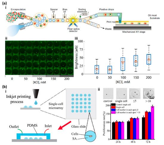
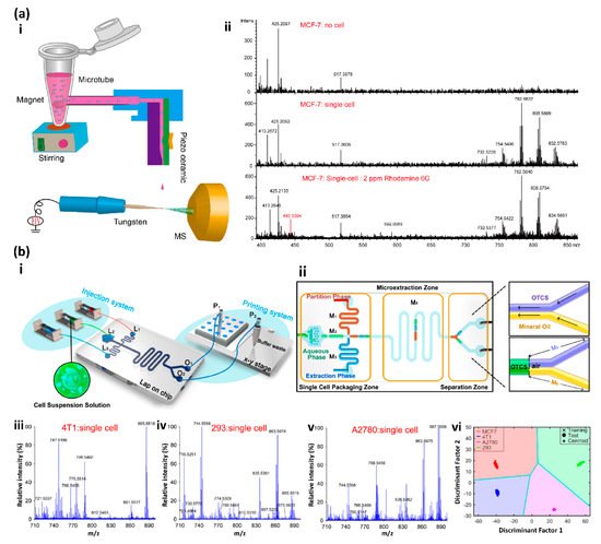
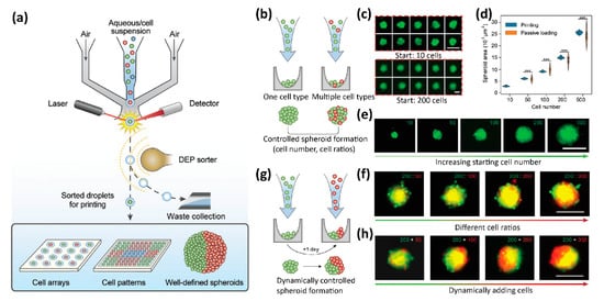
As the key step for single-cell analysis, single-cell isolation has attracted great attention, and a number of techniques have been developed for single-cell isolation and manipulation [1][29]. Single-cell printing could utilize various microfluidic technologies for single-cell isolation and analysis, such as droplet microfluidics [2][3][4][6,33,34], microwell arrays [5][6][35,36], and hydrodynamic traps [7][8][37,38]. Single-cell printing has several distinct advantages. First, single-cell printing can effectively and precisely deposit cells at specific sites in high throughput [9][39]. Second, the printed single cells or colonies can be easily recovered with addressability for subsequent analysis. Third, it is convenient to integrate the highly efficient single-cell printing with other techniques, such as imaging system [10][11][40,41], electric field [12][13][14][42,43,44], and acoustic field [15][16][45,46], and the single-cell encapsulation efficiency can reach more than 90%. Furthermore, bioprinting with the single-cell resolution can not only print 2D single-cell arrays for single-cell analysis but also three-dimensional tissue matrixes and organs for tissue engineering, drug discovery, and toxicology [17][18][47,48]. With the development of single-cell printing in the past decade, various single-cell printing-based single-cell analyses and applications have been performed, ranging from single-cell array-based screening [19][20][21][22][23][24][89,90,93,107,108,109] and single-cell-based mass spectroscopy [25][26][27][28][29][30][110,111,112,113,114,115] to live three-dimensional tissue formation [17][18][31][47,48,116].

2. High Throughput Screening

Recently, Cole et al. developed printed droplet microfluidics (PDM) to print droplets containing single cells and reagents with deterministic control by integrating the fluorescence-activated droplet sorter, which provides the capability of selecting the droplets containing the desired cells and reagents from a set of candidates and printing them on a motorized substrate (Figure 1(a-i)) [21][93]. To demonstrate the ability of the PDM for single-cell analysis, a time-sensitive single-cell calcium release assay was performed. PC3 prostate cancer cells with a green-fluorescing Ca2+ indicator dye were printed as a single-cell array (Figure 1(a-ii)). KCl was used to depolarize the cell membrane to induce intracellular calcium release. The results show that the higher concentration of KCl induced more detectable Ca2+ signals, which was consistent with the bulk experiments (Figure 1(a-iii)) [21][93].
Figure 1. (a-i) Schematic diagram of the fluorescence-activated cell sorter-based printed droplet microfluidics. (a-ii) The intracellular calcium release assay was screened on a single PC3 prostate cancer cell array with different concentrations of KCl. (a-iii) Box plots show the results of the single-cell-based intracellular calcium release assay. Reproduced with permission from [21][93]. (b-i) Schematic diagram of the inkjet printing process to fabricate the single-cell microarray. (b-ii) ATP-induced proliferation experiment indicated that the multi-cell group had a higher proliferation rate than the single cell. Reproduced with permission from [19][89].
More recently, Zhou and co-workers developed a laboratory-made inkjet printing system to construct single-cell arrays (Figure 1(b-i)) [19][89]. This modified inkjet printer can precisely control the number of cells in each printing spot on a hydrophobic substrate for subsequent in-depth research, and the single-cell occupancy reaches as high as 91%. Single-cell arrays of MCF-7 cells with a DMEM medium and sodium alginate were constructed with this modified inkjet printing system, and the real-time single-cell assays showed high activity and proliferation, low levels of ROS, and cell apoptosis, which demonstrated the capability of this inkjet printing system for single-cell study. Interestingly, in the ATP-induced proliferation experiment, they found that extracellular ATP can indeed significantly increase MCF-7 cell proliferation over 72 hours as reported, and the multi-cell group showed a higher proliferation rate than the single-cell group, which indicates that cell communication might also play an important role in cell proliferation (Figure 1(b-ii)) [19][89].

3. Mass Spectrometry-Based Single-Cell Analysis

Mass spectrometry (MS) is a powerful tool to qualitatively and quantitatively detect molecules at the femtomolar sensitivity without a labeling requirement [32][117]. Furthermore, the multiplex detection with high throughput and low sample consumption makes MS the ideal tool for single-cell analysis [25][26][27][28][29][30][110,111,112,113,114,115], especially for single-cell proteomics [33][34][35][8,118,119].
Recently, Lin and co-workers developed an MS-based single-cell analysis strategy by integrating the drop-on-demand inkjet printing with the probe electrospray ionization (ESI) mass spectrometry (Figure 2(a-i)) [28][113]. The free-flying droplets containing single cells were generated from a homemade piezoelectric inkjet printer and precisely printed onto the tungsten probe tip of the ESI needle [30][115]. The high voltage applied on the needle would immediately spray and ionize the droplets to the MS detector. To increase the single-cell-droplet percentage, the cell suspension was stirred on a homemade magnetic stirring machine to maintain the homogeneous distribution during printing, which increased the single-cell-droplet percentage from 37% at the random dispersion to 43.8%. Since lipids are often involved in many vital cell physiological processes [36][120], single-cell cellular surface phospholipids profiling was performed to demonstrate the capability of this system for single-cell analysis. Eight different types of single cells were successfully screened and differentiated by their lipid fingerprints, which were obtained with this system. Furthermore, this system differentiated the single Rhodamine 6G labeled single MCF-7 cell from unlabeled cells (Figure 2(a-ii)), which indicates the capability of cell marker detection [28][113].
Figure 2. (a-i) Schematic illustration of the experiment setup: (a-ii) Detection of the single MCF cell labeled with Rhodamine 6G. Reproduced with permission from [28][113]. (b-i) Schematic illustration of the three-phase single-cell printing (TP-SCP) system. (b-ii) Schematic diagram of the microfluidic chip with the three functional zones. (b-iii–b-v) MS spectra of (b-iii) 4T1 single cells, (b-iv) 293 single cells and (b-v) A2780 single cells. (b-vi) The classification result of the four types of cells. Reproduced with permission from [25][110].
Recently, Zhang and co-workers developed a three-phase droplet-based single-cell printing analysis system (TP-SCP) by combining a microfluidic chip with matrix-assisted laser desorption/ionization mass spectrometry, which eliminates the matrix effect and directly analyzes live single cells in their native state (Figure 2(b-i)) [25][110]. The microfluidic chip of the TP-SCP system has three zones: the single-cell package zone where the droplets containing the single cells in PBS buffer were generated, accompanied by the droplets containing the extraction phase and droplets with partition phase; the microextraction zone where the water-soluble substance in cells was extracted; the separation zone where the extraction phase of the single cells and aqueous phase of cell residual liquid were separated for subsequent MS analysis and collection (Figure 2(b-ii)). To achieve the phase separation, the microchannel M5 was modified to be hydrophobic, while the microchannel M6 was hydrophilic. Cell classification was performed to test the performance of the TP-SCP system for single-cell analysis. The partial main phospholipids of four types of cells (MCF-7, 4T1, 293, and A2780) were profiled at the single-cell resolution with the TP-SCP system (Figure 2(b-iii–b-v)), and both the principal component analysis and linear discriminant analysis algorithms were used to successfully classify the four types of cells with an accuracy rate of 100% (Figure 2(b-vi)) [25][110].

4. 3D Tissue Printing

Printing three-dimensional functional live tissues or organs is one of the most important applications of cell bioprinting, not only for academic research and industrial development but also for clinical practice [9][17][18][37][38][39][40][41][39,47,48,68,69,74,79,121]. However, current bioprinters for the 3D bioprinting of tissues and organs cannot print live functional tissues and organs with single-cell resolution, which is required for real functional tissues and organs [17][18][40][31][47,48,79,116].
Recently, Abate and co-workers developed a high-definition single-cell printing system (HD-SCP), which can reliably print the single cells of interest from a bunch of multiple candidates with high accuracy and speed (Figure 3) [31][116]. The HD-SCP system integrated a miniaturized FACS-based cell sorter in a microfluidic air ejector (Figure 3a). Cells to be printed were labeled with fluorescence dyes for sorting. The miniaturized cell sorter has two functional zones: the fluorescent detection zone to identify the desired single cells and the dielectrophoresis-based sorting zone to sort droplets by deflecting the undesired droplets to the downstream vacuum channel. When the sprayed droplets passed through the detection zone, the fluorescence signals were detected and analyzed in real-time by a four-color detector. Only the droplets containing the desired single cells are be printed to the predefined location, otherwise, the droplets are deflected by the dielectrophoresis sorter and collected by the vacuum channel as waste (Figure 3a). HD-SCP can print single-cell with the accuracy of 10 μm at the speed of about 100 Hz. To demonstrate the capability of HP-SCP for 3D bioprinting, the well-defined spheroids with controlled single cells and morphologies were printed (Figure 3b–h). The fine size of the spheroids can be precisely controlled by the initial number of the printed single cells (Figure 3c–e). Spheroids with two different cell compositions were also printed with HD-SCP (Figure 3b,f–h). When two different cells were printed sequentially at the same time (Figure 3b), the cells tended to aggregate together in the resultant spheroids (Figure 3f), whereas if the red cells were printed to the pre-formed spheroids, which had formed with only green cells for one day (Figure 3g), the multicellular Janus spheroids were formed (Figure 3h) [31][116].
Figure 3. (a) Schematic illustration of the high-definition single-cell printing (HD-SCP) system. (b) Schematic illustration of controlled spheroid formation with HD-SCP. (ce) The size of the spheroids can be precisely controlled by the initial number of the printed single cells. (f) Multicellular spheroids are formed by printing multiple cell types with different cell ratios. (g) Schematic illustration of dynamically controlled spheroid formation with HS-SCP. (h) Bioprinting multicellular Janus spheroids. Scale bars: 100 μm for (c) upper, and 200 μm for (c) lower, (e,f,h). Reproduced with permission from [31][116].

5. Summary and Future Perspective

Cell bioprinting has made remarkable progress in printing three-dimensional multicellular tissues and organs in the past decade [17][18][39][47,48,74]. However, compared to the 3D bioprinting, where the single-cell resolution is not necessarily required, there is little progress in single-cell printing-based single-cell analysis, although the recently developed single-cell bioprinting strategies show the great potential of single-cell analysis in-depth with promising advantages, such as high encapsulation efficiency, precise deposition, and easy recovery. Currently, the most common strategies for single-cell analysis (omics) are based on droplet microfluidics [3][33]. Several issues limit the applications of current single-cell printing for in-depth single-cell analysis. First and foremost, although the reliability and efficiency of the encapsulation of single cells are dramatically enhanced, the overall throughput of the single-cell printing is still low, at a rate of ~2 Hz [42][99]. Second, compared to the popular microfluidic technologies in an isolated system, current printing strategies normally rely on printing the single-cell droplets in an open environment, which may cause interference and deviation in the subsequent analysis. Third, many subsequent analyses after single-cell isolation are performed on instruments that are incompatible with single-cell printing [43][44][2,7].

Despite the above issues, single-cell printing is experiencing rapid development. Several commercial single-cell printers are now available. Throughput, single-cell efficiency, robustness, and reproducibility are among the most important factors to consider when evaluating a single-cell printing technique or product. With the increasing demand in tissue engineering, precision medicine, liquid biopsy, and drug discovery, we expect that single-cell printing will play a more vital role in single-cell analysis in the future.