MetS and EMS: Comparison
Please note this is a comparison between Version 2 by Jason Zhu and Version 1 by Zsofia Daradics.

Obesity and insulin resistance are considered as the main underlying risk factors for metabolic disturbances and are involved in the rise of other risk factors, such as hypertension, hyperglycemia, dyslipidemia. The cluster of such risk factors is referred to as metabolic syndrome, a common condition among both human population and animals. Although there are numerous differences between metabolic dysregulation in humans and horses in terms of clinical manifestations, complications, outcomes, etc, a number of disease mechanisms common in both species can be identified (e.g., root causes of metabolic syndrome, role of liver malfunction). The most important pathological factor associated with metabolic syndrome is the affliction of the cardiovascular system in humans and the development of laminitis in horses. The mechanisms that lead to these potentially life-limiting consequences are not fully comparable, although the changes in these species take place in the vascular system. Inflammatory conditions in adipose tissue and effects on metabolic and biochemical processes show similarities between all species.

  • Human Metabolic Syndrome
  • Equine metabolic syndrome (EMS)
  • obesity

1. Human Metabolic Syndrome (MetS)

The existence of a cluster of metabolic disturbances has been described by Kylin in 1923, reporting the association of hyperuricemia, hyperglycemia, and hypertension.[1] Later, in 1947, Vague described two types of obesity: lower body adiposity and abdominal adiposity, the latter being associated with cardiovascular disease and Type 2 Diabetes Mellitus.[2] In 1988, Reaven linked this above-mentioned cluster of metabolic disturbances to insulin resistance and termed it as Syndrome X.[3] One year later, Kaplan named the combination of upper-body obesity, glucose intolerance, hypertriglyceridemia, and hypertension as ‘The Deadly Quartet’.[4].
The adipose tissue has the central role in the pathophysiology of the syndrome by producing several bioactive molecules named adipokines.[5][6] Obese people are exposed to an increased concentration of circulating non-esterified fatty acids (NEFAs). These conditions induce a pro-inflammatory state, cardiovascular damage, and insulin resistance (IR) of the adipose tissue, liver, and skeletal muscle. Hypertrophic adipocytes become hypoxic and eventually necrotic, and lead to macrophages infiltration and activation, and to the overproduction of pro-inflammatory cytokines (tumor necrosis factor-alpha [TNF-α], interleukin-6 [IL-6], interleukin 1 [IL-1]).[7][8] The continuous delivery of NEFAs to the liver induces ectopic fat and brings about the installation of nonalcoholic fatty liver disease (NAFLD).[5][9] Moreover, NEFAs accumulated in the liver lead to atherogenic dyslipidemia, i.e., hypertriglyceridemia, increased levels of low density lipoprotein (LDL), and decreased concentrations of high density lipoprotein (HDL).[10] Beside hepatic functions, dyslipidemia has also been associated with postprandial phenomenon.[11][12] The development of hypertension is induced via several pathophysiological mechanisms, such as loss of vasodilator effect of insulin, increased secretion of angiotensinogen in the adipose tissue, and activation of the sympathetic nervous system and the renin–angiotensin–aldosterone system associated with the increased renal absorption of sodium and the expansion of the blood volume.[13]
Several diagnostic criteria have been proposed by different organizations to define the combination of risk factors that defines MetS. The first working definition for MetS was released in 1998 by the World Health Organization (WHO) and was focused on glucose metabolism markers, considering IR the major underlying risk factor, plus at least any two of the following additional factors: obesity, impaired glucose regulation, hypertension, hypertriglyceridemia and/or low HDL cholesterol, and microalbuminuria.[14] One year later, in 1999, the European Group for Study of Insulin Resistance (EGIR) proposed the term of IR syndrome instead of MetS as the syndrome includes non-metabolic features as well.[15] In 2004, the International Diabetes Federation (IDF) made abdominal obesity as a compulsory criterion required for the diagnosis.[16] A joint interim statement of the IDF Task Force on Epidemiology and Prevention, National Heart, Lung, and Blood Institute, American Heart Association, World Heart Federation, International Atherosclerosis Society, and International Association for the Study of Obesity released in 2009 attempted to resolve the disparities between the previous definitions, and to harmonize the diagnostic criteria of MetS. The statement declared that none of the five criteria should be obligatory, and the presence of three out of the following five components qualifies a person for MetS: elevated waist circumference (population- and country-specific definitions), triglycerides (TGs) ≥150 mg/dL (drug treatment for elevated TGs is an alternate indicator), HDL-cholesterol <40 mg/dL in men, <50 mg/dL in women (drug treatment for reduced HDL-cholesterol is an alternate indicator), blood pressure ≥130 mmHg (systolic) and/or ≥85 mmHg (diastolic) (antihypertensive drug treatment in a patient with a history of hypertension is an alternate indicator), blood glucose ≥100 mg/dL (drug treatment of elevated glucose is an alternate indicator).[17]
In fact, due to the reported association between chronic diseases, such as T2DM and cardiovascular disease, and the disturbances of the circadian rhythm, recently, Zimmet P et al. (2019) argued that circadian disruption may play a causative role in MetS, and, therefore, proposed the syndrome to be renamed as ‘Circadian Syndrome’.[18]

2. Hyperlipidemias and Hepatic Lipidosis in Horses
The metabolic syndrome in equines (EMS) has been defined as a cluster effect of the following factors: increased adiposity in specific locations (regional adiposity) or generally (obesity), IR, and a predisposition towards laminitis. Additional conditions were hypertriglyceridemia or dyslipidemia, hyperleptinemia, arterial hypertension, altered reproductive cycling in mares, and increased systemic markers of inflammation.[19] Recently in 2019, the European College of Equine Internal Medicine delivered an up-to-date consensus on EMS and stated that this cluster of risk factors (but not a disease per se) holds insulin dysregulation (ID) as central hallmark where hyperinsulinemia is the most important feature that might occur either as a compensatory response to IR or independently of IR.[20] While laminitis is the main clinical consequence of EMS, horses with EMS might be at increased risk of hyperlipidemia, hyperglycemia, and hypertriglyceridemia.[20] Equine hyperlipidemias can develop various forms, which is the reason why different terms are also used to differentiate them based on the severity of these diseases. In this context, four terms are currently used and defined by the level of serum TG concentrations (mg/dL) in the blood of horses: hypertriglyceridemia (serum TG concentration >100 mg/dL, no evidence of clinical disease), hyperlipidemia (concentration between 100 and 500 mg/dL, absence of gross lipemia), severe hypertriglyceridemia (concentration >500 mg/dL, absence of gross lipemia), and hyperlipemia (concentration >500 mg/dL, visible signs of lipemia and fatty infiltration of the liver or multiple organ systems).[21][22][23] Previous scientific evidence have indicated that concentrations above 1200 mg/dL in the blood can cause death but their effect highly depends on the type of horses being affected.[21][23][24][25][26][27] It has been proven that hyperlipidemia usually occurs as a result of a negative energy balance predominantly caused by feed restriction, especially during high-energy requirement periods, such as pregnancy, lactation, or disease-induced anorexia.[21][26] However, other findings have suggested that female, obese, and stressed donkeys present the highest risk of developing hyperlipemia regardless of their status of pregnancy.[28]
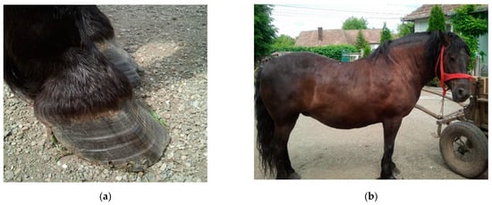
There are several factors which can induce abnormalities of energy metabolism in horses, but the most frequent cause is IR. Various investigations have been carried out to demonstrate that equids with IR are predisposed to hyperlipidemia,[21] though the exact etiology of hyperlipidemia remains unknown. Hyperlipidemia most frequently appears as a primary disease; however, many other experiments and clinical trials were performed to present the existence and development of hyperlipidemia as secondary to various systemic diseases which result in negative energy balance. Among these, disease enterocolitis, dental disease, bacterial infections, pneumonia, colic impactions, parasitism, and laminitis were the most common diseases affecting miniature breeds.[23][29][30] (Figure 1a). The list could be continued with hypovolemia, electrolyte imbalances, hepatorenal insufficiency, or esophageal laceration.[22][24][31]
Figure 1. Ex. A 7-year-old Romanian Draft Horse mare with physical characteristics of equine metabolic syndrome(EMS) and laminitis: (a) divergent growth rings in the hoof, indicating laminitis; (b) general physical aspect of the horse with EMS. Source: author’s private collection.

2.1. Clinical Signs and Diagnosis

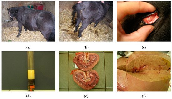
Clinical signs of hyperlipidemias are non-specific, may vary depending on the degree of metabolic disturbance, and may not be related to the loss of liver function. The most common signs include dullness, lethargy, lack of appetite, decreased water intake, and anorexia. These can progress to severe signs, such as depression, colic, cachexia, and coma (Figure 2a). Affected animals typically experience a rapid loss of body condition, diarrhea, fever, ventral edema (Figure 2b),[21][24][32] and hepatic icterus (Figure 2c). Moreover, animals affected with this disease had glossy blood, and opaque lipemic plasma (Figure 2d). A simple laboratory blood test can reveal whether concentrations of all lipids, especially TG, NEFA, or very-low density lipoprotein (VLDL), fall into healthy ranges. The results of biochemical analyses performed in miniature horses and ponies diagnosed with hyperlipemia revealed an impaired function of their liver. Though general blood tests can only reveal lipemia, lipemia is not present in all forms of hyperlipidemias. Moreover, lipemia is usually uncommon in large-breed, standard-size horses affected with severe hypertriglyceridemia.[22] For this reason, more reliable confirmation for a definitive diagnosis of hyperlipidemia can be achieved by the analysis of serum TG concentrations, blood glucose, liver enzyme, and even liver biopsy.[23]
Figure 2. Clinical signs of hyperlipidemias in ponies: (a) profoundly depressed pony with hyperlipemia; (b) diarrhea as a sign of hyperlipemia; (c) hepatic icterus; (d) blood sample from a pony with hyperlipemia showing marked opalescence of the plasma; (e) lipid accumulation in the kidneys due to hepatic dysfunction; (f) damaged fatty liver of hepatic steatosis. Source: author’s private collection.
Hepatic involvement can be detected by the increased concentration of bilirubin and hepatocellular and biliary enzyme levels.[22][27][33][34][35] Constant and long-term increase in serum TG concentrations triggers lipid accumulation in the liver, kidneys, myocardium, and skeletal muscles, leading to the depletion of these organs and death (hepatic failure) (Figure 2e). Serum bile acid level measurements can be used to verify hepatic dysfunction, and avoid death.
It is known that horses and ponies possess a remarkable liver capacity to convert free fatty acids into TGs; however, the ketone body formation pathway in ponies are more developed. After hepatic synthesis, TGs are incorporated into VLDL mediated by lipoprotein lipases, and either became available, following hydrolysis, for energy production, or are stored in the adipose tissue. When the liver’s capability to process incoming free fatty acids is exceeded, ponies release large amounts of mobilized lipid back into the plasma.[27] In horses with hyperlipidemia, hyperactivity of lipoprotein lipases was found to result in an excess production of VLDL. Under stress conditions, massive mobilization of fatty acids from fat tissue is induced as an exaggerated response to the action of catecholamines.[36] In ponies with hyperlipidemia, the activity of lipoprotein lipase, which is responsible for peripheral catabolism of TG, is doubled than that in healthy ponies, likely as a physiological response to the increased concentration of substrate. The largest fraction of TG produced by the liver in ponies with hyperlipidemia is a buoyant fraction of VLDL.[27] In pony breeds, the overproduction of TGs may be precipitated in IR during stress periods.
The cumulative effect of excess VLDL and lipomobilization results in dyslipidemia and fatty liver disease (Figure 2f). Unlike ponies, horses benefit from a highly efficient liver system of TG synthesis and incorporation into VLDL. In addition, lipomobilization under the effect of catecholamines is significantly lower in horses than in ponies, and does not reach the critical points for the development of dyslipidemia and liver lipidosis. Although there are reports of sporadic cases of natural hyperlipidemia in horses, the observed clinical symptoms (inappetence, lethargy, depression) have been attached to primary inflammatory organopathies.[22]

2.2. Prevention and Treatment

Various approaches have been proposed to prevent the development of hyperlipidemias in equids, but the most effective measures are the reduction of stress-inducing factors (stress during transport, changes in management conditions, or parasites) and negative energy balance.[21][22] Stress factors can be controlled by appropriate attention and management.[29][30] In miniature horses, donkeys, ponies, and horses with systemic disease associated with hypophagia and high metabolic demands, nutritional supplementation can prevent hyperlipemia.[32] Another possible way to prevent the development of hyperlipidemia in susceptible animals might be to improve their insulin sensitivity. Since there is a vast amount of evidence on the linkage between obesity and ID, dietary restriction could be an effective solution in order to reduce body weight in predisposed animals.[37] However, special attention should be attributed to this practice because inadequate reduction of energy intake could provoke a negative energy balance and, consequently, hyperlipidemia.[38] Fat feeding could improve the TG clearance in ponies, but the diet might lead to impaired glucose tolerance and IR.[39][40] Conversely, other studies have found that IR and adiponectin concentrations can be decreased more efficiently by a regular nonstructural carbohydrates-rich diet, rather than forage or fat-rich diets.[41][42][43] However, the association of dietary nonstructural carbohydrates intake and insulin regulation is not so obvious; some recent work has indicated that though the adaptation to high nonstructural carbohydrate diets can improve glucose tolerance and tissue insulin sensitivity, it could lead to an exaggerated postprandial insulinemic response.[44][45]
Effective management for treating hyperlipidemias in horses can be reached only if the disease is identified in early stages, and the therapy is started before triglyceridemia becomes severe. The first and most important step is to recognize the animals with a predisposition for hyperlipidemias. After recognition, the correction of the underlying diseases and conditions that caused the negative energy balance are of a great importance. Nutritional supplementation is one of the essential factors that needs to be corrected in the treatments of hyperlipemia. In fact, nutritional support can reverse the existing negative energy balance, increase serum glucose concentrations, stimulate endogenous insulin release, and inhibit the mobilization of peripheral adipose tissue.[21][32] In cases where the affected individual refuses voluntary feed intake, enteral nutrition becomes necessary. Enteral nutritional support is the most natural nutrient delivery approach, making the intestinal mucosa to be partially dependent on the digestion products for energy and nutrients. Positive responses to oral administration of small amounts of simple sugars or high-fructose corn syrup have been reported with a dosage of 5 kcal/kg/d administrated multiple times per day.[21] Other results have shown that enteral nutritional supplementation and treatment of the primary disease often reversed hyperlipemia in miniature horses and donkeys, but less frequently in ponies.[32] If the enteral route of administration is not available for various reasons (gastric reflux, bowel distention, ileus), parenteral administration remains the only solution. However, this is expensive and poses the risk of secondary complications, including hyperglycemia, hypertriglyceridemia, thrombophlebitis, and an increased risk of bloodstream infections.[46] Parenteral solutions with added lipids might be used in horses that need long-term support, or those unable to tolerate carbohydrates.[47] In more severe cases, when more complex nutritional supplementation is required, nasogastric intubation should be used.
Pharmacological attempts have also been carried out to find potential agents to avoid the development of hyperlipidemias, particularly in IR horses. Results have shown that metformin and a synthetic thyroid hormone called levothyroxine can improve insulin sensitivity by controlling blood glucose concentrations through the inhibition of hepatic gluconeogenesis and glycogenolysis.[48][49][50][51] These agents have successfully been used in human medicine as an insulin-sensitizing agent, but their effectiveness in horses is controversial. When administrated orally at high dosage, levothyroxine induced weight loss in horses and reduced intestinal glucose absorption and insulinemic responses to oral carbohydrate ingestion.[52][48][50] Pretreatment of clinically healthy horses with levothyroxine for 14 days prevented the development of IR following endotoxin infusion.[52][21]
Hyperlipidemias can also be treated by insulin therapy, but patients with IR are more difficult to treat, and need special care to prevent hypoglycemia. Subcutaneous administration of insulin can result in periods of hypoglycemia post-administration, but this effect can be alleviated by oral carbohydrates given in parallel. To determine the appropriate dose of insulin and carbohydrates for treatment, blood glucose levels need to be determined first. Insulin therapy for the treatment of hyperlipidemias is still a subject of scientific debates. On one hand, Moore et al. (1994) declared that insulin administration did not improve the medical stage of the affected animals, and proved to be inefficient because the excessive fat mobilization occurred as secondary to IR.[27] On the other hand, Durham et al. (2008), and Waitt et al. (2009) reported that insulin administrated simultaneously with adequate nutritional support alleviated hypertriglyceridemia in equids.[48][53] As an alternative, heparin has been tested and used in the treatment of hyperlipidemias in horses due to its stimulative action upon peripheral utilization of triglycerides and lipoprotein lipase activity. However, the efficiency of this treatment was questioned by other researchers who obtained negative results: heparin administration may induce bleeding complications, thrombocytopenia, and pancreatitis.[23][26][54]

3. Comparison between MetS and EMS

MetS differs from EMS as the most important pathologies that occur are T2DM and cardiovascular diseases in humans and laminitis in horses. In both humans and equids, high energy diet combined with sedentary lifestyle have resulted in obesity and metabolic disturbances. Moreover, the adipose tissue does not only serve as fat storage, but also has endocrine functions in both species. The adipose tissue produces adipokines (adiponectin, leptin, visfatin), and releases inflammatory mediators and pro-inflammatory cytokines.[55] Though mechanisms behind these processes are less documented in equids as compared to humans, the adipose tissue inflammation that affects metabolic and biochemical processes show similarities between both species;[56][57][58][59] metabolic syndrome has been associated with chronic systemic inflammatory state, and concentrations of inflammatory cytokines have been correlated with obesity and IR.[55][57][60] Pro-inflammatory cytokine expression, including IL-6 and TNF-α, have been detected in the lamellar tissue of horses with hyperinsulinemia.[61] Elevated levels of circulating NEFAs have also been associated with IR and physiological disturbances in several tissues.[36][62][63][64][65]
Though hypertriglyceridemia and low HDL-cholesterol concentrations are criteria used to define MetS, obesity and onset of IR are not associated with decreased HDL neither in equids nor cows. VLDL and HDL-cholesterol concentrations are usually greater in obese and IR horses as compared with healthy horses, and this is likely to be attributable to an increased activity of lipoprotein lipases.[66] In the study by Frank et al. (2006), plasma NEFA, VLDL, and HDL-cholesterol levels were 86%, 104%, and 29% greater, respectively, in obese IR horses than in non-obese horses.[67] In humans, the transfer of cholesterol from HDL to VLDL is catalyzed by cholesterol ester transfer protein, a protein that is not active in equine blood.[68][69]
In pathological conditions associated with overproduction of cortisol, catecholamines, and somatotropic hormone, lipolysis is stimulated by the elevated activity of hormon-sensitive lipases, but it is accompanied by reduced activity of lipoprotein lipases in both equids and cows. The conjugate effect of these enzymes results in lipomobilization coupled with a decreased liver capacity to convert NEFAs to TGs, and incorporate TGs in VLDL, leading to a significant increase in serum NEFA concentration. Thus, elevated serum NEFA levels indicate both a marked degradation of lipids in adipose tissues and liver dysfunction. Consequently, the assessment of NEFA is more important for the diagnosis and evaluation of lipomobilization syndrome in horses and cows than the determination of lipoproteins.
General obesity can accompany ID in equids with EMS. Horses and ponies with EMS should be examined for various evidence of laminitis, such as divergent hoof growth rings, or third phalanx rotation. Increased adiposity is evident before laminitis in most animals; however, it has been observed that not all obese horses present ID and laminitis, and lean phenotypes are not necessarily an exception to this rule.[70] Similarly, in human medicine, there is a group of obese people who have metabolically healthy obesity, featured by non-dysfunctional adipose tissue with higher adiponectin levels, lower inflammatory markers, and different fatty acid composition.[71] These people do not present ID and have a supposedly more favorable prognosis for cardiovascular disease, cancer, and all-cause mortality as compared with obese and insulin-dysregulated individuals.[71][72][73] However, if obesity is not addressed in time, a considerable proportion of these individuals could develop ID and MetS.[74]
MetS affects all organ systems involved in the metabolism and is characterized by abdominal obesity, hypertension, dyslipidemia, IR, vascular dysfunction, and inflammation of adipose tissue.[75] Clinical signs of EMS include general or regional adiposity, IR, and increased oxidative burst.[76][77] MetS and obesity have also been associated with neuropathies, such as distal symmetric sensory polyneuropathy, which is a major risk factor for the development of diabetic foot syndrome, accompanied in many cases by peripheral arterial occlusive disease (atherosclerosis).[78][79] Previous studies conducted on laminitic horses suggested that neuropathic changes are rare but have an important contribution to pain response in this disease. However, until now, no direct relationship between laminitis-associated neuropathy and EMS or ID has been described, even though diabetes in humans has been used as a comparison together with other causes of neuropathy.[80][81][82]
In humans, IR induces increased secretion of insulin by the pancreatic beta-cells to regulate glucose concentration in blood. However, compromised beta-cell function results in hyperglycemia and glucotoxicity.[83] Conversely, equids with EMS have compensated hyperinsulinemia. The development of hyperinsulinemia in horses is highly dependent on dietary composition, which is an important factor in the development and management of MetS as well. It has been observed that horses fed on a high cereal diet became obese and presented ID, whereas horses fed on a high fat diet became obese but without alterations of insulin sensitivity.[41] Similarly, a high fat diet was associated with higher insulin sensitivity as compared to a high starch and sugar diet.[43] In a human diet, partial replacement of digestible carbohydrates by unsaturated fats in the daily diet may positively affect the metabolic processes in obese individuals.[84] Simple sugars in a human diet are known to contribute to IR and obesity, whereas unsaturated fats have been proven to improve metabolic profiles and decrease inflammation.[85][86] However, further investigations are needed to better understand the effect of dietary composition on insulin metabolism and inflammatory mediators in horses, which could help to improve dietary management.[87] Negative energy balance is more difficult to control in equids than in humans, especially in female individuals, since their pregnancy and lactation periods automatically alter their energy balance. Previous clinical results have shown that early enteral feeding has a positive effect not only in humans, but also in horses.[21][88][89][90][91] Physical exercises are efficient and safe practices to reduce body weight and adiposity and have shown some insulin sensitivity stimulating effects as well.[92] Therefore, the optimal prevention technique to avoid hyperlipidemias is likely to set up a very well balanced plan combining dietary restriction with physical activity, similar to that proposed for human patients.
T2DM is a major problem in people with metabolic syndrome, and is less likely in equids, though horses may also develop diabetes in some cases of EMS.[93] In humans, the most common risk factors associated with T2DM are a family history of diabetes, hypertension, impaired glucose tolerance, being overweight, physical inactivity, ethnicity, and poor nutrition during pregnancy.[94][95] Risk factors in horses include metabolic syndrome and PPID, but available scientific evidence are scarce.[96][97]

References

  1. Kylin, E. Studien ueber das Hypertonie-Hyperglyka “mie-Hyperurika” miesyndrom. Zent. Inn. Med. 1923, 44, 105–127.
  2. Vague, J. La differenciation sexuelle-facteur determinant des formes de l’obesite. Presse Med. 1947, 30, 339–340.
  3. Reaven, G.M. Role of insulin resistance in human disease. Diabetes 1988, 37, 1595.
  4. Kaplan, N.M. The deadly quartet. Upper-body obesity, glucose intolerance, hypertriglyceridemia, and hypertension. Arch. Intern. Med. 1989, 149, 1514–1520.
  5. Lee, M.-J.; Wu, Y.; Fried, S.K. Adipose tissue heterogeneity: Implication of depot differences in adipose tissue for obesity complications. Mol. Asp. Med. 2013, 34, 1–11.
  6. Hotamisligil, G.S.; Shargill, N.S.; Spiegelman, B.M. Adipose expression of tumor necrosis factor-alpha: Direct role in obesity-linked insulin resistance. Science 1993, 259, 87–91.
  7. Bondia-Pons, I.; Ryan, L.; Martinez, J.A. Oxidative stress and inflammation interactions in human obesity. J. Physiol. Biochem. 2012, 68, 701–711.
  8. Willerson, J.T.; Ridker, P.M. Inflammation as a Cardiovascular Risk Factor. Circulation 2004, 109, II-2–II-10.
  9. Cătoi, A.F.; Pârvu, A.; Mureşan, A.; Busetto, L. Metabolic mechanisms in obesity and type 2 diabetes: Insights from bariatric/metabolic surgery. Obes. Facts 2015, 8, 350–363.
  10. Lorenzatti, A.J.; Toth, P.P. New perspectives on atherogenic dyslipidaemia and cardiovascular disease. Eur. Cardiol. Rev. 2020, 15, 1–9.
  11. Manchanayake, J.; Chitturi, S.; Nolan, C.; Farrell, G.C. Postprandial hyperinsulinemia is universal in non-diabetic patients with nonalcoholic fatty liver disease. J. Gastroenterol. Hepatol. 2011, 26, 510–516.
  12. Hsieh, J.; Hayashi, A.A.; Webb, J.; Adeli, K. Postprandial dyslipidemia in insulin resistance: Mechanisms and role of intestinal insulin sensitivity. Atheroscler. Suppl. 2008, 9, 7–13.
  13. Grundy, S.M. Metabolic syndrome update. Trends Cardiovasc. Med. 2016, 26, 364–373.
  14. Alberti, K.G.; Zimmet, P.Z. Definition, diagnosis and classification of diabetes mellitus and its complications. Part 1: Diagnosis and classification of diabetes mellitus provisional report of a WHO consultation. Diabet. Med. 1998, 15, 539–553.
  15. Balkau, B.; Charles, M.A. Comment on the provisional report from the WHO consultation. European Group for the Study of Insulin Resistance (EGIR). Diabet. Med. 1999, 16, 442–443.
  16. Alberti, K.G.; Zimmet, P.; Shaw, J. Metabolic syndrome—A new world-wide definition. A Consensus Statement from the International Diabetes Federation. Diabet. Med. 2006, 23, 469–480.
  17. Alberti, K.G.M.M.; Eckel, R.H.; Grundy, S.M.; Zimmet, P.Z.; Cleeman, J.I.; Donato, K.A.; Fruchart, J.-C.; James, W.P.T.; Loria, C.M.; Smith, S.C. Harmonizing the metabolic syndrome. Circulation 2009, 120, 1640–1645.
  18. Zimmet, P.; Alberti, K.G.M.M.; Stern, N.; Bilu, C.; El-Osta, A.; Einat, H.; Kronfeld-Schor, N. The Circadian syndrome: Is the metabolic syndrome and much more! J. Intern. Med. 2019, 286, 181–191.
  19. N. Frank; Raymond Geor; Simon Bailey; A. E. Durham; P. J. Johnson; Equine Metabolic Syndrome. Journal of Veterinary Internal Medicine 2010, 24, 467-475, 10.1111/j.1939-1676.2010.0503.x.
  20. Andy E. Durham; Nicholas Frank; Cathy M. McGowan; Nicola J. Menzies‐Gow; Ellen Roelfsema; Ingrid Vervuert; Karsten Feige; Kerstin Fey; ECEIM consensus statement on equine metabolic syndrome. Journal of Veterinary Internal Medicine 2018, 33, 335-349, 10.1111/jvim.15423.
  21. McKenzie, H.C., III. Equine hyperlipidemias. Vet. Clin. N. Am. Equine Pract. 2011, 27, 59–72.
  22. Dunkel, B.; McKenzie, H.C., III. Severe hypertriglyceridaemia in clinically ill horses: Diagnosis, treatment and outcome. Equine Vet. J. 2003, 35, 590–595.
  23. Naylor, J.M.; Kronfeld, D.S.; Acland, H. Hyperlipemia in horses: Effects of undernutrition and Disease. Am. J. Vet. Res. 1980, 41, 899–905.
  24. Field, J. Hyperlipaemia in a Quarterhorse. Comp. Cont. Educ. Pract. 1987, 10, 218–221.
  25. McAuliffe, S. Knottenbelt and Pascoe’s Color Atlas of Diseases and Disorders of the Horse E-Book, 2nd ed.; Elsevier: Amsterdam, The Netherlands, 2014.
  26. Mogg, T.D.; Palmer, J.E. Hyperlipidemia, hyperlipemia, and hepatic lipidosis in American miniature horses: 23 cases (1990–1994). J. Am. Vet. Med. Assoc. 1995, 207, 604–607.
  27. Moore, B.R.; Abood, S.K.; Hinchcliff, K.W. Hyperlipemia in 9 miniature horses and miniature donkeys. J. Vet. Intern. Med. 1994, 8, 376–381.
  28. Reid, S.W.; Mohammed, H.O. Survival analysis approach to risk factors associated with hyperlipemia in donkeys. J. Am. Vet. Med. Assoc. 1996, 209, 1449–1452.
  29. Jeffcott, L.B.; Field, J.R.; McLean, J.G.; O’Dea, K. Glucose tolerance and insulin sensitivity in ponies and Standardbred horses. Equine Vet. J. 1986, 18, 97–101.
  30. Watson, T.D.; Murphy, D.; Love, S. Equine hyperlipaemia in the United Kingdom: Clinical features and blood biochemistry of 18 cases. Vet. Rec. 1992, 131, 48–51.
  31. Ralston, S. Hyperglycemia/hyperinsulinemia after feeding a meal of grain to young horses with osteochondritis Dissecans (OCD) lesions. Pferdeheilkunde Equine Med. 1996, 12, 320–322.
  32. Foreman, J.H. Hyperlipemia and Hepatic Lipidosis in Large Animals. Available online: https://www.msdvetmanual.com/Dig.estive-system/hepatic-Disease-in-large-animals/hyperlipemia-and-hepatic-lipidosis-in-large-animals#v3265716 (accessed on 12 September 2021).
  33. Watson, T.D.; Burns, L.; Packard, C.J.; Shepherd, J. Effects of pregnancy and lactation on plasma lipid and lipoprotein concentrations, lipoprotein composition and post-heparin lipase activities in Shetland pony mares. J. Reprod Fertil. 1993, 97, 563–568.
  34. Gilbert, R.O. Congenital hyperlipaemia in a Shetland pony foal. Equine Vet. J. 1986, 18, 498–500.
  35. Gan, S.I.; Edwards, A.L.; Symonds, C.J.; Beck, P.L. Hypertriglyceridemia-induced pancreatitis: A case-based review. World J. Gastroenterol. 2006, 12, 7197–7202.
  36. Breidenbach, A.; Fuhrmann, H.; Deegen, E.; Lindholm, A.; Sallmann, H.P. Studies on equine lipid Metabolism. Lipolytic activities of plasma and tissue lipases in large horses and ponies. J. Vet. Med. Ser. A 1999, 46, 39–48.
  37. Van Weyenberg, S.; Hesta, M.; Buyse, J.; Janssens, G.P. The effect of weight loss by energy restriction on Metabolic profile and glucose tolerance in ponies. J. Anim. Physiol. Anim. Nutr. 2008, 92, 538–545.
  38. Geor, R.J.; Harris, P. Dietary management of obesity and insulin resistance: Countering risk for laminitis. Vet. Clin. N. Am. Equine Pract. 2009, 25, 51–65.
  39. Hughes, K.J.; Hodgson, D.R.; Dart, A.J. Equine hyperlipaemia: A review. Aust. Vet. J. 2004, 82, 136–142.
  40. Schmidt, O.; Deegen, E.; Fuhrmann, H.; Dühlmeier, R.; Sallmann, H.P. Effects of fat feeding and energy level on plasma Metabolites and hormones in Shetland ponies. J. Vet. Med. A Physiol. Pathol. Clin. Med. 2001, 48, 39–49.
  41. Bamford, N.J.; Potter, S.J.; Baskerville, C.L.; Harris, P.A.; Bailey, S.R. Effect of increased adiposity on insulin sensitivity and adipokine concentrations in different equine breeds adapted to cereal-rich or fat-rich meals. Vet. J. 2016, 214, 14–20.
  42. Pratt, S.E.; Geor, R.J.; McCutcheon, L.J. Effects of dietary energy source and physical conditioning on insulin sensitivity and glucose tolerance in Standardbred horses. Equine Vet. J. 2006, 38, 579–584.
  43. Quinn, R.W.; Burk, A.O.; Hartsock, T.G.; Petersen, E.D.; Whitley, N.C.; Treiber, K.H.; Boston, R.C. Insulin sensitivity in Thoroughbred geldings: Effect of weight gain, diet, and exercise on insulin sensitivity in Thoroughbred geldings. J. Equine Vet. Sci. 2008, 28, 728–738.
  44. Jacob, S.I.; Geor, R.J.; Weber, P.S.D.; Harris, P.A.; McCue, M.E. Effect of age and dietary carbohydrate profiles on glucose and insulin dynamics in horses. Equine Vet. J. 2017, 50, 249–254.
  45. Chameroy, K.A.; Frank, N.; Elliott, S.B.; Boston, R.C. Comparison of plasma active glucagon-like peptide 1 concentrations in normal horses and those with equine Metabolic syndrome and in horses placed on a high-grain diet. J. Equine Vet. Sci. 2016, 40, 16–25.
  46. Krause, J.B.; McKenzie, H.C., III. Parenteral nutrition in foals: A retrospective study of 45 cases (2000–2004). Equine Vet. J. 2007, 39, 74–78.
  47. Magdesian, K.G. Parenteral nutrition in the mature horse. Equine Vet. Educ. 2010, 22, 364–371.
  48. Durham, A.E.; Rendle, D.I.; Newton, J.E. The effect of metformin on measurements of insulin sensitivity and beta cell response in 18 horses and ponies with insulin resistance. Equine Vet. J. 2008, 40, 493–500.
  49. Frank, N.; Sommardahl, C.S.; Eiler, H.; Webb, L.L.; Denhart, J.W.; Boston, R.C. Effects of oral administration of levothyroxine sodium on concentrations of plasma lipids, concentration and composition of very-low-density lipoproteins, and glucose dynamics in healthy adult mares. Am. J. Vet. Res. 2005, 66, 1032–1038.
  50. Rendle, D.I.; Rutledge, F.; Hughes, K.J.; Heller, J.; Durham, A.E. Effects of metformin hydrochloride on blood glucose and insulin responses to oral dextrose in horses. Equine Vet. J. 2013, 45, 751–754.
  51. Tinworth, K.D.; Boston, R.C.; Harris, P.A.; Sillence, M.N.; Raidal, S.L.; Noble, G.K. The effect of oral metformin on insulin sensitivity in insulin-resistant ponies. Vet. J. 2012, 191, 79–84.
  52. Frank, N. Equine Metabolic syndrome. Vet. Clin. N. Am. Equine Pract. 2011, 27, 73–92.
  53. Waitt, L.H.; Cebra, C.K. Characterization of hypertriglyceridemia and response to treatment with insulin in horses, ponies, and donkeys: 44 cases (1995–2005). J. Am. Vet. Med. Assoc. 2009, 234, 915–919.
  54. Cole, R.P. Heparin treatment for aevere hypertriglyceridemia in diabetic ketoacidosis. Arch. Intern. Med. 2009, 169, 1439–1441.
  55. Ouchi, N.; Parker, J.L.; Lugus, J.J.; Walsh, K. Adipokines in inflammation and Metabolic Disease. Nat. Rev. Immunol. 2011, 11, 85–97.
  56. Dandona, P.; Aljada, A.; Bandyopadhyay, A. Inflammation: The link between insulin resistance, obesity and diabetes. Trends Immunol. 2004, 25, 4–7.
  57. Vick, M.M.; Adams, A.A.; Murphy, B.A.; Sessions, D.R.; Horohov, D.W.; Cook, R.F.; Shelton, B.J.; Fitzgerald, B.P. Relationships among inflammatory cytokines, obesity, and insulin sensitivity in the horse. J. Anim. Sci. 2007, 85, 1144–1155.
  58. Vick, M.M.; Murphy, B.A.; Sessions, D.R.; Reedy, S.E.; Kennedy, E.L.; Horohov, D.W.; Cook, R.F.; Fitzgerald, B.P. Effects of systemic inflammation on insulin sensitivity in horses and inflammatory cytokine expression in adipose tissue. Am. J. Vet. Res. 2008, 69, 130–139.
  59. Yudkin, J.S.; Stehouwer, C.D.; Emeis, J.J.; Coppack, S.W. C-reactive protein in healthy subjects: Associations with obesity, insulin resistance, and endothelial dysfunction: A potential role for cytokines originating from adipose tissue? Arterioscler. Thromb. Vasc. Biol. 1999, 19, 972–978.
  60. Treiber, K.; Carter, R.; Gay, L.; Williams, C.; Geor, R. Inflammatory and redox status of ponies with a history of pasture-associated laminitis. Vet. Immunol. Immunopathol. 2009, 129, 216–220.
  61. de Laat, M.A.; Clement, C.K.; McGowan, C.M.; Sillence, M.N.; Pollitt, C.C.; Lacombe, V.A. Toll-like receptor and pro-inflammatory cytokine expression during prolonged hyperinsulinaemia in horses: Implications for laminitis. Vet. Immunol. Immunopathol. 2014, 157, 78–86.
  62. Corcoran, M.P.; Lamon-Fava, S.; Fielding, R.A. Skeletal muscle lipid deposition and insulin resistance: Effect of dietary fatty acids and exercise. Am. J. Clin. Nutr. 2007, 85, 662–677.
  63. Eckardt, K.; Taube, A.; Eckel, J. Obesity-associated insulin resistance in skeletal muscle: Role of lipid accumulation and physical inactivity. Rev. Endocr. Metab. Disord. 2011, 12, 163–172.
  64. Einstein, F.H.; Huffman, D.M.; Fishman, S.; Jerschow, E.; Heo, H.J.; Atzmon, G.; Schechter, C.; Barzilai, N.; Muzumdar, R.H. Aging per se Increases the Susceptibility to Free Fatty Acid–Induced Insulin Resistance. J. Gerontol. Ser. A 2010, 65A, 800–808.
  65. Treiber, K.H.; Kronfeld, D.S.; Hess, T.M.; Byrd, B.M.; Splan, R.K.; Staniar, W.B. Evaluation of genetic and Metabolic predispositions and nutritional risk factors for pasture-associated laminitis in ponies. J. Am. Vet. Med. Assoc. 2006, 228, 1538–1545.
  66. Geelen, S.N.; Jansen, W.L.; Sloet van Oldruitenborgh-Oosterbaan, M.M.; Breukink, H.J.; Beynen, A.C. Fat feeding increases equine heparin-released lipoprotein lipase activity. J. Vet. Intern. Med. 2001, 15, 478–481.
  67. Frank, N.; Elliott, S.B.; Brandt, L.E.; Keisler, D.H. Physical characteristics, blood hormone concentrations, and plasma lipid concentrations in obese horses with insulin resistance. J. Am. Vet. Med. Assoc. 2006, 228, 1383–1390.
  68. Carr, M.C.; Brunzell, J.D. Abdominal obesity and dyslipidemia in the Metabolic syndrome: Importance of type 2 diabetes and familial combined hyperlipidemia in coronary artery Disease risk. J. Clin. Endocrinol. Metab. 2004, 89, 2601–2607.
  69. Watson, T.D.; Packard, C.J.; Shepherd, J. Plasma lipid transport in the horse (Equus caballus). Comp. Biochem. Physiol. B Comp. Biochem. 1993, 106, 27–34.
  70. McCue, M.E.; Geor, R.J.; Schultz, N. Equine Metabolic syndrome: A complex Disease influenced by genetics and the environment. J. Equine Vet. Sci. 2015, 35, 367–375.
  71. Perreault, M.; Zulyniak, M.A.; Badoud, F.; Stephenson, S.; Badawi, A.; Buchholz, A.; Mutch, D.M. A Distinct fatty acid profile underlies the reduced inflammatory state of Metabolically healthy obese individuals. PLoS ONE 2014, 9, e88539.
  72. Calori, G.; Lattuada, G.; Piemonti, L.; Garancini, M.P.; Ragogna, F.; Villa, M.; Mannino, S.; Crosignani, P.; Bosi, E.; Luzi, L.; et al. Prevalence, Metabolic features, and prognosis of Metabolically healthy obese Italian individuals: The Cremona study. Diabetes Care 2010, 34, 210–215.
  73. Phillips, C.M. Metabolically healthy obesity: Definitions, determinants and clinical implications. Rev. Endocr. Metab. Disord. 2013, 14, 219–227.
  74. Engelsen, C.D.; Gorter, K.J.; Salomé, P.L.; Rutten, G.E. Development of Metabolic syndrome components in adults with a healthy obese phenotype: A 3-year follow-up. Obesity 2013, 21, 1025–1030.
  75. Schlaich, M.; Straznicky, N.; Lambert, E.; Lambert, G. Metabolic syndrome: A sympathetic Disease? Lancet Diabetes Endocrinol. 2015, 3, 148–157.
  76. Carter, R.A.; Geor, R.J.; Burton Staniar, W.; Cubitt, T.A.; Harris, P.A. Apparent adiposity assessed by standardised scoring systems and morphometric measurements in horses and ponies. Vet. J. 2009, 179, 204–210.
  77. Carter, R.A.; Treiber, K.H.; Geor, R.J.; Douglass, L.; Harris, P.A. Prediction of incipient pasture-associated laminitis from hyperinsulinaemia, hyperleptinaemia and generalised and localised obesity in a cohort of ponies. Equine Vet. J. 2009, 41, 171–178.
  78. Noor, S.; Zubair, M.; Ahmad, J. Diabetic foot ulcer—A review on pathophysiology, classification and microbial etiology. Diabetes Metab. Syndr. Clin. Res. Rev. 2015, 9, 192–199.
  79. Papanas, N.; Ziegler, D. Risk factors and comorbidities in diabetic neuropathy: An update. Rev. Diabet. Stud. 2015, 12, 48–62.
  80. Jones, E.; Viñuela-Fernandez, I.; Eager, R.A.; Delaney, A.; Anderson, H.; Patel, A.; Robertson, D.C.; Allchorne, A.; Sirinathsinghji, E.C.; Milne, E.M.; et al. Neuropathic changes in equine laminitis pain. Pain 2007, 132, 321–331.
  81. Zamboulis, D.E.; Senior, M.; Clegg, P.D.; Milner, P.I. Expression of purinergic P2X receptor subtypes 1, 2, 3 and 7 in equine laminitis. Vet. J. 2013, 198, 472–478.
  82. Driessen, B.; Bauquier, S.H.; Zarucco, L. Neuropathic pain management in chronic laminitis. Vet. Clin. N. Am. Equine Pract. 2010, 26, 315–337.
  83. Chang-Chen, K.J.; Mullur, R.; Bernal-Mizrachi, E. Beta-cell failure as a complication of diabetes. Rev. Endocr. Metab. Disord. 2008, 9, 329–343.
  84. Rajaie, S.; Azadbakht, L.; Khazaei, M.; Sherbafchi, M.; Esmaillzadeh, A. Moderate replacement of carbohydrates by dietary fats affects features of Metabolic syndrome: A randomized crossover clinical trial. Nutrition 2014, 30, 61–68.
  85. Imamura, F.; Micha, R.; Wu, J.H.Y.; de Oliveira Otto, M.C.; Otite, F.O.; Abioye, A.I.; Mozaffarian, D. Effects of saturated fat, polyunsaturated fat, monounsaturated fat, and carbohydrate on glucose-insulin homeostasis: A systematic review and meta-analysis of randomised controlled feeding trials. PLoS Med. 2016, 13, e1002087.
  86. Shivappa, N.; Steck, S.E.; Hurley, T.G.; Hussey, J.R.; Hébert, J.R. Designing and developing a literature-derived, population-based dietary inflammatory index. Public Health Nutr. 2013, 17, 1689–1696.
  87. Ragno, V.M.; Zello, G.A.; Klein, C.D.; Montgomery, J.B. From Table to Stable: A Comparative Review of Selected Aspects of Human and Equine Metabolic Syndrome. J. Equine Vet. Sci. 2019, 79, 131–138.
  88. Carr, E.A. Enteral/Parenteral Nutrition in Foals and Adult Horses Practical Guidelines for the Practitioner. Vet. Clin. N. Am. Equine Pract. 2018, 34, 169–180.
  89. Golenz, M.R.; Knight, D.A.; Yvorchuk-St Jean, K.E. Use of a human enteral feeding preparation for treatment of hyperlipemia and nutritional support during healing of an esophageal laceration in a miniature horse. J. Am. Vet. Med. Assoc. 1992, 200, 951–953.
  90. Magdesian, K.G. Nutrition for critical gastrointestinal illness: Feeding horses with diarrhea or colic. Vet. Clin. N. Am. Equine Pract. 2003, 19, 617–644.
  91. Lewis, S.J.; Andersen, H.K.; Thomas, S. Early Enteral Nutrition Within 24 h of Intestinal Surgery Versus Later Commencement of Feeding: A Systematic review and Meta-analysis. J. Gastrointest. Surg. 2008, 13, 569.
  92. Borghouts, L.B.; Keizer, H.A. Exercise and insulin sensitivity: A review. Int. J. Sports Med. 2000, 21, 1–12.
  93. Durham, A.E.; Hughes, K.J.; Cottle, H.J.; Rendle, D.I.; Boston, R.C. Type 2 diabetes mellitus with pancreatic β cell dysfunction in 3 horses confirmed with minimal model analysis. Equine Vet. J. 2009, 41, 924–929.
  94. Fletcher, B.; Gulanick, M.; Lamendola, C. Risk factors for type 2 diabetes mellitus. J. Cardiovasc. Nurs. 2002, 16, 17–23.
  95. Wu, Y.; Ding, Y.; Tanaka, Y.; Zhang, W. Risk factors contributing to type 2 diabetes and recent advances in the treatment and prevention. Int J. Med. Sci. 2014, 11, 1185–1200.
  96. Durham, A.E. Endocrine Disease in Aged Horses. Vet. Clin. N. Am. Equine Pract. 2016, 32, 301–315.
  97. Durham, A.E. Therapeutics for Equine Endocrine Disorders. Vet. Clin. N. Am. Equine Pract. 2017, 33, 127–139.
More
ScholarVision Creations