Mitochondrial Retinopathies: Comparison
Please note this is a comparison between Version 2 by Nora Tang and Version 3 by Nora Tang.

The retina is an exquisite target for defects of oxidative phosphorylation (OXPHOS) associated with mitochondrial impairment. Retinal involvement occurs in two ways, retinal dystrophy (retinitis pigmentosa) and subacute or chronic optic atrophy, which are the most common clinical entities. Both can present as isolated or virtually exclusive conditions, or as part of more complex, frequently multisystem syndromes. In most cases, mutations of mtDNA have been found in association with mitochondrial retinopathy. The main genetic abnormalities of mtDNA include mutations associated with neurogenic muscle weakness, ataxia and retinitis pigmentosa (NARP) sometimes with earlier onset and increased severity (maternally inherited Leigh syndrome, MILS), single large-scale deletions determining Kearns–Sayre syndrome (KSS, of which retinal dystrophy is a cardinal symptom), and mutations, particularly in mtDNA-encoded ND genes, associated with Leber hereditary optic neuropathy (LHON). However, mutations in nuclear genes can also cause mitochondrial retinopathy, including autosomal recessive phenocopies of LHON, and slowly progressive optic atrophy caused by dominant or, more rarely, recessive, mutations in the fusion/mitochondrial shaping protein OPA1, encoded by a nuclear gene on chromosome 3q29.

  • retina
  • mitochondrial disorders
  • mitochondrial DNA
  • retinitis pigmentosa

1. Mitochondrial Bioenergetics
This review is focused on retinopathy caused by primary genetic mitochondrial disorders, although mitochondrial dysfunction may be an important pathogenetic component of other retinopathies. Present in virtually all eukaryotes, mitochondria are double-membraned organelles with a central role as the powerhouses of the cell [1]. They are in fact the major source of the high-energy phosphate molecule, adenosine triphosphate (ATP), synthesized by the mitochondrial respiratory chain (MRC) through the process of oxidative phosphorylation (OXPHOS) [2]. ATP is required for all active processes in the cell, and ATP deficiency leads to cellular dysfunction and ultimately cell death. ATP is synthesized by mitochondrial oligomycin-sensitive, proton (H+)-dependent ATP synthase, (also termed as complex V, cV), which is a multi-subunit structure embedded in the IMM and strictly linked to the MRC. Since cV can also act in reverse mode, i.e., as an ATPase which hydrolyses ATP into ADP + Pi, and this is in fact the usual way it is measured spectrophotometrically, it is also called oligomycin-sensitive mitochondrial ATPase. The MRC and the H+-ATPase/synthase stores the energy liberated through respiration, i.e., the stepwise flow of electrons extracted by oxidative catabolism from nutrient-derived substrates, as ATP and transfers it through the MRC complexes cI or cII, to cIII and eventually to cIV, due to mobile electron shuttles, Coenzyme Q (from cI-cII to cIII) and cytochrome c (from cIII to cIV). In turn, cIV (cytochrome c oxidase, COX), fixates the electrons to molecular O2, the final “electron sink” of the pathway, converting it into H2O. The energy liberated by this electronic current is exploited by cI, cIII, and cIV to pump protons across the inner mitochondrial membrane (IMM). As a result, energy is conserved generating a mitochondrial membrane potential, MtMP, which is composed of a proton concentration gradient (∆pH) and an electrostatic gradient, more negative outside, but more positive inside, the IMM (∆Ψ). The MtMP provides the proton-motive force (∆p) [3][4] that drives the rotational engine of H+-ATP synthase, by exploiting the protons flowing from the outside to the inside of the IMM through the subunit A channel, to ultimately cause the condensation of ADP and Pi into ATP. In addition to ATP production through OXPHOS, ∆p can also be dissipated into heat, a crucial function, especially in homeothermic animals, which is controlled by uncoupling proteins [5]. In addition, the MtMP provides energy for numerous other processes, such as trafficking of Ca2+, Fe2+, and other important ions, reactive oxygen and nitrous oxygen species (ROS and NOS) production, mitochondrial protein translocation, etc. A host of other pathways residing within mitochondria can modify and influence OXPHOS, and OXPHOS abnormalities can in turn generate signals triggering either homeostatic pathways, e.g., mitochondrial biogenesis, or execution programs, e.g., mitophagy, which eliminates some impaired parts, or the whole spent organelle, or apoptosis, which eliminates the dysfunctional cell altogether [6]. This complex metabolic network is largely based on the repertoire of the entire mitochondrial proteome, summing up to approximately 1500 proteins in mammals [7]. Mitochondria contain their own DNA (mitochondrial DNA, mtDNA), which in mammals is a 16.5 kb circular molecule of double-stranded DNA that encodes 13 polypeptides and 24 RNAs (22 transfer tRNAs and two ribosomal rRNAs), essential for in situ protein synthesis. This is also needed because the genetic code of mtDNA differs in different phyla, including vertebrates, from the universal code, which makes the two genomes, nuclear and mitochondrial, reciprocally untranslatable. MtDNA polypeptides are all subunits of four OXPHOS complexes (c), namely cI (n. 7), cIII (n. 1), cIV (n. 3) and cV (n. 2), where they interact with over 70 nucleus-encoded protein subunits and several non-protein prosthetic groups [8]. Additional nucleus-encoded factors are essential for mtDNA maintenance and expression, as well as for MRC formation and activity.
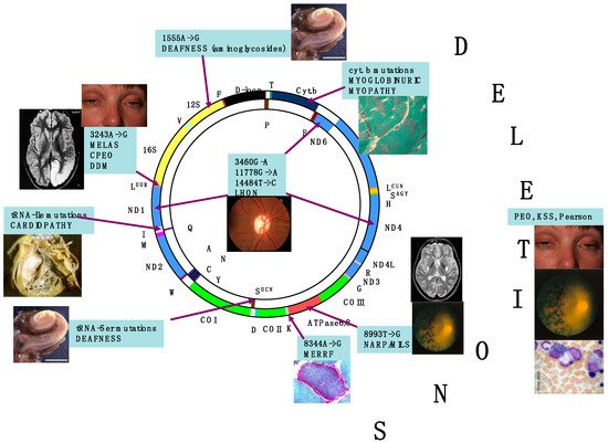
In contrast with mitochondrion-related nuclear DNA (nDNA) genes, present in a diploid organization in somatic cells, and transmitted by mendelian inheritance, mtDNA is present in thousands of copies in each cell, (polyplasmy) and, in sexuate organisms, its transmission from one generation to the next occurs exclusively through the female gamete. Uniparental maternal inheritance is also the way transmissible deleterious mtDNA mutations, leading to disease, are passed to the offspring. As a result, genetic defects affecting mtDNA or OXPHOS-related nDNA genes can impair ATP synthesis, determine mitochondrial dysfunction, and cause human disease [9]Figure 1 depicts the major mtDNA-related syndromes against a scheme of the molecule.
Figure 1. Morbidic map of mtDNA mutations. The scheme of the circular 16.5 kb human mtDNA is depicted, with several clinical and molecular syndromes associated with mutations of the molecule. Genes encoding subunits belonging to the same MRC complex have identical colours. In yellow are indicated the genes encoding the 12S and 18S ribosomal RNAs. Different colours and aminoacids expressed in the single-letter code designate the tRNA-encoding genes. The D-loop control region is in black. LHON is associated predominantly with three-point mutations, usually homoplasmic, in different genes of cI, but other, rare mutations have also been reported. Deletions, associated with KSS and other syndromes including adult-onset progressive external ophthalmoplegia and neonatal Pearson’ syndrome, usually affects the “major” arc occupying approximately two thirds of the mtDNA circle, included between the D-loop and the WANCY cluster where the origin of light strand replication is contained.
Mitochondrial disorders are in fact clinical entities associated with the ascertained or allegedly genetic defects of mitochondrial OXPHOS [10]. Tissue and organ functions where energy demand is high, such as neurons and muscle fibres, critically depend on adequate ATP supply. This explains why primary mitochondrial disorders usually cause neurodegeneration and/or muscle weakness, in children and adults [11]. However, specific mitochondrial syndromes may involve any other organ, either individually or in combination with brain and muscle dysfunction. The double genetic contribution and the complexity of the OXPHOS-related biochemical network account for the extreme clinical and genetic heterogeneity of mitochondrial disorders. In addition, many pathogenic mtDNA mutations are heteroplasmic, that is, they co-exist with a variable percentage of wild-type (wt) mtDNA, and this proportion can differ from tissue to tissue and in each individual [12], partly dictating the clinical outcome and the tissue-specific involvement in different subjects. Because of polyplasmy, the transmission of mutant vs. wt-mtDNA is largely dependent on the stochastic distribution of the organelles during mitosis in somatic cells, as well as meiotic divisions in female germ cells, and on the capacity of different tissues to work out effective selection against OXPHOS-defective cells. Thus, the same mutation, for instance the 3243G > A transition in the gene encoding mt-tRNALeu(UUR), can be associated with a virtually disease-free individual when the percentage of heteroplasmy in critical tissues is low or cause a range of different clinical syndromes, from relatively benign hearing loss and type 2 diabetes, to late-onset myopathy, to a devastating juvenile or infantile syndrome known as mitochondrial encephalomyopathy with lactic acidosis and stroke-like episodes (MELAS) [13]. Although individually rare, when taken as a group primary mitochondrial disorders are a frequent category of monogenic diseases (~1 affected individual in 4300 live births in Europe) [14]. However, the number of individuals carrying a mtDNA mutation in a percentage below the clinical threshold is probably much higher, around 1 in 500 live births.

2. Retina Is a Preferential Target of Mitochondrial Dysfunction and Diseases

Retinal dystrophy with features resembling retinitis pigmentosa and maculopathies, as well as optic neuropathy with retinal ganglion cells (RGCs) loss and atrophy of the optic nerve are very frequent in human mitochondrial disorders [15].

3. Functional Anatomy of the Retina

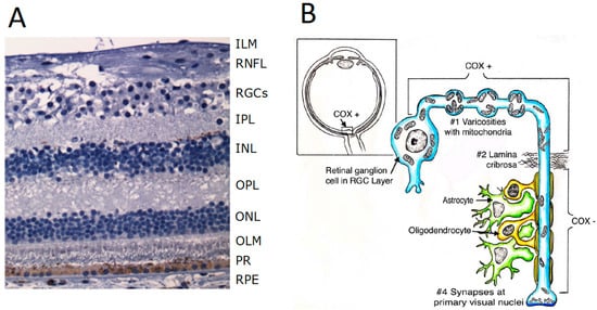
The retina is an extra-encephalic extension of the central nervous system (CNS) [16], composed of light (photo)receptors, rods, cones and a subset of retinal ganglion cells (RGCs) expressing the photopigment melanopsin (mRGCs) [17], which establish synaptic connections with several neuronal cells, eventually converging on the RGC layer as the final output projecting to the brain and ultimately the occipital cortex, where vision is formed. In fact, from RGCs, unmyelinated axons form the retinal fibre layer gathering on the optic disc, eventually forming the myelinated optic nerve, once they have passed the lamina cribrosa. Most axons through the chiasm and optic tracts reach the lateral geniculatus nucleus (LGN) serving the image-forming visual pathway; others project to the suprachiasmatic nucleus of the hypothalamus serving the non-imaging forming function, mainly deputed to photoentraining the circadian rhythms. As shown in Figure 2A, from the outer to the inner areas, the retina is composed of ten layers: (1) retinal pigmented epithelium (RPE), (2) photoreceptors layer, (3) outer limiting membrane, (4) outer nuclear layer containing the cell bodies of rods and cones, (5) outer plexiform layer composed of synapses between dendrites of horizontal cells from the inner nuclear layer and photoreceptor cells, (6) inner nuclear layer with cell bodies of horizontal cells, bipolar cells, amacrine cells, interplexiform neurons, Müller cells, (7) inner plexiform layer composed of synapses between axons of bipolar cells and dendrites of RGCs, (8) retinal ganglion cells (RGCs plus mRGCs photoreceptors) layer, (9) retinal nerve fibres layer (RNFL), and (10) inner limiting membrane. The RPE is in tight functional conjunction with photoreceptors, i.e., cones responsible for colour vision, and rods, deputed to detect dim light and contrast sensitivity serving central vision, which transduce light into action potentials, then elaborated by the retinal circuitry to convey visual information to the RGCs axons ultimately forming the optic nerve.
Figure 2. Anatomy of the retina and its connections. (A) A section of normal retina. ILM: inner limiting membrane; RNFL: retinal nerve fibres layer; RGCs: retinal ganglion cells; IPL: inner plexiform layer; INKL: inner neuronal layer; OPL: outer plexiform layer; ONL: outer neuronal layer; OLM: outer limiting membrane; PR: photoreceptors (inner segments); RPE: retinal pigmentary epithelium (modified from Maresca A, Carelli V, 2021 [18]). (B) The structure of the eyeball is outlined in the upper left inset and a retinal ganglion cell is outlined with the part of the axon (proximal to the lamina cribrosa, unmyelinated), as well as the initial part of the axon after the emersion from the lamina cribrosa, where it is myelinated by oligodendrocytes of the optic nerve. Notice the high number of mitochondria in the cell body and unmyelinated portion of the axon, often organized in mitochondrial “varicosities” compared with the few mitochondria present in the Ranvier nodes in the myelinated part of the axon (modified from Carelli V et al., 2004 [19]).

4. Physiology of the Photoreceptors

As light passes through the lens and the vitreous, it hits the retina from the inside of the eye, thus penetrating through the entire retinal thickness to eventually reach rods and cones at the outer edge. In the central foveal region of the retina, the inside layers are pulled aside from the fovea, a minute area in the centre of the retina, occupying a total surface of 1 mm2, deputed to provide visual acuity and detailed vision. The central fovea, only 0.3 mm in diameter, is composed almost entirely of slender cones. Additionally, the blood vessels, RGCs, inner nuclear layer, and plexiform layers are all displaced on the sides, letting light reach unimpeded the cones. The surrounding macular area is the on-ly region of the retina characterized by multilayered RGCs, functional to provide the highest visual discrimination, the so-called visual acuity.
The major functional segments of either a rod or cone are: (1) the outer segment, conical in shape in the cone, rod-like in the rod, (2) the inner segment, (3) the nucleus, and (4) the synaptic body. The light-sensitive photochemical is found in the outer segment. In the rods is rhodopsin; in the cones there is one of three colour pigments, functioning almost exactly the same as rhodopsin except for differences in spectral sensitivity.
Each outer segment of rods and cones are packed with approximately 1000 piled up discs, each being an infolded shelf of cell membrane. Both rhodopsin and the colour pigments are incorporated into the membranes of the discs. The pigments constitute about 40 percent of the entire mass of the outer segment. The inner segment of the rod or cone contains cytoplasmic organelles, especially mitochondria, which provide energy. The synaptic body is the portion of the rod or cone that connects with subsequent neuronal cells, the horizontal and bipolar cells, the next stages in the retinal circuitry.
When exposed to light, the resulting photoreceptor potential causes increased negativity of the membrane potential, which is a state of hyperpolarization. This is exactly the opposite to the decreased negativity (the process of “depolarization”) that occurs in almost all other sensory receptors. When rhodopsin is decomposed by light, it decreases the rod membrane conductance for Na+ ions in the outer segment of the rod, resulting in the hyperpolarization of the entire rod membrane. In the light cGMP accumulates and the cGMP-gated Na+ channels are closed, thus reducing the inward Na+ current. Na+ ions are continuously pumped outward through the membrane of the inner segment. Thus, more sodium ions now leave the rod than leak back in, creating increased negativity inside the membrane, in proportion to the amount of light energy striking the rod. The photochemicals in the cones have almost exactly the same chemical composition as that of rhodopsin in the rods. The only difference is that the protein portions, or cone-specific photopsins, are slightly different from the scotopsin of the rods. The retinal portion of all the visual pigments is exactly the same in the cones as in the rods. The colour-sensitive pigments of the cones, therefore, are combinations of retinal and photopsins. Only one of three types of colour pigments is present in each cone, thus making the cones selectively sensitive to different colours: blue, green, or red, with peak absorbencies at 445, 535, and 570 nm, respectively.
The different neuronal cell types of the retina are as follows:
  • The photoreceptors—rods and cones—which transmit signals to the outer plexiform layer, where they synapse with bipolar cells and horizontal cells.
  • The horizontal cells, which transmit signals horizontally in the outer plexiform layer from the rods and cones to bipolar cells.
  • The bipolar cells, which transmit signals vertically from the rods, cones, and horizontal cells to the inner plexiform layer, where they synapse with ganglion cells and amacrine cells.
  • The amacrine cells, which transmit signals in two directions, either directly from bipolar cells to RGCs or horizontally within the inner plexiform layer from axons of the bipolar cells to dendrites of the ganglion cells or to other amacrine cells.
  • The ganglion cells, or RGCs, which transmit output signals from the retina through the optic nerve into the brain serving both the visual and non-visual pathways (mRGCs). The first leads to formed vision, whereas the second is instrumental to photoentrain circadian rhythms.
Finally, the interplexiform cell transmits inhibitory signals in the retrograde direction from the inner plexiform layer to the outer plexiform layer, thus controlling lateral spread of visual signals by the horizontal cells in the outer plexiform layer and the degree of contrast in the visual image.
Both rods and cones release glutamate at their synapses with the bipolar cells. Amacrine cells secrete at least eight types of neurotransmitters, including γ-aminobutyric acid (GABA), glycine, dopamine, acetylcholine, and indolamine, all of which normally function as inhibitory transmitters.
Virtually all the retinal neurons, including the photoreceptors and except the RGCs, conduct their visual signals by electrotonic conduction. This allows graded conduction of signal strength directly related to the intensity of illumination. Thus, the signal is not all or none, as would be the case for action potentials. The only retinal neurons operating by action potentials are the RGCs.
Each retina contains about 100 million rods and 3 million cones, yet the number of RGCs is only about 1.2 million. Thus, an average of 60 rods and 2 cones converge on each ganglion cell and the corresponding optic nerve axon connecting to the brain. As fovea is approached, fewer rods and cones converge on each RGC axon, and the rods and cones also become slender. These effects progressively increase the acuity of vision in the central retina. In the central fovea there are only slender cones—about 35,000 of them—and no rods. Additionally, the number of RGC axons leading from this part of the retina is almost exactly equal to the number of cones. This explains the high degree of visual acuity in the central retina in comparison with peripheral retina.
The peripheral retina is much more sensitive than the fovea to weak light, which occurs partly because rods are 30 to 300 times more sensitive to light than cones are. In addition, as many as 200 rods converge on a single RGC axon in the more peripheral portions of the retina, and thus signals from the rods summate to give even more intense stimulation of the peripheral RGCs.

References

  1. Wallace, D.C. Mitochondria, bioenergetics, and the epigenome in eukaryotic and human evolution. Cold Spring Harb. Symp. Quant. Biol. 2009, 74, 383–393.
  2. Wallace, D.C. A mitochondrial paradigm of metabolic and degenerative diseases, aging, and cancer: A dawn for evolutionary medicine. Annu. Rev. Genet. 2005, 39, 359–407.
  3. Dzbek, J.; Korzeniewski, B. Control over the contribution of the mitochondrial membrane potential (ΔΨ) and proton gradient (ΔpH) to the protonmotive force (Δp). J. Biol. Chem. 2008, 283, 33232–33239.
  4. Mitchell, P. Chemiosmotic coupling in oxidative and photosynthetic phosphorylation (1966). Biochim. Biophys. Acta Bioenerg. 2011, 1807, 1507–1538.
  5. Ježek, P.; Holendová, B.; Garlid, K.D.; Jabůrek, M. Mitochondrial Uncoupling Proteins: Subtle Regulators of Cellular Redox Signaling. Antioxid. Redox Signal. 2018, 29, 667–714.
  6. Montava-Garriga, L.; Ganley, I.G. Outstanding Questions in Mitophagy: What We Do and Do Not Know. J. Mol. Biol. 2020, 432, 206–230.
  7. Calvo, S.E.; Mootha, V.K. The mitochondrial proteome and human disease. Annu. Rev. Genom. Hum. Genet. 2010, 11, 25–44.
  8. Wallace, D.C. Why do we still have a maternally inherited mitochondrial DNA? Insights from evolutionary medicine. Annu. Rev. Biochem. 2007, 76, 781–821.
  9. Gorman, G.S.; Chinnery, P.F.; DiMauro, S.; Hirano, M.; Koga, Y.; McFarland, R.; Suomalainen, A.; Thorburn, D.R.; Zeviani, M.; Turnbull, D.M. Mitochondrial diseases. Nat. Rev. Dis. Primers 2016, 2, 16080.
  10. Zeviani, M.; Di Donato, S. Mitochondrial disorders. Brain 2004, 127, 2153–2172.
  11. DiMauro, S.; Schon, E.A.; Carelli, V.; Hirano, M. The clinical maze of mitochondrial neurology. Nat. Rev. Neurol. 2013, 9, 429–444.
  12. DiMauro, S.; Schon, E.A. Mitochondrial respiratory−chain diseases. N. Engl. J. Med. 2003, 348, 2656–2668.
  13. Schon, E.A.; DiMauro, S. Mitochondrial mutations: Genotype to phenotype. Novartis Found. Symp. 2007, 287, 214–225.
  14. McFarland, R.; Taylor, R.W.; Turnbull, D.M. A neurological perspective on mitochondrial disease. Lancet Neurol. 2010, 9, 829–840.
  15. Carelli, V.; Barboni, P.; Sadun, A.A. Mitochondrial ophthalmology. In Mitochondrial Medicine; DiMauro, S., Hirano, M., Schon, E., Eds.; Informa Healthcare: Oxford, UK, 2006.
  16. Miller, N.R.; Newman, N.J.; Biousse, V.; Kerrison, J.B. (Eds.) Walsh & Hoyt’s Clinical Neuro-Ophthalmology, 6th ed.; Lippincott Williams & Wilkins: Philadelphia, PA, USA, 2005; ISBN 0-7817-4814-3.
  17. La Morgia, C.; Carelli, V.; Sadun, A.A. Retina and melanopsin neurons. Handb. Clin. Neurol. 2021, 179, 315–329.
  18. Strauss, O. The retinal pigment epithelium in visual function. Physiol. Rev. 2005, 85, 845–881.
  19. Steinberg, R.H.; Linsenmeier, R.A.; Griff, E.R. Three light-evoked responses of the retinal pigment epithelium. Vis. Res. 1983, 23, 1315–1323.
More
Video Production Service