Novel Antibacterial Approaches for Eradicating Dental Biofilm: Comparison
Please note this is a comparison between Version 1 by Ali Abdulkareem and Version 2 by Lindsay Dong.

Periodontitis is a multifactorial chronic inflammatory disease that affects tooth-supporting soft/hard tissues of the dentition. The dental plaque biofilm is considered as a primary etiological factor in susceptible patients; however, other factors contribute to progression, such as diabetes and smoking. Current management utilizes mechanical biofilm removal as the gold standard of treatment. Antibacterial agents might be indicated in certain conditions as an adjunct to this mechanical approach. Studies suggest efficacy in the use of adjunctive antimicrobials in patients with grade C periodontitis of young age or where the associated risk factors are inconsistent with the amount of bone loss present. Meanwhile, alternative approaches such as photodynamic therapy and probiotics showed limited supportive evidence, and more studies are warranted to validate their efficiency.

  • antibacterial
  • biofilms
  • periodontal debridement
  • bacterial resistance
  • photodynamic therapy
  • periodontal disease
  • probiotics
  • periodontitis

1. Introduction

Periodontitis is an inflammatory disease initiated by dysbiosis of the subgingival microbiome, with aberrant immune response, causing collateral damage to the tooth-supporting tissues and ultimately leading to tooth loss [1][2][15,16]. Dental plaque biofilm is considered as the primary etiologic factor for the majority of dental/periodontal diseases [3][17]. The gold standard of treatment for periodontitis is mechanical debridement of subgingival biofilm. Indeed, suppression of pathogenic microorganisms has for a long time been a keystone in regeneration and repair of periodontal tissues, which can be challenging using mechanical debridement alone, which must be complemented by patient-based plaque control programs [4][5][6][18,19,20]. For many decades, attempts have been made to improve the efficacy of mechanical treatment by introducing different adjuncts such as the use of antimicrobials/antibiotics at different dosages and routes of administration. However, the structural complexity of dental biofilm provides a shelter for many pathogenic microorganisms, making delivery to individual bacteria challenging [7][21]. In addition, due to the non-specificity of these drugs, they may target useful commensal species which counteract pathogenic biofilm development [8][22].

2. Structure of Biofilm

Dental plaque biofilm is formed on oral surfaces and composed of microorganisms embedded within an intercellular matrix [9]. The formation of dental biofilm is a complex process that passes through several sequential steps. Briefly, the attachment of salivary glycoproteins on clean tooth surfaces to form the acquired pellicles is the initial step. Several planktonic bacteria in saliva, such as Actinomyces spp. and Streptococcus spp., attach to binding proteins on the surface of acquired pellicles. These pioneer bacteria utilize their appendages such as fimbria and fibrils to enhance their firm adherence to the acquired pellicles and start to excrete extracellular polymeric substances (EPS) to act as a ground substance for the biofilm. Moreover, they provide specific binding sites for the adhesion of the subsequent bacterial colonization. After the sequential competitive adhesion and colonization of bacteria, the dental biofilm expands and matures within days [10].
The microorganisms in dental biofilms are mainly bacterial cells, with over 600 species of bacteria having been identified within the biofilm [9]. These bacteria are arranged in microcolonies forming about 15% to 20% of the biofilm volume. The organization of the microcolonies is not even within the layers of the biofilm. The microorganisms are well-organized in deep layers forming a dense layer of microbes, while the superficial layers contain loosely organized microbes. Consequently, dental biofilms appear as an irregular mass and may blend with the surrounding medium [11]. Each microcolony contains multiple species of bacteria. The proximity of bacterial cells allows gene exchange and quorum sensing between the cells [10]. The latter is a communication mechanism that is induced with increasing density of bacteria in the biofilm resulting in regulation of gene expression, thereby controlling certain biological processes such as symbiosis, virulence, stress adaptation, and biofilm formation [12].
The backbone of the biofilm is the extracellular matrix where the microorganisms are embedded. This matrix is porous and contains water channels that act as routes for supplying bacteria with nutrients and disposing of waste products [10]. The matrix is composed of inorganic as well as organic materials. These are mainly derived from gingival crevicular fluid, saliva and bacterial products such as EPS [11]. EPS act as selective barriers surrounding biofilms that trap nutrients from the outside environment and shelter the bacteria from it. Additionally, EPS can repel harmful agents and protect resident bacteria from outside attack [10].
Dental plaque biofilm is formed on oral surfaces and composed of microorganisms embedded within an intercellular matrix [23]. The formation of dental biofilm is a complex process that passes through several sequential steps. Briefly, the attachment of salivary glycoproteins on clean tooth surfaces to form the acquired pellicles is the initial step. Several planktonic bacteria in saliva, such as Actinomyces spp. and Streptococcus spp., attach to binding proteins on the surface of acquired pellicles. These pioneer bacteria utilize their appendages such as fimbria and fibrils to enhance their firm adherence to the acquired pellicles and start to excrete extracellular polymeric substances (EPS) to act as a ground substance for the biofilm. Moreover, they provide specific binding sites for the adhesion of the subsequent bacterial colonization. After the sequential competitive adhesion and colonization of bacteria, the dental biofilm expands and matures within days [24].
The microorganisms in dental biofilms are mainly bacterial cells, with over 600 species of bacteria having been identified within the biofilm [23]. These bacteria are arranged in microcolonies forming about 15% to 20% of the biofilm volume. The organization of the microcolonies is not even within the layers of the biofilm. The microorganisms are well-organized in deep layers forming a dense layer of microbes, while the superficial layers contain loosely organized microbes. Consequently, dental biofilms appear as an irregular mass and may blend with the surrounding medium [25]. Each microcolony contains multiple species of bacteria. The proximity of bacterial cells allows gene exchange and quorum sensing between the cells [24]. The latter is a communication mechanism that is induced with increasing density of bacteria in the biofilm resulting in regulation of gene expression, thereby controlling certain biological processes such as symbiosis, virulence, stress adaptation, and biofilm formation [26].
The backbone of the biofilm is the extracellular matrix where the microorganisms are embedded. This matrix is porous and contains water channels that act as routes for supplying bacteria with nutrients and disposing of waste products [24]. The matrix is composed of inorganic as well as organic materials. These are mainly derived from gingival crevicular fluid, saliva and bacterial products such as EPS [25]. EPS act as selective barriers surrounding biofilms that trap nutrients from the outside environment and shelter the bacteria from it. Additionally, EPS can repel harmful agents and protect resident bacteria from outside attack [24].

3. Management of Dental Biofilm

3.1. Periodontal Debridement: The Gold Standard for Periodontal Therapy

The objective of periodontal therapy is removal of the causative factor, i.e., dental biofilm from tooth surfaces. In the majority of cases, patients’ oral hygiene measures are adequate to resolve gingivitis. This may be accomplished by mechanical debridement to remove hard deposits from teeth which enhance retention of dental biofilm [13]. In periodontal pockets, subgingival debridement (SD) is pivotal for the removal of hard and soft sub-gingival deposits. To date, SD is the most effective method in the treatment of periodontitis [13]. It aims to remove the bulk of the dental biofilm, together with calculus which acts as a plaque-retentive factor and absorb bacterial toxins, thereby lowering the periodontal pathogens levels at subgingival sites, hence promoting recovery of periodontal health [13] by maintaining the level of periodontal pathogens to a threshold compatible with periodontal health [14]. This can be seen clinically through the reduction of inflammation and probing pockets depth (PPD), and the gain in clinical attachment levels (CAL) after SD using either hand or machine-driven instruments [15]. Therefore, periodontal debridement, with or without adjuncts, is still the gold standard modality for the treatment of periodontal diseases. However, long-term success is only ensured when the patients practice oral hygiene measures regularly [16].

3.2. Adjunctive Systemic and Local Antimicrobials/Antibiotics in Periodontics

Although periodontitis is not related to specific bacteria, a number of periodontal pathogens have been identified. One of the goals of periodontal therapy is to move from a ‘pathogenic’ to a ‘healthy’ biofilm [17]. SD does not always produce the desired clinical improvement in all subjects or for the same subject in the long term [18][19]. This could be attributed to the operator’s lack of skill, the presence of inaccessible areas such as multi-rooted teeth, and/or the colonization by tissue invading species such as Porphyromonas gingivalis and Aggregatibacter actinomycetemcomitans that cannot be completely eradicated by SD alone [20][21]. Thus, other forms of treatment modalities such as antibiotics/antimicrobials have been proposed as adjunctive therapy [22][23]. These therapeutic agents have diverse mechanisms of action (

3.1. Periodontal Debridement: The Gold Standard for Periodontal Therapy

The objective of periodontal therapy is removal of the causative factor, i.e., dental biofilm from tooth surfaces. In the majority of cases, patients’ oral hygiene measures are adequate to resolve gingivitis. This may be accomplished by mechanical debridement to remove hard deposits from teeth which enhance retention of dental biofilm [29]. In periodontal pockets, subgingival debridement (SD) is pivotal for the removal of hard and soft sub-gingival deposits. To date, SD is the most effective method in the treatment of periodontitis [29]. It aims to remove the bulk of the dental biofilm, together with calculus which acts as a plaque-retentive factor and absorb bacterial toxins, thereby lowering the periodontal pathogens levels at subgingival sites, hence promoting recovery of periodontal health [29] by maintaining the level of periodontal pathogens to a threshold compatible with periodontal health [30]. This can be seen clinically through the reduction of inflammation and probing pockets depth (PPD), and the gain in clinical attachment levels (CAL) after SD using either hand or machine-driven instruments [31]. Therefore, periodontal debridement, with or without adjuncts, is still the gold standard modality for the treatment of periodontal diseases. However, long-term success is only ensured when the patients practice oral hygiene measures regularly [32].

3.2. Adjunctive Systemic and Local Antimicrobials/Antibiotics in Periodontics

Although periodontitis is not related to specific bacteria, a number of periodontal pathogens have been identified. One of the goals of periodontal therapy is to move from a ‘pathogenic’ to a ‘healthy’ biofilm [33]. SD does not always produce the desired clinical improvement in all subjects or for the same subject in the long term [34,35]. This could be attributed to the operator’s lack of skill, the presence of inaccessible areas such as multi-rooted teeth, and/or the colonization by tissue invading species such as Porphyromonas gingivalis and Aggregatibacter actinomycetemcomitans that cannot be completely eradicated by SD alone [36,37]. Thus, other forms of treatment modalities such as antibiotics/antimicrobials have been proposed as adjunctive therapy [38,39]. These therapeutic agents have diverse mechanisms of action (
Figure 1); either by inhibiting cell wall synthesis, acting on cell membrane, inhibiting RNA/DNA synthesis, interfering with metabolic pathways, and inhibiting protein synthesis [24].
Antibiotics 11 00009 g002 550
); either by inhibiting cell wall synthesis, acting on cell membrane, inhibiting RNA/DNA synthesis, interfering with metabolic pathways, and inhibiting protein synthesis [40].
Figure 1. Mode of action of antimicrobial agents.
If suitable regimes can be identified where appropriate use of adjunctive antibiotics has shown a clinical benefit, this could result in a decreased need for repeated non-surgical and/or surgical periodontal interventions [25][26][27]. This has many advantages for the patient, such as reducing hard tissue trauma to non-responsive sites as well as avoiding the high emotional and financial costs of surgical intervention [28]. Furthermore, smoker subjects with deep PPD might specifically benefit from adjunctive antibiotic prescription in the non-surgical phase; however, the correct action is smoking cessation for such patients [29].

Mode of action of antimicrobial agents.
If suitable regimes can be identified where appropriate use of adjunctive antibiotics has shown a clinical benefit, this could result in a decreased need for repeated non-surgical and/or surgical periodontal interventions [48,49,50]. This has many advantages for the patient, such as reducing hard tissue trauma to non-responsive sites as well as avoiding the high emotional and financial costs of surgical intervention [51]. Furthermore, smoker subjects with deep PPD might specifically benefit from adjunctive antibiotic prescription in the non-surgical phase; however, the correct action is smoking cessation for such patients [52].

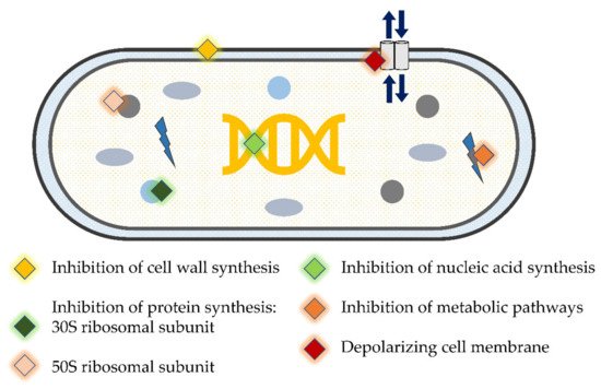
4. Using Antimicrobials/Antibiotics as Adjunct to Periodontal Therapy

A number of studies using various antimicrobials against periodontal pathogens have been carried out. It is apparent that the antimicrobials can kill periodontal pathogens in in vitro biofilm models. However, some studies indicated that amoxicillin (AMX) + metronidazole (MET) were not efficient in reducing the bacterial count [30][31]. Nevertheless, most of the studies consistently reported that the combination of these two antibiotics was superior to using either of them alone, particularly against red complex bacteria [32][33][34][35]. Similar results with regard to these bacteria were obtained with other antibiotics including azithromycin (AZM) [32][35], minocycline [36], and active organic ingredients of mouthrinses [37][38][39]. However, it is important to acknowledge that owing to greater tolerance to antimicrobials, the minimum inhibitory concentration calculated in in vitro studies would purportedly be lower and would bear little relevance to in vivo situations [18][40]. Furthermore, the majority of these studies used laboratory strains in their biofilm models, which apparently differ from clinical strains in their behavior and resistance to antimicrobials [41].

Many studies have been conducted to evaluate the additional benefits of using antibiotics within the course of periodontal therapy. Results from some of these studies have concluded that antibiotics are important adjuncts to SD in specific situations [42][43][44]. The additional clinical benefits of antibiotics are more pronounced in molar sites than in non-molar sites [45]. Following SD, the administration of broad-spectrum antibiotics for three to seven days improves microbiological outcomes compared to SD alone [44]. In regenerative periodontal therapy, better clinical outcomes could be achieved when systemic antibiotics are prescribed for patients [46]. On the other hand, many studies have reported that the use of antibiotics as an adjunct to periodontal therapy has no additional clinical benefits. Following periodontal surgery, the adjunctive use of AMX alone [47] or in combination with MET [48] for more than one week postoperatively provides no additional clinical improvements after one year. Similarly, the use of AZM as an adjunct to SD for the treatment of periodontitis seems to have no role in improving clinical outcomes compared to SD alone [49] despite the reduction in the levels of periodontal pathogens in deep periodontal pockets [50]. Precaution in prescribing antibiotics for patients with periodontal disease is advised and should be limited to certain conditions. To date, available evidence suggest prescribing adjunctive antimicrobials in patients with grade C periodontitis of young age or where the associated risk factors are inconsistent with the amount of bone loss present. 

A number of studies using various antimicrobials against periodontal pathogens have been carried out. It is apparent that the antimicrobials can kill periodontal pathogens in in vitro biofilm models. However, some studies indicated that amoxicillin (AMX) + metronidazole (MET) were not efficient in reducing the bacterial count [66,67]. Nevertheless, most of the studies consistently reported that the combination of these two antibiotics was superior to using either of them alone, particularly against red complex bacteria [68,69,70,71]. Similar results with regard to these bacteria were obtained with other antibiotics including azithromycin (AZM) [68,71], minocycline [72], and active organic ingredients of mouthrinses [73,74,75]. However, it is important to acknowledge that owing to greater tolerance to antimicrobials, the minimum inhibitory concentration calculated in in vitro studies would purportedly be lower and would bear little relevance to in vivo situations [34,76]. Furthermore, the majority of these studies used laboratory strains in their biofilm models, which apparently differ from clinical strains in their behavior and resistance to antimicrobials [77].

Many studies have been conducted to evaluate the additional benefits of using antibiotics within the course of periodontal therapy. Results from some of these studies have concluded that antibiotics are important adjuncts to SD in specific situations [93,94,95]. The additional clinical benefits of antibiotics are more pronounced in molar sites than in non-molar sites [42]. Following SD, the administration of broad-spectrum antibiotics for three to seven days improves microbiological outcomes compared to SD alone [95]. In regenerative periodontal therapy, better clinical outcomes could be achieved when systemic antibiotics are prescribed for patients [96].
On the other hand, many studies have reported that the use of antibiotics as an adjunct to periodontal therapy has no additional clinical benefits. Following periodontal surgery, the adjunctive use of AMX alone [97] or in combination with MET [98] for more than one week postoperatively provides no additional clinical improvements after one year. Similarly, the use of AZM as an adjunct to SD for the treatment of periodontitis seems to have no role in improving clinical outcomes compared to SD alone [99] despite the reduction in the levels of periodontal pathogens in deep periodontal pockets [100].
Precaution in prescribing antibiotics for patients with periodontal disease is advised and should be limited to certain conditions. To date, available evidence suggest prescribing adjunctive antimicrobials in patients with grade C periodontitis of young age or where the associated risk factors are inconsistent with the amount of bone loss present. 

5. Novel Antibacterial Agents and Strategies to Overcome Bacterial Resistance in Dental Biofilm: Pros and Cons

Interest in seeking novel alternative adjuncts to SD was raised due to limitations of conventional SD methods [51][52] and drawbacks of antimicrobials/antibiotics. Antimicrobial photodynamic therapy (aPDT) and laser are among the suggested methods that have been thoroughly investigated. Additionally, probiotics emerged as another promising approach to prevent and treat periodontal disease [53][54]. The use of lasers to debride periodontal pockets and ablate subgingival deposits has gained some traction in dental practice [55]. The utilization of a low-level laser light in combination with a photosensitizer, e.g., toluidine blue, is known as aPDT. The principle of this technique is based on light exposure of a photosensitizer releasing highly reactive oxygen radicals which destroy bacteria in periodontal pockets [56]. Additionally, photonic energy is presumed to enhance tissue healing by bio-stimulatory effects; further improvement of clinical parameters is therefore expected, such as the reduction of PPD and bleeding on probing, as well as CAL gain [57]. Concomitant improvement in clinical and microbiological parameters when aPDT was used as adjunct to SD was reported in several studies [58][59][60][61]. This was consistent with the results from a current systematic review and meta-analysis that have shown positive effects on the clinical outcomes of using aPDT together with laser, with a high impact on key periodontal pathogens, particularly the red complex [62]. However, other trials showed only a reduction of periodontal pathogens without significant difference in clinical parameters when compared to SD only [63][64]. In addition, results from other studies indicated that neither microbiological nor clinical parameters were improved following the application of laser or aPDT [65][66][67][68][69][70][71] (
Interest in seeking novel alternative adjuncts to SD was raised due to limitations of conventional SD methods [108,109] and drawbacks of antimicrobials/antibiotics. Antimicrobial photodynamic therapy (aPDT) and laser are among the suggested methods that have been thoroughly investigated. Additionally, probiotics emerged as another promising approach to prevent and treat periodontal disease [110,111]. The use of lasers to debride periodontal pockets and ablate subgingival deposits has gained some traction in dental practice [112]. The utilization of a low-level laser light in combination with a photosensitizer, e.g., toluidine blue, is known as aPDT. The principle of this technique is based on light exposure of a photosensitizer releasing highly reactive oxygen radicals which destroy bacteria in periodontal pockets [113]. Additionally, photonic energy is presumed to enhance tissue healing by bio-stimulatory effects; further improvement of clinical parameters is therefore expected, such as the reduction of PPD and bleeding on probing, as well as CAL gain [114]. Concomitant improvement in clinical and microbiological parameters when aPDT was used as adjunct to SD was reported in several studies [115,116,117,118]. This was consistent with the results from a current systematic review and meta-analysis that have shown positive effects on the clinical outcomes of using aPDT together with laser, with a high impact on key periodontal pathogens, particularly the red complex [119]. However, other trials showed only a reduction of periodontal pathogens without significant difference in clinical parameters when compared to SD only [120,121]. In addition, results from other studies indicated that neither microbiological nor clinical parameters were improved following the application of laser or aPDT [122,123,124,125,126,127,128] (
Table 1
and
Table 2
).
Table 1.
Efficacy of laser and antimicrobial photodynamic therapy as adjuncts to nonsurgical periodontal therapy on microbiological and clinical parameters.
Author, Year Study Design, Follow-Up Study Population Clinical/Microbiological Parameters aPDT Treatment Modalities
Author, Year Study Design, Follow-Up Study Population Strain of Probiotic Mode/Frequency of Administration Clinical/Microbiological Parameters
Improvement in microbiological and clinical parameters §
Moreira et al., 2015 [61]Moreira et al., 2015 [118] Split-mouth RCT, 3-months Patients with generalized AgP (n = 20)
Improvement in microbiological and clinical parameters §
Invernici et al., 2018 [72]Invernici et al., 2018 [134] Parallel arm RCT, 3-months
Chronic periodontitis patients (n = 41)
  • PI, BOP, PPD, REC, CAL
  • 40 bacterial species using the checkerboard DNA–DNA hybridization technique
SD + Diode laser (670 nm)/phenothiazine chloride (10 mg/mL) photosensitizer
Bl (HN019) 1 × 10⁹ CFU Lozenges (10 mg) 2×/day for 30-days
  • PI, BOP, PPD, CAL, REC
  • 40 subgingival bacterial species were identified using the checkerboard DNA-DNA hybridization technique
Gandhi et al., 2019 [59]Gandhi et al., 2019 [116]
Invernici et al., 2020 [73]Invernici et al., 2020 [133]Split-mouth, RCT, 9-months Periodontitis patients (n = 26) Parallel arm RCT, 3-months
Chronic periodontitis patients (n = 30)
  • PPD, PI, GI, CAL
  • Count of
  • Pg, Aa
SD + Diode laser (810 nm)/ICG photosensitizer
Bl (HN019) 1 × 10⁹ CFU Lozenges 2×/day in the morning and before bedtime for 30-days
  • PI, BOMP
Annaji et al., 2016 [60]Annaji et al., 2016 [117] Split-mouth RCT, 3-months Patients with AgP (n = 15)
  • PI, BOP, RAL, PPD
  • Culture method to identify
  • Pg, Aa, Pi
SD+ Diode Laser (810 nm)
Wadhwa et al., 2021 [58]Wadhwa et al., 2021 [115] Split-mouth RCT, 6-months Chronic periodontitis patients (n = 30) Total viable anaerobic count SD + Diode laser (810 nm)/ICG photosensitizer
Improvement in microbiological parameters only §
  • In vitro assay for adhesion of
  • Bl
  • and
  • Pg
  • to BEC
  • Antimicrobial activity of
  • Bl
  • against Fn, Pg, Pi, and Aa
Improvement in clinical parameters only §
Laleman et al., 2020 [74]Laleman et al., 2020 [135] Parallel arm RCT, 6-months Chronic periodontitis patients (n = 39) Lr (DSM 17,938 and ATCC PTA 5289) 2 × 10⁸ CFU each Five probiotic drops applied to residual pocket immediately after SD. Then each patient instructed to use lozenges 2×/day after brushing for 3-months
  • PPD, REC, CAL, FMPS, FMBS
  • PCR was used to quantify
  • Pg, Pi, Fn, Aa
Tekce et al., 2015 [75]Tekce et al., 2015 [137] Parallel arm RCT, 12-months Chronic periodontitis patients (n = 30) Lr (DSM 17,938 and ATCC PTA 5289) 2 × 10⁸ CFU each Lozenges 2×/day after brushing for 3-weeks Muzaheed et al., 2020 [63]Muzaheed et al., 2020 [120] Parallel arm RCT, 3-months Periodontitis patients (n = 45)
  • PI, CAL, PPD, GI
  • Culture method to identify
  • Pg, Aa, Td, Pi, Fn
SD + Diode laser (660 nm)/methylene-blue (0.005%) photosensitizer
Chondros et al., 2009 [64]Chondros et al., 2009 [121] Parallel arm RCT, 6-months Periodontitis patients (n = 24)
  • PPD, REC, CAL, FMPS, FMBS
  • Quantification of
  • Pg, Aa, Td, Pi, Tf, Fn, Pm, Cr, En, Ec, Cs by PCR
SD + Diode Laser (670 nm)/phenothiazine chloride (10 mg/mL) photosensitizer
No improvement in microbiological and clinical parameters §
  • PI, GI, BOP, PPD, CAL
  • Total viable cell count and the proportions of obligate anaerobic bacteria were determined
Improvement in microbiological parameters only §
Dhaliwal et al., 2017 [76]Dhaliwal et al., 2017 [136] Parallel arm RCT, 3-months Chronic periodontitis patients (n = 30) Sf (T-110 JPC), 30 × 107 CFU, Cb (TO-A HIS), 2 × 10⁶ CFU, Bm (TO-A JPC), 1 × 10⁶ CFU and Ls (HIS), 5 × 10⁷ CFU Bifilac lozenges 2×/day or 21-days
  • PI, GI, PPD, CAL
  • Microbiologic count of
  • Aa, Pg, Pi
Teughels et al., 2013 [77]Teughels et al., 2013 [138] Parallel arm RCT, 3-months Chronic periodontitis patients (n = 30) Lr (DSM17938 and ATCC PTA5289) 9 × 10⁸ CFU each Lozenges 2×/day for 3-months
  • PPD, BOP, REC, GI, PI
  • PCR was used to quantify
  • Tf, Pg, Aa, Fn, Pi
Chitsazi et al., 2014 [70]Chitsazi et al., 2014 [127] Split-mouth RCT, 3-months Patients with AgP (n = 24)
  • PPD, CAL, REC, BOP, PI, GI
  • Quantification of
  • Aa by PCR
No improvement in microbiological and clinical parameters SD + Diode Laser (670–690 nm)
§ Rühling et al., 2010 [71]Rühling et al., 2010 [128] Parallel arm RCT, 3-months Periodontitis patients (n = 54)
  • PI, PPD, CAL, BOP
Pudgar et al., 2021 [78]Pudgar et al., 2021 [139]
  • Quantification of
  • Pg, Aa, Td, Pi, Tf, Fn by PCR
Parallel arm RCT, 3-months
SD + Diode Laser (635 nm)/5% tolonium chloride photosensitizer
Chronic periodontitis patients (n = 40) Lb (CECT7480) and Lp (CECT7481), 6.0 × 10⁹ CFU/mL each One lozenge/day
  • GBI, PI, PPD, CAL, BOP, REC
  • Culture method and MALDI TOF MS for
  • Pi, Pm, Fn, Ec, Cr, Ca, Pg, Tf, Aa
Queiroz et al., 2015 [68]

Queiroz et al., 2014 [69]
Queiroz et al., 2015 [125]

Queiroz et al., 2014 [
Morales et al., 2018 [79126] ]Morales et al., 2018 [Parallel arm RCT, 3-months 140Periodontitis smoker patients (n = 20) ]
Parallel arm RCT, 9-months
  • PI, BOP, PPD, CAL, REC
Chronic periodontitis patients (n = 47)
  • 40 bacterial species using the checkerboard DNA–DNA hybridization technique
SD + Diode Laser (660 nm)/phenothiazine chloride (10 mg/mL) photosensitizer
Lrh (SP1) 2 × 10⁷ CFU One sachet in water (150 mL) and ingest it once a day after brushing for 3-months
  • PPD, PI, BOP, CAL
  • Culture method and PCR to cultivate and identify
  • Pg, Tf, Aa,
Tabenski et al., 2017 [66]Tabenski et al., 2017 [123] Parallel arm RCT, 12-months Periodontitis patients (n = 45)
  • API, PBI, BOP, PPD, CAL
  • molecular-biological testing system to identify
  • Pg, Aa, Td, Tf + TML and TBL
SD + Diode Laser (670 nm)/phenothiazine chloride photosensitizer
Hill et al., 2019 [65]Hill et al., 2019 [122] Split-mouth RCT, 6-months Periodontitis patients (n = 20)
  • BOP, PPD, RAL, REC
  • Quantification of
  • Pg, Aa, Td, Pi, Tf by PCR
SD + Diode laser (808 nm)/ICG photosensitizer
Pulikkotil et al., 2016 [67]Pulikkotil et al., 2016 [124] Split-mouth RCT, 3-months Periodontitis patients (n = 20)
  • BOP, PPD, CAL
  • Quantification of
  • Aa by PCR
SD + LED lamp (red spectrum, 628 Hz)/methylene blue photosensitizer
NSPT: nonsurgical periodontal therapy, aPDT: antimicrobial photodynamic therapy, RCT: randomized clinical trial, AgP: aggressive periodontitis, SD: subgingival debridement, PI: plaque index, PPD: probing pocket depth, CAL: clinical attachment level, RAL: relative attachment level, BOP: bleeding on probing, GI: gingival index, REC: recession, FMPS: full-mouth plaque score, FMBS: full-mouth bleeding score, PCR: polymerase chain reaction, ICG: indocyanine green, Pg: Porphyromonas gingivalis, Aa: Aggregatibacter actinomycetemcomitans, Td: Treponema denticola, Pi: Prevotella intermedia, Fn: Fusobacterium nucleatum, Tf: Tannerella forsythensis, Pm: Peptostreptococcus micros, Cr: Campylobacter rectus, En: Eubacterium nodatum, Ec: Eikenella corrodens, Cs: Capnocytophaga species, TML: total marker load, TBL: total bacterial load, API: approximal plaque index, PBI: papillary bleeding index.
§
Outcomes of PDT at endpoint as compared to control arm.
Table 2.
Efficacy of probiotics as an adjunct to nonsurgical periodontal therapy on microbiological and clinical parameters.
RCT: randomized clinical trial, Lr: Lactobacillus reuteri, PPD: probing pocket depth, CAL: clinical attachment level, BOP: bleeding on probing, Lb: Lactobacillus brevis, Lp: Lactobacillus plantarum, GBI: gingival bleeding index, PI: plaque index, REC: recession, FI: furcation involvement, MALDI TOF MS: matrix assisted laser desorption/ionization time-of-flight mass spectrometry, Pi: Prevotella intermedia, Pm: Parvimonas micra, Fn: Fusobacterium nucleatum, Ec: Eikenella corrodens, Cr: Campylobacter rectus, Ca: Capnocytophaga ochracea, Pg: Porphyromonas gingivalis, Tf: Tannerella forsythia, GI: gingival index, Lr: Lactobacillus Rhamnosus, PCR: polymerase chain reaction, Ba: Bifidobacterium animalis, FMPS: full-mouth plaque scores, FMBS: full-mouth bleeding scores, So: Streptococcus oralis, Su: Streptococcus uberis, Sr: Streptococcus rattus, Bl: Bifidobacterium animalis subsp. Lactis, BOMP: bleeding on marginal probing, BEC: buccal epithelial cells, Sf: Streptococcus faecalis, Cb: Clostridium butyricum, Bm: Bacillus mesentericus, Ls: Lactobacillus sporogenes.
§ Outcomes of probiotic treatment at endpoint as compared to control arm. In summary, a high degree of heterogeneity can be recognized in the selected studies regarding laser and aPDT such as wavelengths, single vs. multiple applications, light dose and type of photosensitizer. In addition, a carry-over effect could compromise the results from studies which followed a split-mouth design. Furthermore, applying a photosensitizer within periodontal pockets could disturb and remove the biofilm mechanically by a flushing effect. Similarly, studies on probiotics also showed heterogeneity, mainly through using different probiotic strains which were administrated in various modes, dosage, and frequency. Generally, the majority of trials on aPDT and probiotics in combination with SD have evaluated their efficacy only in the short term, i.e., three-months, and few studies extended up to 12-months. Additionally, these studies exhibited differences in the microbiological techniques and bacterial species investigated. Despite promising results being observed in some trials, the level of evidence supporting the use of these modalities as adjuncts to periodontal treatment is still low. Therefore, further well-designed, controlled, randomized clinical trials with a longer follow-up period are required to draw confirmatory conclusions.
Outcomes of probiotic treatment at endpoint as compared to control arm.
In summary, a high degree of heterogeneity can be recognized in the selected studies regarding laser and aPDT such as wavelengths, single vs. multiple applications, light dose and type of photosensitizer. In addition, a carry-over effect could compromise the results from studies which followed a split-mouth design. Furthermore, applying a photosensitizer within periodontal pockets could disturb and remove the biofilm mechanically by a flushing effect. Similarly, studies on probiotics also showed heterogeneity, mainly through using different probiotic strains which were administrated in various modes, dosage, and frequency. Generally, the majority of trials on aPDT and probiotics in combination with SD have evaluated their efficacy only in the short term, i.e., three-months, and few studies extended up to 12-months. Additionally, these studies exhibited differences in the microbiological techniques and bacterial species investigated. Despite promising results being observed in some trials, the level of evidence supporting the use of these modalities as adjuncts to periodontal treatment is still low. Therefore, further well-designed, controlled, randomized clinical trials with a longer follow-up period are required to draw confirmatory conclusions.
Video Production Service