Algae taxa are notably diverse regarding pigment diversity and composition, red seaweeds (Rhodophyta) being a valuable source of phycobiliproteins (phycoerythrins, phycocyanin, and allophycocyanin), carotenes (carotenoids and xanthophylls), and chlorophyll a. These pigments have a considerable biotechnological potential, which has been translated into several registered patents and commercial applications.
1. Phycobiliproteins
Most plants and algae filo contain chlorophyll
a and chlorophyll
b to harvest light energy. As chlorophyll
a is active at wavelengths 430 and 680 nm, and chlorophyll
b at wavelengths 450 and 660 nm, these plants and algae are photosynthetically active within this range. However, since red seaweeds only have chlorophyll
a, light is only harvested within the blue and red region of the visible spectrum, and there would be an absorption gap in the spectra region in between
[1][17]. In order to fill this gap and optimize light harvests, red seaweeds assemble phycobilissomes (PBS) in the thylakoid membrane. PBS are highly efficient, supramolecular complexes, with an absorption range of 500–660 nm, which capture solar energy and transfer it to photosystems. The role of PBS as the main light-harvesting chromoproteins was discovered in 1883 by Theodor Wilhelm Engelmann
[2][18], through the study of the Cyanobacteria
Oscillatoria. Nowadays, however, it is known that the PBS role extends beyond light-harvesting, it being also acknowledged to hold an important task as photo-protectors against high irradiances, as well as a nutrient source in times of nitrogen and phosphorus insufficiency
[3][19].
1.1. Distribution, Properties and Structure
Phycobilissomes can be classified into three types according to their morphology (hemi-ellipsoidal, hemi-discoidal, and bundle shaped)
[4][1] and are composed by phycobiliproteins (PBP). PBP present an intrinsic brilliant color being highly fluorescent
[5][20], and can be found not only in Rhodophyta (comprising up to 50% of all water-soluble proteins)
[6][21] but also in Cyanobacteria (comprising up to 60% of all water-soluble proteins)
[7][22] and in Cryptomonads (phylum Cryptophyta)
[8][23].
PBP are classified in different families, according to absorption properties, and present a distinctive color conveyed by the chromophores: the red–pink phycoerythrin (PE, λ max = 540–570 nm), the blue phycocyanin (PC, λ max = 610–625 nm), the blue–green allophycocyanin (APC, λ max = 650–660 nm), and the blue–pink phycoerythrocyanin (PEC, λ max = 560–600 nm, not present in red seaweeds)
[9][1][10][4,17,24], along with hydrophobic linker peptides
[4][1]. All these PBP are generally composed of an α and a β subunit, and among these, PE also holds a γ subunit
[11][25]. These subunits may contain about 160 to 180 amino acid residues, and are connected with prosthetic group chromophores, which are essentially linear tetrapyrrole groups that bind an apoprotein to cysteine residues through a thioether bond
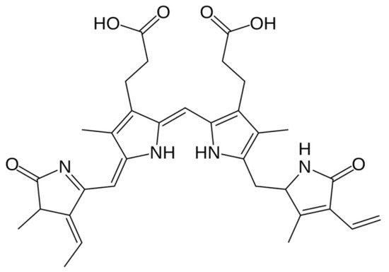
[9][12][4,26]. The protein to which this attachment occurs determines the PBP absorption spectrum, and the prosthetic group of the chromophore. A phycobilin can be categorized as phycoerythrobilin (pink–red compound), phycocyanobilin (blue compound), phycourobilin (yellow compound), and phycoviolobilin (purple compound) (
Figure 1)
[9][4]. PE and PEC are rich in blue-absorbing phycoerythrobilin, phycourobilin, and phycoviolobilin, whereas PC and APC contain phycocyanobilin only
[13][27].
Figure 1. Molecular structure of a phycobilin (phycoerythrobilin).
All PBP are tasked with (1) capturing incident light, and (2) participating in the energy transfer chain. This energy transfer is unidirectional, highly efficient (greater than 90%)
[14][28], and allows red seaweeds to harvest a much wider range of light wavelengths than the other groups of seaweeds
[1][17] and thus, optimize their photosynthetic efficiency.
In fact, PBP are essential in guaranteeing the growth and adaptation of red algae, by optimizing their light-harvesting abilities in the deeper layers of the water column, where only the blue–green spectrum of the incident light prevails
[9][4]. Generally, red algae growing under low light have the highest amount of PBP per cell and the highest number of PBS per square micrometer, over red algae growing under high light
[15][16][6,29], but other factors such as nutrient cycles and diurnal/annual photoperiod can also play a role in shaping the relative pigment content
[16][17][29,30].
1.2. Biotechnological Potential and Applications
PBPs have noteworthy spectroscopic properties, such as high absorption coefficient, high excitation and emission spectra, high quantum yield, low interference, high quenching stability, and water solubility [9][18][4,31]. Therefore, they have been widely considered in several and well documented applications, namely, in biomedical research, clinical diagnostics, therapeutic science, and cosmeceutical and pharmaceutical industries [4][14][19][20][1,28,32,33].
However, the primary commercial interest in PBP stems from the fact that these proteins offer health benefits as antioxidants and free-radical scavengers
[21][22][23][24][5,37,38,39], having a therapeutic and nutraceutical effect
[25][26][27][28][40,41,42,43], as well as being effective neuroprotective, anti-bacterial
[22][37], anti-viral
[29][44], anti-inflammatory
[30][45], anti-allergic
[31][46], anti-tumoral
[22][32][33][34][35][37,47,48,49,50], anti-ageing
[31][46], anti-Alzheimer, hepatoprotective
[24][39], immunomodulatory
[36][51], and hypocholesterolemia agents
[9][3][27][4,19,42].
In addition to their antioxidant power, PBPs are non-toxic and non-carcinogenic natural dyes. Therefore, they are also earning crescent interest over synthetic colorants within the food and cosmetic industry
[37][38][6][9,10,21], following consumer demands in their pursue for a healthier lifestyle.
1.3. Extraction and Purification Methods
PBPs are not easy and straightforward obtained. Traditionally, the methods to obtain PBP extracts present a challenge by themselves since, as mentioned, PBP are located within the phycobilissome inside the chloroplast, and thus, the algae must be pretreated with appropriate solvents, and the cells must be homogenized and disrupted using suitable methods, to release the contents within [2][18]; this must be achieved while avoiding any step or method that involves high temperatures, as these pigments are highly thermosensitive.
There are different methods to perform the extraction, whose choice is a critical step to attain a maximum PBP recover, as it has a significant impact on activity and purity of the obtained pigment [4][1]. Specific factors include biomass conditioning (fresh versus dry algae), biomass/solvent ratio, cellular disruption method, solvent, extraction time and number of steps taken [2][18], storage method prior to extraction [39][53], and even factors unconnected to the methodology itself, such as the harvest season [2][18], and species where the pigment was extracted from [40][54].
Traditionally, the abundance and diversity of the different PBP in an organism has been commonly estimated by means of light absorption assessment at distinct wavelengths, performed upon the PBP extracted from the biological matrix [41][42][55,56]. However, authors such as Saluri et al. [43][57], who lists several other works that reply on this approach to assess the PBP R-PE (a subtype of PE obtained from Rhodophyta [14][28]) content in red seaweed species, defend a more reliable method being needed, so that the most promising algae species regarding R-PE content can be targeted. According to the authors, the traditional method of absorbance reading is both quick and widely used; however, it can produce misleading results whenever impurities remain present in the samples, and it is unreliable overall, regardless of whether it is assessed upon crude extracts or following their purification. An example offered in order to circumvent this is found in Saluri et al. [43][57]’s work, which describes an alternative approach of employing the High Performance Liquid Chromatography technique of Size Exclusion Chromatography (HPLC-SEC) method with fluorescence and photo-diode array detectors, not only to separate PBP from interfering compounds, but also to reliably quantify their yields.
1.4. Production and Commercialization
As the production of PBP from natural sources requires a high investment in large-scale cyanobacteria or algae cultures, alternative approaches to obtain this pigment for biotechnological purposes has been investigated. Specifically, the production of recombinant PBP in Escherichia coli, while retaining all the qualities of a pigment obtained from native organisms, has been studied by a number of authors [20][44][45][33,60,61].
Nevertheless, a few examples can be found on a commercial level, where there are a few companies profiting from PBP commercialization in the form of natural dyes yet featuring prominently microalgae as the source for their products. For example, the commercially available blue colorant Linablue® Spirulina Extract is sold as a food product decoration component, and several companies promote their microalgae-based natural dyes to be introduced in cosmetic products, namely, phycocyanin from Arthrospira sp.
1.5. Phycoerythrin
Phycoerythrin (PE), the red pigment, owes part of its name to “erythros”, which means “red” in Greek. PE is a photosynthetic pigment, present in red macroalgae
[46][35], red microalgae
[8][23] and cryptomonads
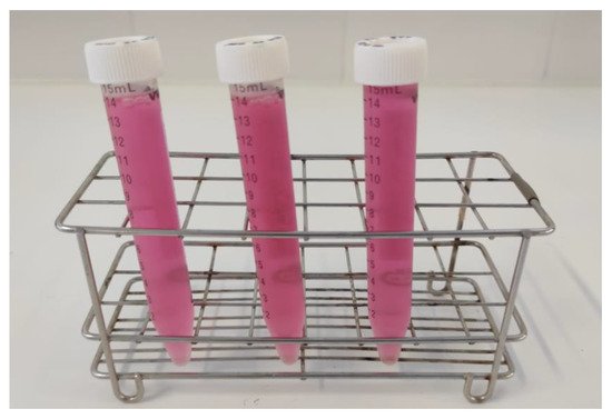
[47][64]. The original source where they are found determines their further classification into R-PE (PE obtained from Rhodophyta, λ max = 545–565 nm,
Figure 23 and
Figure 34), C-PE (PE obtained from Cyanobacteria, λ max = 540–570 nm), and B-PE (PE obtained from Bangiales, a specific family of filamentous red macroalgae, λ max = 546–565 nm)
[9][14][4,28]; besides their origin, these compounds have among them slight variations in their absorption characteristics
[48][62]. PE stands as the major soluble protein produced by the cell of red algae kept growing under low light but high nutrient load
[49][65].
Figure 23. Crude extract of R-PE obtained from the red macroalgae Ceramium ciliatum.
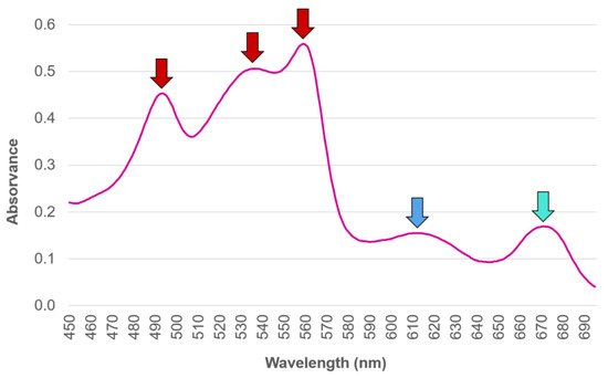
Figure 34. Typical absorption spectra of phycobilin’s (R-phycoerythrin and R-phycocyanin) extracted from the red macroalgae Gracilaria gracilis. The red arrows represent the absorption maxima that characterizes the spectrum of R-phycoerythrin, whereas the blue and the blue–green arrows represent the plateau where the absorption maxima of R-phycocyanin and allophycocyanin can be found.
PE share the qualities all PBP have, PE is also widely applied as a natural dye in the food and cosmetic industries. Additionally, as it is able to emit a strong yellow fluorescence, PE extracted from the microalgae Porphyridium sp. (phylum Rhodophyta) has been extensively explored and tested in the formulation of foods with “special effects”, such as visually appealing cake decorations, soft drinks, and alcoholic beverages that fluoresce under UV light or specific pH values
[38][10].
1.6. Phycocyanin
Phycocyanin (PC), the blue pigment, owes part of its name to “cyan” in English, which in turn derives from “kyanos” in Greek. Each of these words mean, interestingly, their own distinct shade of blue: blue-green and dark blue, respectively. PC is classified according to its source, and thus Rhodophyta contain R-Phycocyanin (R-PC), Cyanobacteria, and Cryptophyta contain C-Phycocyanin (C-PC), and Bangiophyceae contain B-Phycoyanin (B-PC)
[50][109]. These pigments have slight variations among them, in their absorption characteristics
[27][48][42,62].
PC shares the same applications as PE, with the added fact that it is the most frequently used natural blue pigment in the food industry, to color products such as jelly and bubble gum; in fact, other alternatives, likewise approved as natural blue food colorant, are yet scarce
[51][110]. PC has an extra advantage and versatility over other natural pigments, which lies within its health-promoting abilities, and not as much as a food colorant; this is due to its lower stability under heat and light when compared to gardenia and indigo natural pigments, for example
[52][111].
1.7. Allophycocyanin
Allophycocyanin (APC), the blue-green pigment, owes its name to “allos” (other) and “kyanos” (blue) in Greek. This pigment is situated in the core of phycobilissomes (PBS), where assembled trimers (αβ)
3 and phycocyanobilin’s bind to a α or β phycobilissome (PBP) subunit
[12][26].
Research into APC seems to be yet quite scarce when compared with research targeted on the other PBP found in red seaweeds, and it seems to be more focused on understanding mechanisms of structure
[53][54][55][107,141,142] and synthesis
[20][56][33,143]. Nevertheless, regarding extraction methods, diethyl ether gave good results, but methanol and ethanol were deemed improper in APC extraction of
Furcelaria lumbricalis [57][74]. On the other hand, APC was extracted from a number of Rhodophyta species using phosphate buffer
[58][70] and potassium phosphate buffer
[59][96].
2. Carotenoids
2.1. Distribution, Properties and Structure
Carotenoids are a family of long conjugated isoprenoid pigments that have a cosmopolitan presence among the vegetal kingdom, as well as in photosynthetic organisms and fungi
[60][146], whereas the animal kingdom lacks the ability to produce them, probably due to the evolutionary loss of the required genes needed to encode the enzymes for carotenoid biosynthesis
[61][147]. These accessory pigments are part of a light-harvesting antenna complex in the thylakoid membrane of chloroplasts
[60][146], and contribute to the photosynthetic process by enhancing light harvest in the blue spectrum, and thus extending the light-absorption range
[61][147]. Additionally, they have a key role as photo-protective and antioxidant agents, by shielding organisms from excess light energy, by performing the thermal dissipation of any energy surplus in the photosynthetic apparatus, and by acting as direct quenchers of reactive oxygen damage
[62][63][64][148,149,150].
Being red-to-yellow isoprenoid compounds, with eight isoprene units composed of 40 carbon atoms
[65][151], carotenoids have further chemical differences that earned them the main classification as carotenes (pure hydrocarbons) or xanthophylls (oxygenated carotenes)
[62][148]. Examples of carotenes include α-carotene, β-carotene, and lycopene, while examples of xanthophylls include lutein, zeaxanthin, fucoxanthin, and astaxanthin
[66][152].
2.2. Biotechnological Potential and Applications
Carotenoids enhance the nutritional value of a myriad of natural sources, such as fruit and vegetables, eggs, fish, algae, fungi, and yeasts [67][68][157,158]. In the human diet, approximately 50 carotenoids can be found [66][152]. Additionally, carotenoids are highly acknowledged in the cosmetic industry, by improving skin health by increasing dermal defense against UV [63][149]. They act as antioxidant agents, working connected with other reducing agents such as polyphenols and vitamins (C and E) [69][159]. Carotenoids also aid in enhancing immune defenses, and have a role in reducing the incidence of chronic diseases and diseases associated with aging, such as inflammatory diseases, cardiovascular diseases, bone/skin/eye disorders, diabetes, and cancer [62][63][70][148,149,160], while also promoting mental and metabolic health in both pregnancy and early life [70][160]. A number of carotenoids combat vitamin A deficiencies, due to their ability to be converted into retinoids that exhibit vitamin A activity [68][158]. Additionally, they are sources of color, odor, and taste [71][161].
2.3. Carotenes
Carotenes are photosynthetic pigments that are involved in primary light absorption
[72][170], but also in the seaweed photoprotective system
[73][74][171,172].
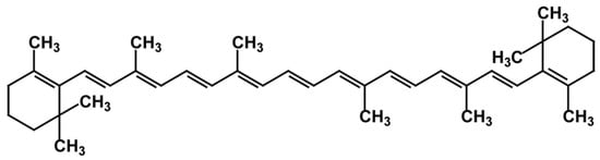
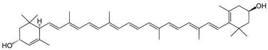
The position of a double bond (and so a hydrogen) in the cyclic group at one end differs between the two major isomers of carotene, α-carotene, and β-carotene. The molecule of β-carotene has two rings known as β-rings that are made up of nine carbon atoms, while α-carotene has a β-ring at one end of the molecule chain and a ε-ring on the other end (
Figure 45). Thus, carotenes are tetraterpenoids considered lipophilic hydrocarbons, due to the absence of oxygen
[75][175].
Figure 45.
Molecular structure of the β-carotene.
A variety of methods have been used to extract carotenoids. Liquid–liquid extraction is the most common extraction method. However, this technique has several drawbacks, such as low efficiency of carotene extraction and time consumption and has high solvent requirements
[76][178]. In contrast, many more recently developed extraction procedures have been reported and reviewed by other authors
[76][77][78][79][80][81][178,179,180,181,182,183]. These techniques include ultrasound assisted extraction (UAE), microwave assisted extraction (MAE), enzymatically assisted extraction (EAE), pressurized liquid extraction (PLE), also known as accelerated solvent extraction (ASE), and supercritical fluid extraction (SFE)
[76][77][78][79][80][81][178,179,180,181,182,183]. However, the high temperatures associated with the UAE and MAE techniques can cause carotenes to degrade, while the SFE technique can be costly due to the need for dried samples and solvents
[76][178]. Subcritical fluid extraction, which operates at lower temperatures and pressures than SFE, has recently been demonstrated to be a promising technique to extract carotene from seaweeds
[82][83][84][184,185,186].
2.4. Xanthophylls
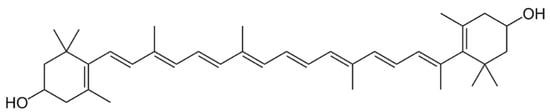
Non-provitamin A carotenoid, lutein, and zeaxanthin are structural isomers. Lutein is a polyisoprenoid with 40 carbon atoms with cyclic structures at both ends of its conjugated chain chemically (
Figure 6). As a result, it has a structure similar to zeaxanthin, but differs in the location of the double bond in one ring, resulting in three chiral centers compared to zeaxanthin’s
[60][85][146,197]. Regarding the zeaxanthin chemical structure, is constituted by a polyene chain with 11 conjugated double bonds and ionone rings (
Figure 7). The hydroxyl group on the ionone rings might connect to the fatty acids during esterification
[86][198].
Figure 56.
Molecular structure of lutein.
Figure 67.
Molecular structure of zeaxanthin.
Due to its anti-inflammatory potential, zeaxanthin is considered a tool against tumor and cancer development, and its application on chemotherapy can be beneficial for the patients
[87][203]. This molecule, as a photoprotective agent, can be also employed on eye diseases, such as cataracts, or to mitigate macular degeneration
[88][89][204,205].
For another perspective, lutein is already widely employed in industries such as cosmetics, pharma, and food, due to its color and biotechnological applications
[90][91][92][206,207,208]. In fact, several investigations have shown that lutein has anticancer properties and prevents age-related macular degeneration and cataracts
[90][93][206,209]. Oral lutein supplementation, for example, was found to minimize the impact of ultraviolet (UV) irradiation by reducing initial inflammatory reactions and the hyper-proliferative rebound caused by UV rays
[94][210].
3. Chlorophyll
3.1. Distribution, Properties and Structure
Red seaweeds have only one type of chlorophyll, chlorophyll
a, which is present in all oxygenic photosynthetic organisms. Chlorophyll
a is located in the photosystem cores and in the light-harvesting antennas on the chloroplasts
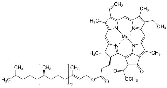
[95][225]. This molecule is chemically characterized by a porphyrin ring, comprised by a hetero polycyclic planner structure surrounding a central Mg
2+ ion, that bonds with nitrogen atoms coordinately positioned around it; a long phytol tail is attached to the polycyclic structure by ester linkage
[96][227] (
Figure 78). Chlorophyll
a is crucial to the algae’s autotrophic system, converting light energy, carbon dioxide, and water into chemical energy. This reaction is essential for the development and growth of all photosynthetic organisms, including algae
[97][228].
Figure 78.
Molecular structure of chlorophyll a
.
3.2. Biotechnological Potential and Applications
Chlorophylls are naturally strong antioxidants acting as free radical scavengers
[98][229], and have antimutagenic effects
[99][230]. In addition, the study of Lee et al.
[100][231] demonstrated that chlorophyll and chlorophyll derivatives extracted from the red seaweed
Grateloupia elliptica have anti-obesity potential. This potential was analyzed by in vitro assays with 3T3-L1 adipocytes, where chlorophyll suppressed lipid accumulation by down-regulating adipogenic protein expression at the intracellular level without being cytotoxic to cells
[100][231].
3.3. Extraction and Purification Methods
The extraction of chlorophylls from red seaweeds is mainly carried out by two methods: the conventional method of extraction by organic solvent (alcoholic or acetone-based solutions) or by cell disruption (mechanical or ultrasonication techniques). However, innovative green techniques demonstrate a high potential to be exploited to extract chlorophyll from red seaweeds, such as microwave-assisted extraction or green solvent extraction methods
[101][102][103][104][105][232,233,234,235,236]. Heating of the red seaweeds reduces chlorophyll recovery and bioavailability, unlike green and brown seaweeds; however, heating decreases chlorophyll micellarization
[101][232]. In addition, during extraction and handling the extract, light is also critical to the quality of chlorophyll, as chlorophyll immediately reacts to the light intensity by absorbing energy
[101][232].
Purification methods based on liquid chromatography are regularly applied. The main technique for purification and characterization of chlorophyll is the HPLC–MS/MS (High Performance Liquid Chromatography–Mass Spectrometry detector and selector) or using a UV/V spectrophotometry detector, due to the two well-known light absorbance peaks that chlorophyll
a has (372 and 642 nm)
[106][107][108][109][110][237,238,239,240,241]. However, there are methods that facilitate the isolation of pigments after extraction and before purification/characterization of chlorophyll. These preparatory methods are thin-layer chromatography or column chromatography, which reduce time and effectively separate the chlorophyll
[107][111][238,242].
3.4. Production and Commercialization
Chlorophyll
a (chemical reference CAS 479-61-8) from red seaweeds is being studied as pigments/colorant for textile, food (EFSA approved food additive with code E140), animal feed, skin care, and cosmetic industry
[112][113][114][115][116][117][245,246,247,248,249,250].