Klotho and Mesenchymal Stem Cells: Comparison
Please note this is a comparison between Version 1 by Erika Rangel and Version 2 by Vivi Li.

Chronic kidney disease (CKD) and acute kidney injury (AKI) are public health problems, and their prevalence rates have increased with the aging of the population. They are associated with the presence of comorbidities, in particular diabetes mellitus and hypertension, resulting in a high financial burden for the health system. Studies have indicated Klotho as a promising therapeutic approach for these conditions. Klotho reduces inflammation, oxidative stress and fibrosis and counter-regulates the renin-angiotensin-aldosterone system. In CKD and AKI, Klotho expression is downregulated from early stages and correlates with disease progression. Therefore, the restoration of its levels, through exogenous or endogenous pathways, has renoprotective effects. An important strategy for administering Klotho is through mesenchymal stem cells (MSCs).

  • chronic kidney disease
  • acute kidney injury
  • Klotho
  • mesenchymal stem cells

1. Introduction

The Klotho gene was first introduced and described in 1997 by Kuro, et al., as an anti-aging gene. They reported a mutant Klotho-deficient animal model, which presented phenotypes similar to age-related events in humans, such as reduced lifespan, alongside vascular calcification and cardiovascular disease [1][2][1,2]. Named after the Greek goddess of fate in mythology [1], Klotho is a 140 kDa protein and it has a high homology with β-glucosidases [1]. This review comprises information about αKlotho—one of this protein family’s isoforms—referred to in the present study as “Klotho”.
In humans, Klotho is present in two distinct isoforms—anchored on the membrane protein or as a soluble protein. Membrane-anchored Klotho is a single-pass transmembrane protein, of which the large extracellular domain is composed of two repeated sequences of 440 amino acids, named K11 and K12. These sequences can be proteolytically cleaved by different enzymes, leading to cleaved Klotho—one of the soluble Klotho forms [1][3][4][5][6][7][1,3,4,5,6,7]. The soluble form of this protein may be also produced from alternative splicing from its gene, leading to the formation of secreted Klotho, although this form of Klotho has not been detected in vivo to date [8][9][10][8,9,10] and studies indicate that it might not actually be secreted, because it is molecularly degraded [10].
In adult humans, the membrane-anchored form of Klotho is expressed especially in the parathyroid glands, sinoatrial node, choroid plexus and, mainly, in distal tubules in the kidneys [11]. Soluble Klotho, on the other hand, is present in systemic circulation and it has pleiotropic roles, acting as an endocrine factor with both renal and extrarenal effects [12]. It can be detected in the blood, urine and cerebrospinal fluid [9][13][14][15][9,13,14,15]. Both isoforms are of pivotal importance for homeostasis, as will be further addressed. In short, they impact on the balance of phosphate and other ions [16][17][18][16,17,18], for example, through the regulation of the absorption of different molecules such as calcium [8][19][8,19], as well as ion channels such as transient receptor potential V5 (TRPV5), sodium-phosphate cotransporter (NaPi2a) and, indirectly, sodium-chloride cotransporter (NCC) [20][21][22][20,21,22]. Klotho is also important for the cardiovascular system [23] and it participates in other biological events [24], as it will be discussed next.
Concerning its importance in embryo development, some data with animal models have indicated that Klotho is expressed since the early stages of life. Mangos, S., et al., for instance, have reported that this protein is detected in zebrafish within 24 h postfertilization (hpf) in the brain and in the pronephric ducts, which are the primitive tubules [25]. Likewise, similar findings were obtained in rodent models [26][27][26,27]. In adult zebrafish, it was observed that the expression of Klotho is maintained in the mesonephric kidney [25].
The levels of Klotho decrease with aging [28] and in the case of kidney diseases, such as chronic kidney disease (CKD) [29] and acute kidney injury (AKI). This decline is associated, for example, with the loss of renal mass and 1,25(OH)2D synthesis [3]—which, in physiology, enhances Klotho expression in the kidneys, as albuminuria, angiotensin II and proinflammatory molecules lead to the reduction of its expression [30][31][30,31].
Klotho is, in this way, an essential factor to be investigated in both the physiology and pathology of renal diseases.

2. Klotho and Chronic Kidney Disease

2.1. Chronic Kidney Disease

Chronic Kidney Disease (CKD) is a multifactorial disease [7], defined by the Kidney Disease Improving Global Outcomes Work Group (KDIGO) in 2012 as the presence of either a reduction in kidney function and/or albuminuria, that is, the abnormal excretion of albumin in the urine, for at least three months [32][33][32,33]. This illness is considered a public health issue, with prevalence rates of about 8–16% worldwide [34], and this percentage continues to rise, especially due to the aging population and the increasing incidence of type-2 diabetes, for instance [2]. Despite its several etiologies, some of the risk factors for CKD are diabetes, a family history of CKD, heart disease, high blood pressure and obesity. As early CKD might not cause any symptoms, measurement of both the serum creatinine level and protein in the urine are important methods for CKD diagnosis [35]. A key aspect of CKD is the progressive deterioration of renal function, which often leads to end-stage kidney disease (ESKD) [36], resulting in renal failure—mainly treated with either dialysis or renal transplant—and even to extrarenal complications, such as cardiovascular disease (CVD) [35]. Hence, this disorder is associated with morbidity and mortality; furthermore, it represents a high socioeconomic concern for health systems [33].
Although there are tests to detect CKD, as mentioned above, there is a scarcity of biomarkers to diagnose this disease early and precisely and avoid its progression to ESKD and other complications [37][38][37,38]. Thus, some potential early biomarkers for CKD have been studied over the years, as reviewed by Shabaka, A., et al. [37], such as Dickkopf-3 (DKK-3), a glycoprotein associated to the degree of tubulointerstitial fibrosis, of which high levels in the urine indicate an elevated risk for a reduction in eGFR within a year [39]. Neutrophil gelatinase-associated protein (NGAL) [40] is another example of a potential biomarker for CKD. These and other possible biomarkers, however, do not represent increased advantages in CKD diagnosis when compared to the traditional markers analyzed. Therefore, the study of new molecules for diagnostic purposes is still necessary [37].
Regarding therapeutic approaches, there are non-pharmacological options for the treatment of CKD, such as weight reduction and blood pressure control. Still considering the study of Shabaka, A., et al. [37], though, CKD is a complex disease, so the use of some drugs alongside non-pharmacological treatments is also important. Some examples of pharmacological treatment that can be used are sodium-glucose-cotransporter 2 inhibitors, such as empagliflozin—which induce both renal and extrarenal benefits for patients [37]—and renin-angiotensin-aldosterone system (RAAS) inhibitors, such as blockers for angiotensin II receptors and inhibitors for angiotensin-converting enzymes [37]. These are relevant therapeutic options, due to the fact that they are able to reduce the loss of eGFR, through the decline of intraglomerular pressure. Moreover, new drugs are under development, such as a mineralocorticoid receptor antagonist and potassium-lowering therapies [37]. Considering the complexity of CKD, however, the development of new therapeutic strategies is of pivotal importance.
Some of the main characteristics of CKD are chronic inflammation, hypoxia and oxidative stress, which contribute to structural and functional changes in the kidneys, resulting in glomerular, tubular and vascular injuries [33]. Thus, this disease is responsible for disturbances in mineral metabolism [41], with hyperphosphatemia and mineral-bone disorders [2] being some of its consequences.
During the progression of CKD, proinflammatory factors—interleukin 6 (IL-6) and tumor necrosis factor (TNF) [7], for example—show increased levels in the kidneys. Another significant proinflammatory molecule increased in CKD is the nuclear factor κB (NF-κB), a transcription factor related to the upregulation of cytokine expression [1]. There is also an activation of macrophages, alongside T-cell recruitment. As a consequence, these cell types and tubular epithelial cells produce profibrotic molecules. As such, transforming growth factor β (TGF-β) is one of the most influential mediators in the fibrosis process in CKD, since it stimulates the accumulation of matrix proteins and the epithelial-to-mesenchymal transition (EMT), inhibits matrix degradation and regulates myofibroblast activation [42][43][44][45][42,43,44,45]. In this context, injured tubular epithelial cells undergo a dedifferentiation process and lose their transport function and polarity. Furthermore, they synthesize the extracellular matrix. The final result of this microenvironment is the development of renal fibrosis [7].
In spite of the above description, the mechanisms relating to CKD have not yet been fully elucidated. Strong evidence, however, has pointed out the involvement of Klotho in this process, as will be addressed next.

2.2. Klotho in Chronic Kidney Disease

It has been observed in several studies that there is a decrease in Klotho levels (mRNA and protein) [2] both in animal models and individuals with CKD and renal failure [7]. The sustained Klotho imbalance in its soluble and membrane-anchored forms is associated with a decline in renal function, even in early CKD, when urinary excretion of Klotho is present in patients with this disease [33][38][33,38]. Klotho expression and levels become lower during CKD progression [38], as the estimated glomerular filtration rate (eGFR) decreases [34]. These changes in eGFR, as part of the natural history of CKD, are reflected by soluble Klotho and because of this, among other reasons, this protein could be used as an indicator for the evolution of CKD [33], for the degree of renal insufficiency in general and even for extrarenal complications [36]. Some evidence implies that the depletion of Klotho in murine models is positively correlated with persistent and increased inflammation [7]. Furthermore, it has been demonstrated that CKD is also associated with a decrease in Klotho expression [2]. Although the consequences of Klotho deficiency are not fully understood yet, it has been evidenced that renal and vascular cells senescence are some of the outcomes of this situation [2]. Likewise, a reduction in Klotho levels is associated with CKD inflammation and increased albumin excretion in patients, alongside a higher risk for some extrarenal complications, such as CV diseases and mortality. Interestingly, the restoration of Klotho levels in rodent animals through the administration of soluble Klotho, or the activation of endogenous protein, for instance, promotes the reduction of renal fibrosis, EMT and a decrease in oxidative stress and the inflammatory burden [2]. In conjunction with these data, further analyses identified that the overexpression of Klotho can lead to the enhancement of phosphaturia and to a decrease in vascular calcification in vivo, as well as to an improvement in renal function [36].
In regard to this topic, studies conducted with rodents have strongly suggested that the administration of soluble Klotho is a safe approach [19][46][19,46], although the complete spectrum of effects promoted by Klotho is still being evaluated. Likewise, in a rodent model of glomerulonephritis, which overexpresses exogenous Klotho, there is evidence of the improvement of proteinuria and serum creatinine levels. Moreover, there is also evidence of a reduction in renal cellular senescence—through a decline in β-galactosidase activity—as well as a restoration of mitochondrial activity in the cortex and the attenuation of mitochondrial damage, through cytochrome C enzyme activity reestablishment and a reduction in mitochondrial DNA damage, respectively [47]. The same study also reported a decrease in both oxidative stress and apoptosis in renal tissue. It is important to mention that viral gene delivery of Klotho, on the other hand, has not been proven to be safe in clinical studies yet [48], although it has been shown to be effective in preclinical studies, as will be further addressed in this review. Taken together, these results suggest that Klotho is a sensitive biomarker for CKD and renal function in general due to the fact that this protein level is reduced since the early stages of CKD and accompanies the decrease in eGFR. In addition, the reduction in Klotho is associated, as shown by different studies and as discussed above, with some of the characteristics of CKD, such as cellular senescence, albuminuria and cardiovascular disease.
Thus, Klotho deficiency is not only a biomarker for CKD, but also a pathogenic factor in the development, progression and complication of this disorder [38]. Importantly, preclinical data have strongly suggested that the increase in Klotho levels is safe and can mitigate fibrosis, vascular calcification, proteinuria, creatinine levels and oxidative stress, among other biological responses that are unbalanced in CKD. Therefore, further clinical studies are still necessary in order to shed light on Klotho efficiency and safety on CKD treatment, but current data strongly point to this molecule as a potential therapeutic target and its restoration levels as an approach for the treatment of CKD. Although the exact mechanisms through which Klotho influences CKD have not yet been well elucidated, several studies have addressed this issue, as discussed below.

2.2.1. Klotho and FGF-23

Anchored on the membrane form of Klotho is a co-receptor for fibroblast growth-factor 23 (FGF-23), a hormone that is produced by cells residing in bone, namely osteocytes, to target a distant organ, the kidney. The Klotho/FGF-23 complex is responsible for, among other biological responses, the activation of extracellular signal-regulated kinase (ERK)1/2 and serum/glucocorticoid-regulated kinase (SGK)1. These signaling pathways downregulate the expression of the main sodium phosphate cotransporter in proximal tubules, NaPi-2a, on the membrane of tubular cells, resulting in phosphaturia, that is, renal phosphate excretion in urine [7]. Experiments with mice suggest that the phosphaturic effect promoted by FGF-23 depends on Klotho, although the molecular mechanisms of this regulation are not yet fully understood [22][49][50][22,49,50]. FGF-23 also reduces the levels of 1,25-dihydroxyvitamin D3 (1,25-(OH)2VD3), leading to decreased intestinal reabsorption of phosphate [38]. Hence, the Klotho/FGF-23 axis is important for the ion balance and homeostasis [36]. A study conducted on 152 patients with CKD has suggested that reduced Klotho levels aggravate phosphaturia [33]. Furthermore, according to the literature, imbalance in the FGF-23-Klotho pathway and its consequent hyperphosphatemia is connected to the progression of CKD [2]. At the same time, some investigations indicate that a deficiency of Klotho limits the regulation of FGF-23 [7].
Concerning other minerals, some studies propose that, in distal tubules, Klotho/FGF-23 complexes are responsible for the modulation of calcium and sodium reabsorption, through the activation of ERK1/2, SGK1 and with-no-lysine kinase 4 (WNK4) signaling cascades. These results show that, in the context of low levels of Klotho, FGF-23 might be one of the explanations for CVD risks in patients with CKD [7].
Additionally, it is a point of interest that soluble Klotho regulates several processes, including anti-oxidation, anti-senescence, Wnt signal transduction and the anti-renin-angiotensin system (RAAS) [38]. It also inhibits fibrosis and apoptosis [51][52][53][51,52,53] and it affects mineral homeostasis through the regulation of FGF-23 and parathyroid hormone (PTH) secretion and phosphorus excretion by the kidneys, as will be discussed in Section 2.2.2 Klotho/FGF/PTH.
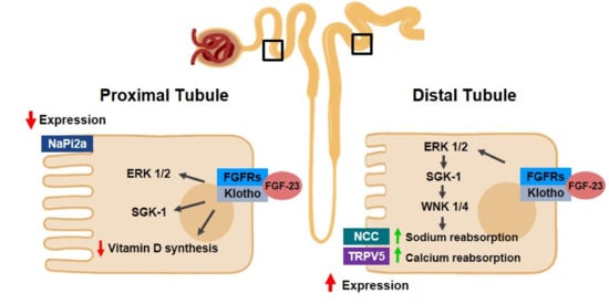
In summary, these findings support the relevance of Klotho/FGF-23 in CKD progression, as indicated in Figure 1.
Figure 1. Klotho and FGF23 in mineral homeostasis in kidneys. In proximal tubules, the Klotho/fibroblast growth-factor (FGF) 23/fibroblast growth factor receptors (FGFRs) complex activates extracellular signal-regulated kinase (ERK) 1/2, serum/glucocorticoid-regulated kinase (SGK)-1 and with no lysine kinase (WNK) 1/4 pathways, which results in the reduction of the expression of sodium phosphate co-transporter (NaPi2a), leading to phosphaturia. In distal tubules, in turn, the same complex and signaling pathways are activated and this results in an increase in sodium chloride cotransporter (NCC) and transient receptor potential cation channel subfamily V member 5 (TRPV5) channels, which contributes to increases in both sodium and calcium reabsorption, respectively.

2.2.2. Klotho/FGF/PTH Axis

Populational studies have pointed out that, in CKD patients, the increase in FGF-23 and PTH, accompanied by the decrease in 1,25 dihydroxyvitamin D3, anticipate hyperphosphatemia [54], which is often observed in these patients and is usually related to a higher mortality risk among them [55][56][57][55,56,57]. Furthermore, hyperphosphatemia is frequently detected only when renal illness is irreversible and progressing to ESKD. It is important to mention that, alongside Vitamin D, PTH regulates not only calcium metabolism, but also phosphate metabolism [55][58][55,58], inducing phosphaturia [55][59][55,59]. Moreover, it stimulates the production of Vitamin D by the kidneys.
Data in the literature indicate that Klotho modulates PTH synthesis and release directly, and also through the regulation of the active form of Vitamin D and FGF-23 in plasma [12]. In addition, it has been proposed that there is a decrease in PTH production stimulated by FGF-23, when the expression of both FGF-23 and Klotho is normal in the parathyroid glands [60]. Interestingly, studies involving epidemiological data and animal models [61][62][61,62] have demonstrated that the decrease in Klotho expression at the beginning of CKD can lead to the overproduction of FGF-23, which results in secondary hyperparathyroidism, a common complication for patients with CKD [63], which contributes to other important comorbidities detected in this condition, such as CVD. It is worth mentioning that patients with CKD have high levels of FGF-23 in the blood [64] and lower expression of Klotho and fibroblast growth factor receptor (FGFR) one in the parathyroid glands [65], although Hofman-Bang, J., et al., have described the higher expression of Klotho in the latter organ [66].
Other models have been suggested to explain the relationship between Klotho and PTH. Imura, et al., for instance, proposed binding between Klotho and Na/K-ATPase in low Ca2+ levels, which would bring this transporter to the cell membrane and trigger the release of PTH, due to the change in the electrochemical gradient, although the exact signaling pathway involved in this model has not yet been completely elucidated [67].
A high level of FGF-23 is observed in patients and animal models with CKD [68], alongside a reduction in Klotho levels in the parathyroid glands [65][69][70][65,69,70]. The inhibition of PTH synthesis by FGF-23 is therefore lost [60]. In advanced stages of CKD, the low levels of Klotho and FGFR observed in the parathyroid glands lead to the inhibition of the suppressive activity promoted by FGF-23/Klotho signaling. Importantly, FGF-23—and Klotho as well—have been also considered important modulators for phosphate homeostasis controlled by the bone-kidney axis [55]. These data suggest that Klotho restoration may be an interesting approach to avoid the development of secondary hyperparathyroidism in CKD [12]. The administration of FGF-23 in animals with CKD, on the other hand, did not reduce the levels of PTH, which might result from low expression of both Klotho and FGFR1 in the parathyroid glands [60][71][60,71].
The levels of FGFR and Klotho, however, may differ among experimental models and stages of CKD, accompanied by differences in levels of calcium in the blood [12]. This fact highlights the need for a better illustration of how this axis works.
Razzaque, et al., demonstrated that FGF23-deficient mice develop hyperphosphatemia and a phenotype similar to aging and to Klotho-deficient mice [72], including vascular calcification related to hyperphosphatemia [1][73][1,73]. Hence, FGF-23 and Klotho are associated with phosphate homeostasis [55]. Interestingly, this phenotype can be reversed using interventions to reverse hyperphosphatemia [72][74][75][76][72,74,75,76]. These data suggest a link between phosphate levels and aging [55].
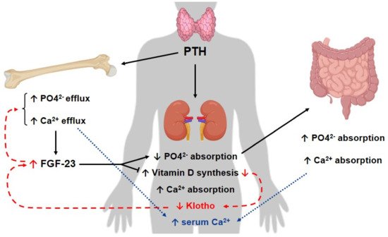
In short, studies have shown that both Klotho and FGF-23 [12] are able to regulate PTH synthesis. The hyperphosphatemia is believed to maintain the elevation of PTH levels in CKD and the dysregulation in the FGF-23/Klotho/PTH axis might lead to the progression of secondary hyperparathyroidism in CKD [12][38][12,38], as illustrated in Figure 2.
Figure 2. Klotho/FGF and PTH axis in CKD. In kidneys, parathyroid hormone (PTH) leads to an increase in calcium (Ca2+) absorption and Vitamin D synthesis, whereas it diminishes phosphorus absorption. On the other hand, PTH stimulates both phosphorus and calcium efflux in bones. In turn, PTH-stimulated Vitamin D production increases the gastrointestinal reabsorption of these minerals. As a result, both gastrointestinal calcium reabsorption and its efflux from bones contribute to a rise in calcium excretion. Phosphorus (PO42−) is also eliminated as a consequence. In chronic kidney disease (CKD) (red dotted line), there is a reduction in Klotho expression, alongside a decrease in Vitamin D levels and an increase in fibroblast growth factor (FGF-)23 levels. It is important to mention that the decrease of Vitamin D is related to the decrease of Klotho in the kidneys, which then leads to a rise in FGF-23 levels in the bones. As a consequence, this hormone diminishes Vitamin D production. As a result of this axis dysregulation, the inhibition of PTH synthesis promoted by these components is lost, which leads to a rise in the levels of this hormone, which also contributes to the elevation of these molecules. Secondary hyperparathyroidism associated with CKD might be a result of the described imbalance in this axis for CKD patients.
The instability in mineral metabolism, as described above, is a hallmark and also an initiator of the development of mineral bone disease in CKD [48], which contributes to higher mortality due to CVD and morbidity for these patients [77][78][79][80][81][77,78,79,80,81]. The FGF-23/Klotho axis, then, might be a target for new therapies for these patients [82].

2.2.3. CKD and Cardiovascular Disease

Cardiovascular disease is an important and frequent morbidity in patients with renal illness and it is related to the mortality seen in these persons [83].
Preclinical and clinical studies have pointed out the relevance of Klotho, and also FGF-23, for the cardiovascular system and how it can be related to CVD in CKD individuals and mortality among the elderly and in hemodialysis patients [8][84][8,84]. The table below—Table 1—summarizes some of the results found regarding the topic.
Table 1. Klotho and the Klotho/FGF-23 axis in CKD-associated cardiovascular disease. Summary of some of the studies analyzed in this review, concerning the role of Klotho and the Klotho/FGF-23 axis in cardiovascular disease, an important and common morbidity and cause of mortality in CKD patients.
Author/Year Model Used Study Design Conclusion
Karalliedde, J., et al., 2013 [85] Patients with diabetes type 2, presenting systolic hypertension and albuminuria Single-Center, Double-Blind Randomized Controlled Trial Inhibition of RAAS led to an increase in soluble Klotho levels.
Saito, Y., et al., 2000 [86] Rats with atherosclerosis Preclinical Study Klotho adenoviral delivery resulted in the mitigation of vascular endothelial dysfunction and reduction of blood pressure values
Kuro-o, M., et al.,1997 [1] Klotho-deficient mice Preclinical Study The animals presented artery calcification, cardiac fibrosis and hypertrophy. Klotho might participate in the signaling pathways involved in these processes.
Xie, J., et al., 2015 [87] Klotho-deficient mice Preclinical Study The increase in soluble Klotho levels attenuated cardiac remodeling in CKD animals. Decrease in this protein level is proposed to be an independent factor for cardiomyopathy in CKD.
Ding, et al., 2019 [88] Mice with angiotensin-II infusion Preclinical Study Klotho was related to the decrease of cardiac FGF-23 expression in vitro and in vivo; moreover, it prevented cardiac remodeling and dysfunction in this model.
Memmos, et al., 2019 [84] 79 patients on dialysis Prospective Cohort Study Low levels of Klotho are correlated with an increased risk of cardiovascular disease and reduced overall survival in these patients. It might contribute to cardiovascular disease in individuals with CKD.
Brandenburg, V.M., et al., 2015 [89] 2948 patients Multicenter Longitudinal Study In individuals with normal kidney function, Klotho does not act as a predictive marker of cardiovascular and mortality risk.
Pan, H.C., et al., 2018 [90] 168 patients with diabetes type 2 Prospective Study Low levels of Klotho are associated with cardiovascular outcomes, such as coronary disease. In these patients, Klotho level is a predictor for vascular events.
Gutierrez, O.M., et al., 2009 [91] 162 patients with CKD Cross-Sectional Study FGF-23 is correlated with vascular dysfunction, such as left ventricular mass index and hypertrophy in these individuals.
In addition to the previously mentioned results, Faul, C., et al. evaluated the relation between FGF-23 and the pathogenesis of left ventricular hypertrophy (LVH) in humans, in patients from both the Chronic Renal Insufficiency Cohort (CRIC) study—a prospective cohort study conducted with CKD individuals [92]—and studies conducted by their group (Faul C and coworkers) [64]. A direct induction of hypertrophy in cardiomyocytes in vitro and in vivo (LVH) in mouse models has been demonstrated after FGF-23 administration. In mice lacking Klotho, which is in turn an accepted model for constitutively high FGF-23 levels, the development of LVH has been observed in a dose-dependent way. It is important to mention that the group indicated through molecular experiments that, although Klotho is the coreceptor for FGF-23, it is not expressed in murine heart preparations or neonatal rat ventricular myocytes (NRVMs). On the other hand, FGFR isoforms, from FGFR1 to FGFR4, were detected in vitro (NRVMs) and in vivo (murine heart); it is known that FGF-23 can bind these receptors [93][94][93,94], indicating that hypertrophy induced by FGF-23 in these sites is Klotho-independent. Moreover, the inhibition of FGFR with the intraperitoneal administration of PD173074 in a nephrectomy rat model ameliorated the severity of LVH, although improvements in CKD and hypertension were not observed. In brief, this study determined a causal association for elevated FGF-23 and the physiopathology of LVH, which could shed light on the high rate of LVH in CKD patients [64]. Regarding the onset of LVH, the same patients from this study were re-analyzed a few years later and it was found that high levels of FGF-23 were associated with an increased risk of LVH. Taken together, the results indicate that LVH can be preceded by high FGF-23 levels in CKD patients, both with or without hypertension [64].
A similar result was observed in a study conducted with nephrectomy CKD- induced rats, in which FGF-23 caused the hypertrophic growth of myocytes in vitro and induced LVH in mice, through the FGFR pathway [95].
In addition to the previously mentioned data, Klotho deficiency is also associated with vascular calcification (VC) in CKD, although the pathogenesis of this clinical outcome is not completely understood yet. It has been reported in a preclinical study involving mice and human aorta samples, for example, that the increase of Klotho in vitro led to the inhibition of VC [96]. Furthermore, the same research group from the previously mentioned study came up with a mechanism believed to modulate the expression of Klotho. They proposed that the mammalian target of rapamycin (mTOR) pathway is involved in the reduction of Klotho expression in a high-phosphate-concentration scenario and that the administration of rapamycin, an mTOR inhibitor, upregulates the levels of both membrane-anchored and soluble Klotho. This results in a decline in VC in vitro promoted by rapamycin, which was not observed in Klotho-knockout mice, indicating the pivotal role of this protein in the attenuation of VC evaluated in these models [96].
It is also interesting to mention that, as discussed in Section 2.2.2 Klotho/FGF/PTH axis, the lack of Klotho contributes to mineral disease in CKD for several reasons. Among them, Klotho deficiency results in a higher phosphate concentration in the organism, which leads to calcification of tissues as well [97]. Importantly, this imbalance in phosphate levels might also be related to the dysregulation in the PTH axis in CKD, believed to be associated with the lack of Klotho in the parathyroid glands [60][70][98][60,70,98].
Moreover, angiotensin II (Ang II) is also a special point of interest, because of its direct association with CVD, a possible complication in chronic kidney disease, and due to its pathological role, through several different mechanisms, as well as in CKD, as will be discussed. Reports have shown that Ang II and aldosterone suppress the expression of Klotho in the kidneys and in kidney cell lines [7][38][7,38]. Moreover, experiments with long-term infusion of angiotensin II in rats have shown a downregulation of renal Klotho mRNA, even with a low dose of Ang II. On the other hand, the overexpression of Klotho through in vivo gene transfer attenuates damages induced by Ang II in the kidneys [38]. Further analysis also indicated that losartan, an angiotensin type I receptor antagonist, blocked the reduction in Klotho levels induced by Ang II (in vivo and in vitro) and upregulated the expression of this protein in mice, improving structural alteration in the kidneys by nephropathy with cyclosporine. At the same time, one of the roles of soluble Klotho is anti-angiotensin activity in animal models. According to these results, evidence supports the association between Klotho and renoprotection from damages induced by Ang II [38].
The exact mechanisms by which Ang II reduces Klotho expression or contributes to kidney fibrosis are not completely understood yet, but experiments with free-radical scavenging have indicated the inhibition of Klotho expression by both Ang II and oxidative stress, which suggests that oxidative stress is one of these mechanisms. Furthermore, in a mouse model, inhibition of 1,25-(OH)2VD3 synthesis is correlated with increased renin expression, whereas its injection suppresses renin. Furthermore, a study conducted on vitamin D receptor (VDR)-null mice showed that they expressed higher levels of renin and Ang II. The same study has proved that, in mouse kidney and in HEK 293 cells, active vitamin D3 upregulates Klotho expression, reversing the decrease in Klotho’s mRNA by aldosterone. This demonstrates that supplementation with active vitamin D3 might have a positive effect for the upregulation of Klotho expression. Taken together, these results indicate that 1,25-(OH)2VD3 is a negative regulator of RAAS [38].
Additionally, Miao, J., et al., 2021 illustrated in mice a reverse in both mitochondrial loss of mass and the production of reactive oxidative stress with Klotho administration. Correspondingly, it is known that RAAS activation can lead to mitochondrial damage [99]. The inhibition of RAAS by Klotho, as shown in some prior studies [100], could protect mitochondrial function, contributing to the delay of age-fibrosis caused by RAAS. Although this effect has not yet been confirmed in the CKD context, this result suggests that it might be a possibility [99].
A current challenge in regard to RAAS inhibition is that the synthesis of Ang II is not affected by RAAS inhibitors (angiotensin-converting enzyme (ACE) inhibitors or Angiotensin II receptor type I (AT1) receptor blocker, for example) and there is also a stimulation of renin secretion as a consequence [99][101][99,101]. It is suggested, however, that multiple genes of RAAS are regulated by Wnt/β-catenin signaling. In this context, the inhibition of the Wnt/β-catenin signaling pathway has been proposed as a strategy for the inhibition of RAAS [41][99][101][102][41,99,101,102]. In accordance with this notion, Wnt-1, a component of the Wnt/β-signaling pathway, might be linked to kidney fibrosis, since studies with cultured tubular cells have shown that this molecule induced upregulation of fibronectin, a marker for fibrosis. This effect, though, was blocked by treatment with Klotho [99]. A point of interest is that Wnt/β-catenin activation may lead to Klotho expression. Experiments indicate that Klotho is an endogenous antagonist of Wnt and could prevent the activation of β-catenin through binding to other components of this signaling cascade, such as Wnt1, Wnt4 and Wnt7 [99]. This is one potential renoprotective mechanism for Klotho, considering the indirect inhibition of RAAS and the direct inhibition of Wnt/β-catenin. All in all, the information previously presented highlights the association between Klotho and cardiovascular disease in CKD—a common complication that often contributes to mortality in patients with CKD. This event has been observed through different mechanisms, as mentioned above, such as the increase in Klotho resulting from RAAS inhibition and the renoprotection conferred by this protein against renal damage induced by RAAS, whereas the activation of RAAS, on the other hand, is related to reduced levels of Klotho, for example. Moreover, decreased levels or a lack of Klotho are shown to be associated with vascular calcification, LVH and cardiomyopathy. FGF-23, in turn, is also related to endothelial dysfunction and LVH. Taken together, these data indicate the necessity of further research regarding Klotho and the Klotho/FGF-23 axis in CKD patients, since the results of present studies indicate the potential of these molecules as therapeutic targets to prevent mortality in these individuals, considering their involvement in the pathophysiology of a relevant complication in CKD.

3. Acute Kidney Injury

Acute kidney injury (AKI) is a disease with a sudden onset [103][112], marked by renal dysfunction that develops from a few hours to within seven days, according to the KDIGO Acute Kidney Injury Work Group. This disease is characterized by renal and extrarenal complications in organs such as the heart and the brain [104][105][113,114], due to the imbalance in electrolytes [106][115] and the accumulation of waste products [107][108][109][116,117,118]. In the kidneys, it can vary from minor renal deterioration to ESKD, especially in patients with CKD history, leading to dialysis [103][110][111][112,119,120]. Hence, this condition is associated with both high mortality and morbidity [107][112][113][116,121,122]. The worldwide prevalence of AKI is increasing and prior data in the literature have shown, for instance, that individuals who survive AKI can have a 28% rate of mortality in the first year after the onset of the disease [114][123], as well as a 50% increase in the risk of mortality during the period of approximately 10 years of follow-up [115][116][124,125]. These patients might face other long-term outcomes, such as a higher risk of CKD [117][126], although the exact mechanisms for this process are not yet well elucidated [118][127]. Furthermore, patients in intensive care units (ICU) have a 50–70% rate of AKI [107][119][120][116,128,129]; therefore, this disease is considered one of the most worrying issues for hospitalized individuals in ICUs [108][121][122][123][117,130,131,132]. As expected, acute kidney injury also has an impact on costs in the health system [124][125][133,134]. This syndrome has diverse etiologies, such as other prior conditions, such as sepsis, acute or chronic illnesses [126][135], ischemia-reperfusion injury (IRI) and even the use of nephrotoxic drugs [106][115]. Likewise, the aging of the population represents another important trend to a higher incidence of AKI [127][136]. The standard diagnosis for AKI includes an increase in either serum creatinine of 0.3 mg/dL or more within forty-eight hours, in serum creatinine by 1.5 times within seven days or a urine volume inferior to 0.5 mL/kg/h for six hours [108][128][117,137]. A reduction in the glomerular filtration rate (eGFR) [129][138] is also a common consequence of this condition. There are a variety of pathological processes associated with AKI, although the exact mechanisms involved in its physiopathology are not yet completely understood [130][131][139,140]. Proximal tubule cells are the main cells affected after a nephrotoxic insult [132][141] and inflammation is an important response for the development of AKI [133][134][135][142,143,144]. Proinflammatory molecules are released by renal and endothelial cells, resulting in an infiltration of inflammatory cells [136][145]. As a consequence, there is damage to renal tubules, characterized by cell death via necrosis and apoptosis, cytoskeleton disruption [106][115] and oxidative stress [137][146], for instance. Moreover, after a severe injury in the kidneys, tubular fibrosis and a senescent-like phenotype in tubular cells [138][147] might occur. The upregulated production of profibrotic factors, such as TGF-B, leads to the activation and proliferation of fibroblasts [139][140][148,149], stimulating the production of extracellular matrix and tubulointerstitial inflammation [140][141][142][149,150,151]. There are also other proinflammatory molecules that contribute to the progression of AKI, such as NF-κB [143][144][145][152,153,154]. As previously mentioned, severe AKI is considered an independent and important risk factor for the course of CKD [115][116][146][147][124,125,155,156]; as such, patients who experience AKI are more likely to present either CKD or ESKD [148][149][157,158]. Likewise, patients with CKD might also have transient states of renal dysfunction corresponding to AKI [148][157]. In general, the renal impairment caused by this disease is reversible, although long-term outcomes might exist, as previously discussed [150][159]. In addition, AKI is associated with prolonged hospitalization. There is, however, a lack of efficient therapies for AKI currently and few biomarkers that are representative of the early stage of the disease have been used in clinical practice [151][152][153][154][160,161,162,163]. Thus, both prevention and treatment of this condition are of pivotal importance [106][136][115,145]. In this context, Klotho has been suggested to be possibly related to AKI, as will be addressed next.

3.1. Klotho in Acute Kidney Injury

Studies have demonstrated that Klotho production is reduced in different models of AKI, such as in cisplatin-induced AKI [137][146] and AKI induced by IRI [155][164], which contributes to kidney damage during this disease [46][136][156][46,145,165]. Hu et al. reported, for instance, in a preclinical and clinical study, that in rodents with IRI-induced AKI, the levels of Klotho were reduced in the kidneys, urine and blood. Moreover, they also showed that a decrease in this protein level occurred before the reduction of other early biomarkers for kidney injury [46]. In AKI patients, researchers also detected a reduction of urinary Klotho levels, compared to healthy individuals [46].
In order to evaluate the role of Klotho in AKI, the same research group induced different levels of this protein in mice. A higher resistance to injury was found in rodents with a higher expression of Klotho; hence, these animals displayed fewer kidney alterations, which indicates that the overexpression of Klotho might mitigate AKI, whereas its deficiency accentuates the disease [46]. Concerning the restoration of Klotho levels, in rats, the administration of recombinant Klotho led to less renal damage in comparison to the group with no such treatment. Furthermore, it has been shown that the earlier the injection of Klotho after ischemia, the more effective this approach is to improve kidney conditions in AKI [46]. Other studies conducted with animal models also point out the relevance of this strategy in improving renal fibrosis and pathogenesis of AKI [137][146].
Taken together, these results provide evidence that there is indeed a deficiency of Klotho in AKI and that this contributes to renal damage. Moreover, they also highlight that this protein is both an early biomarker [38][46][38,46] and a contributor to AKI pathogenesis [137][146], and it is thus possible to study it as a potential therapeutic tool, considering that renal injuries are attenuated upon administration.
The exact mechanisms through which Klotho influences AKI and is downregulated are not well elucidated yet, though; some of these will be described below. It is important to note that there are several different models for the study of AKI; cisplatin-induced renal injury is a widely accepted one [136][145] and Klotho is reduced in this model [155][164]. This protein level, however, is also reduced in other models of AKI, such as in IRI [46][137][46,146], AKI induced by LPS and folic acid [157][110].

3.1.1. Klotho, Inflammation and AKI

During the development and progression of AKI, there is a dysregulation in cellular processes; some of them are related to Klotho. Data have indicated that the downregulation of Klotho in AKI is associated with cellular senescence and, importantly, that this process might be induced as a response to oxidative stress [158][166], which is a contributor to inflammation. Moreover, reports have shown that Klotho protects the kidneys, having an anti-oxidative [159][160][167,168] role, since it can stimulate the expression of antioxidant enzymes [161][169]. These data are supported by the fact that a deficiency of Klotho was also shown to be associated with increased levels of oxidative stress [162][170] in IRI models of AKI. Furthermore, experiments involving H2O2 as an insult similar to IRI in rodents have highlighted that co-incubation of cells with Klotho reduced the release of lactate dehydrogenase (LDH) [163][171]. It has been reported by Sun, M., et al., 2019, for instance, in a study involving septic mice with AKI, that Klotho has a renal protective role and the mechanism for this process is related to the maintenance of mitochondrial integrity and protection against oxidative stress [164][172].
Furthermore, Bi, F., et al., 2018, observed that Klotho is able to suppress lipopolysaccharide (LPS) AKI through the degradation—via deglycosilation—of toll-like receptor (TLR) 4 [165][173].
Regarding NF-κB in studies conducted in mice, TWEAK and TNF-α were responsible for the downregulation of Klotho, and it has been reported that the mechanism involves NF-κB [157][110]. Furthermore, data also suggest that NF-κB is able to suppress Klotho expression through association with histone deacetylase (HDAC) 1 and nuclear receptor corepressor (NCoR), which interacts with Klotho promoters and may repress its transcription in inflammatory conditions. Moreover, in the same study, the researchers present further evidence of the importance of the anti-inflammatory effects of Klotho in AKI [166][174]. Klotho silencing leads to an aggravated inflammatory response in a rhabdomyolysis model, causing higher expression of TNF-α and IL-1β, when compared to control mice injected with siRNA-control. Interestingly, Klotho is of pivotal importance for the renoprotective effects of nicotinamide, the active form of vitamin B3, which prevents NF-κB and corepressors recruitment to Klotho promoter, therefore attenuating inflammation and rhabdomyolysis-induced AKI and preserving Klotho expression [166][174].
Hence, Klotho is seen as a potential anti-inflammatory molecule in AKI [160][162][165][168,170,173], due to its association with NF-κB and its protective role against oxidative stress, for example. However, it is worth mentioning that there is a scarcity of data in the literature relating to the exact mechanisms through which Klotho is associated with inflammation in AKI, which highlights the importance of further studies regarding this topic.

3.1.2. Klotho and Non-Inflammatory Mechanisms in AKI

In addition to the inflammatory aspects mentioned above, there are also non-inflammatory events associated with Klotho and AKI.
Concerning its anti-fibrotic function, Klotho is an endogenous Wnt antagonist, blocking, as a result, the activation of β-catenin. Through the inhibition of this cascade, as previously mentioned, by binding to Wnt ligands (such as Wnt1 and Wnt4), increased levels of Klotho can reduce fibrosis in kidneys and ameliorate renal function [100][167][100,175]. In animal models of AKI, such as unilateral ureteral obstruction, restoration of Klotho can avoid renal fibrosis [168][169][176,177]. Prior studies have also shown that, in mice, Klotho inhibits Smad signaling induced by TGF-β; as a result, it interrupts fibrotic signaling. There are other mechanisms through which Klotho exerts an anti-fibrotic function in the kidneys, such as the inhibition of HDAC. This inhibition is causally affected by Klotho and contributes to bone morphogenetic protein 7 (BMP-7) restoration, a protein that has a renal protection role by promoting the repair and proliferation of cells from renal tubules cells after injuries [170][178]. The downregulation of BMP-7 worsens renal complications [171][172][179,180]. Furthermore, data from an UUO mice model of AKI demonstrated that the administration of soluble Klotho was able to suppress fibrosis, through binding to the type II receptor of TGF-β, which inhibits TGF-β signaling [169][177].
Moreover, studies have indicated that there is cycle arrest in AKI [138][147]. Researchers have demonstrated a positive correlation between G2/M cell cycle arrest and the synthesis of cytokines related to the fibrotic process in tubular cells, through a c-jun NH(2)-terminal kinase (JNK) signaling pathway [138][147]. Cell cycle arrest may also lead cells to senescence. Klotho, in turn, was shown to be protective against cell senescence after AKI induction [173][181]. Studies have shown, for instance, the attenuation of apoptosis and senescence in cultured endothelial cells after Klotho recombinant protein administration, through mitogen activated-kinase kinase (MAPK) and ERK signaling pathways [173][181]. Furthermore, in vivo studies with mice show that the overexpression of Klotho can abrogate senescence phenotypes in injured renal tissue. In animals with a high expression of Klotho, there is also a reduction in mitochondrial DNA damage, which has been attributed by researchers to this protein. Moreover, researchers have observed a decrease in oxidative stress when Klotho is overexpressed [47].
There is also evidence of increased Wnt signaling pathway activation in animal models that are deficient in Klotho. This event is associated, in vitro and in vivo, with cellular senescence [174][182]. Thus, Klotho has been associated with the inhibition of the Wnt/β-pathway in AKI, and it has therefore been considered as an anti-fibrotic molecule.
In mouse models for AKI induced through IRI, in turn, studies have demonstrated that the delivery of Klotho leads to improvements in apoptosis, histological damage and creatinine values. It has been reported that there is an increase in HSP (heat shock protein) 7o expression according to Klotho levels in this animal model, which contributes to the amelioration of apoptosis [175][183]. Hence, Klotho has also been shown to be involved with the reduction of both senescence and apoptosis, along with the improvement of renal parameters, in different models of study involving AKI.
Moreover, autophagy is activated in several models of AKI, such as in unilateral ureteral obstruction [176][177][184,185], IRI [178][179][186,187] and cisplatin-induced AKI [180][181][182][183][184][188,189,190,191,192], and different studies have shown that lower activity of this biological process can lead to vulnerability in ischemia and nephrotoxicity [176][180][181][182][183][184][185][186][184,188,189,190,191,192,193,194]. Likewise, balanced autophagy activity in the kidneys protects them from several renal insults [187][188][189][190][195,196,197,198]. Interestingly, Klotho has been shown to be associated with autophagy levels [191][192][193][194][199,200,201,202], although the exact molecular mechanism of this association is not yet completely understood.
Experiments with transgenic mice overexpressing Klotho have shown a positive correlation between higher autophagic flux and higher Klotho levels; at the same time, renal cells can become more vulnerable to oxidative stress when autophagy is suppressed. Furthermore, in cell culture experiments, autophagy inhibitors resulted in a decrease of Klotho’s induction of autophagy and cytoprotective effects against H2O2. It can inferred by these results that autophagy is one of the processes through which Klotho induces protection in renal cells, since its activity is upregulated by this enzyme [195][203].
Further analysis has also shown that autophagy contributes to the maintenance of collagen balance in renal cells, according to experiments involving autophagy inducers and inhibitors to evaluate the expression of collagen type I (Col I) in OK cells. Likewise, transfection of OK cells with Klotho resulted in a decrease in Col I accumulation, both extracellular and intracellular. As this effect was abolished in part with the use of autophagy inhibitors, it has been suggested that this regulation of Col I promoted by Klotho might depend on autophagy flux [195][203].
Moreover, there is evidence that due to the collagen degradation stimulated by autophagy [176][196][197][184,204,205] and the induction of autophagy by Klotho, the amelioration of fibrosis in the kidneys promoted by Klotho might be associated with autophagy. Experiments in cell cultures with autophagy inhibitors have shown a decrease in Col I degradation by Klotho [195][203].
Taken together, these results suggest that autophagy is one of the mechanisms through which Klotho protects the kidneys.
Video Production Service