Microbial contamination, including the carryover of infectious microbes, is a global public health concern. An alternative technique that serves as a powerful, rapid, and highly sensitive method for the specific and non-specific detection, monitoring, enumeration, and characterization of microorganisms is flow cytometry (FCM).
1. Introduction
Microbial resistance against extrinsic factors is related to their fast adaptability and the formation of microbial biofilms, which can protect spoilage microorganisms and bacterial pathogens from chemical and physical actions [1][2][3][4][2,3,5,6]. Frequently detected pathogens include Salmonella spp., Listeria monocytogenes, Escherichia coli, Shigella spp., Vibrio spp., Campylobacter jejuni, and Yersinia spp., whereas frequent spoilage microorganisms are, for instance, Acinetobacter spp., Pseudomonas spp. or Botrytis spp. [5][6][7][8][7,8,9,10].
Effective qualitative and quantitative monitoring and detection tools are required to minimize the contamination risk. The gold standard among detection tools is still the conventional plating method, with its high sensitivity and selectivity [9][10][12,13]. However, plating is time-consuming, labor-intensive, and detects only viable and cultivable microbes [5][11][7,14]. Complementarily, there are several rapid and culture-independent approaches that overcome these limitations of plating. Among the most widely-known detection methods are molecular methods such as polymerase chain reaction (PCR) or enzyme-linked immunosorbent assay (ELISA) methods [5][10][12][13][7,13,15,16]. Some molecular methods are vulnerable to interference from inhibitory compounds (i.e., the lipid content) or can affect complex matrices such as food. For highly sensitive methods such as PCR, contamination can easily lead to false results. In addition, PCR may be unable to distinguish between viable and non-viable cells [10][14][13,17].
Flow cytometry (FCM) allows a culture-independent quantitative count of microbial cells. In addition, flow cytometry provides information on the physiological and structural characteristics of microbial cells and their viability and can therefore be used as an additional characterization tool. Rapid and reliable detection, quantification and characterization of foodborne pathogens are of great interest to the food industry in order to minimize foodborne diseases [15][19]. The rapid techniques used to detect foodborne pathogens can be categorized into immunological, biosensor, and nucleic acid-based methods [16][20]. Fluorescence in situ hybridization (FISH) is a nucleic acid-based method and is mainly applied in the medical and diagnostic field [15][19].
2. Non-Specific State-of-the-Art Flow Cytometric Applications for Detection and Monitoring
2.1. FCM Principle and Detection Mechanisms
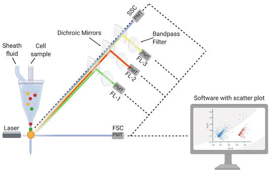
In principle, FCM allows the analysis of the chemical and physical characteristics of any suspended single particle. The optical system of an FCM is illustrated in
Figure 1. Usually, it contains the following: a flow chamber, a source of light (i.e., a laser or mercury lamp), dichroic mirrors to bring the light beam into focus, bandpass filters for the detection of different wavelengths, detectors (i.e., photodiodes (PD) and photomultiplier tubes (PMT)) for the detection and amplification of the signals, as well as a data processing unit
[17][18][19][20][36,37,38,39]. After transferring the particles into a laminar flow of sheath fluid, scattered light and fluorescence signals are utilized one by one at the interrogation point of the laser beam. To differentiate cells regarding their morphology (i.e., particle size or granularity), forward- (FSC) or side-scattered light (SSC) is detected, respectively. Aside from scattered light, fluorescence appears when fluorochromes or particles labeled with them emit light, which is then excited by a beam of an appropriate wavelength. Some cells can emit fluorescence without fluorochromes, which is called autofluorescence. This phenomenon can be either beneficial for analysis or can impede other fluorescence signals. Most of the time, autofluorescence alone is not sufficient to detect and distinguish between cell populations. Thus, FCM protocols include a staining step with one or more fluorescent dyes before sample analysis
[14][21][17,40].
Figure 1. Principle of a flow cytometer (FCM). Forward-scattered light (FSC); side-scattered light (SSC); photomultiplier tubes (PMT, fluorescence detectors); detectors at a specific wavelength (FL-1, FL-2, and FL-3); (made in ©BioRender—biorender.com, Toronto, ON, Canada (accessed on 8 June 2021)).
2.2. Food-Related FCM Applications
3.2. Food-Related FCM Applications
For food-related research, FCM is mainly used for the performance testing of food preservation or disinfection approaches, i.e., sodium hypochlorite or peracetic acid disinfection, ultraviolet light (UV-C), supercritical CO
2 pasteurization, ohmic heating applications, non-thermal inactivation technologies, including pulsed electric fields and cold atmospheric pressure plasma treatment, as well as natural preservatives such as essential oils
[22][23][24][25][26][27][28][29][30][31][47,54,55,56,57,58,59,60,61,62]. The most commonly investigated microorganism was
E. coli [2][24][25][26][28][29][30][32][33][34][35][3,55,56,57,59,60,61,63,64,65,66].
A study by Coronel-Leon et al.
[36][67] tested the antimicrobial effect of the surfactant N
α-lauroyl L arginine ethylester monohydrochloride as a food additive and used FCM to understand the inactivation mechanisms better and to indicate the presence of sublethally injured cells. The most popular cell target for viability staining is membrane integrity, in which DNA-intercalating dyes are applied. Moreover, esterase activity is a suitable detection target, as potential sublethal injured cells after inefficient inactivation procedures are observed. For this purpose, FDA or cFDA are combined with PI
[37][28][44,59]. Tamburini et al.
[29][60] concluded that FCM was the most suitable viability assessment method compared to PCR, plate counts, and fluorescence microscopy.
FCM is not only a suitable tool for detecting sublethally injured cells but also for cells in the viable but non-culturable (VBNC) state. This is important as environmental stresses present during food processes, such as temperature change, pH, or the absence of nutrients, can introduce cells into the VBNC state. With culture-based techniques, only viable and culturable cells are detected. VBNC cells, however, are able to resuscitate and become culturable again
[38][68]. Thus, FCM viability staining, in combination with plate count, can be conducted to detect the VBNC state of cells.
34. Specific State-of-the-Art Flow-FISH Methods and Applications for Monitoring and Detection
3.1. Principle of FISH
4.1. Principle of FISH
DeLong, Wickham, and Pace
[39][96] were the first to describe FISH for microorganisms. The method based on the use of fluorescently-labeled oligonucleotide probes that target a specific region of rRNA (16S/23S in Bacteria/Archaea or 18S/28S in Eukarya) enables the specific identification of microorganisms from the domain to the subspecies level
[39][40][41][42][96,97,98,99]. It is now a well-established technique
[43][100]. In addition to oligonucleotide probes, fluorescently labeled antibodies can also be used for the identification of microbial cells, but the low cost of oligonucleotide probes and the availability of a large number of rRNA sequences, as well as the associated possibility of the in silico design of oligonucleotide probes, have led to the preferred use of oligonucleotide probes
[44][101].
In contrast to culture-dependent methods, microorganisms that are difficult to cultivate can be identified. FISH visualizes whole cells, and since abundant structures in living cells are targeted, it is possible to distinguish between viable and dead cells, which is the main advantage over other molecular techniques such as PCR-based methods
[45][41][1,98]. Additionally, the direct observation of cells within their native environment is possible
[46][47][102,103]. Flow-FISH, a combination of FISH and flow cytometry, was described in the early 1990s by R.I. Amann et al.
[48][104]. The advantage of Flow-FISH is that the method enables the rapid analysis of larger sample volumes, while being more convenient since manual counting is omitted
[49][105].
In general, FISH consists of four preparation steps: (i) fixation and permeabilization, (ii) probe hybridization with the target sequence, (iii) washing of excess and unbound probes, and (iv) observation of cells with epifluorescence microscopic techniques, scanning microscopy, or flow cytometry (Flow-FISH)
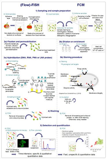
[41][43][50][98,100,106]. Sampling, pre-preparation, and hybridization steps, compared to a typical FCM protocol for quantitative analysis, are illustrated in
Figure 2.
Figure 2. Standard sample preparation protocols and detection mechanisms of specific flow fluorescence in situ hybridization (FISH) and non-specific flow cytometry (FCM). LNA, locked nucleic acids; PNA, peptide nucleic acids.
3.2. Flow-FISH in Food Microbiology
4.2. Flow-FISH in Food Microbiology
Flow-FISH in food microbiology is used to detect microorganisms in food products or for biofilm studies on abiotic surfaces, e.g., food contact surfaces. For food products, the range of food products examined is wide, from vegetables, meat, fish products, and dairy products to vinegar, wine, beer, and water.
FISH DNA or PNA probes are used for biofilm characterization, and the analyzed biofilms are either natural biofilms or those formed under laboratory conditions. FISH was used by Almeida, Azevedo, Santos, Keevil, and Vieira
[51][158] to characterize and quantify the biofilm formation of
S. enterica,
L. monocytogenes, and
E. coli on different surfaces (e.g., glass, stainless steel) using PNA probes. Stainless steel was also used as a surface to capture biofilm formation of various
Arcobacter species using FISH in a study by Šilhová, Moťková, Šilha, and Vytřasová
[52][161]. Bragança, Azevedo, Simões, Keevil, and Vieira
[53][159] screened natural biofilms in a drinking water distribution system for the occurrence of
H. pylori using PNA-FISH and evaluated the composition of natural biofilms from conveyors in breweries.


