COVID-19 and Rheumatoid Arthritis Crosstalk: Comparison
Please note this is a comparison between Version 2 by Beatrix Zheng and Version 1 by Saikat Dewanjee.

COVID-19 and RA share similar immune-inflammatory features of disease pathogenesis executed by analogous mechanistic pathways. However, the treatment of RA patients in the COVID-19 setting itself stands as a challenging task. Implementation of individualized clinical surveillance of RA patients considering the disease severity and appropriate risk-benefit study referring to the recommendations of using anti-rheumatic drugs in the COVID-19 setting by different professional rheumatology associations would stand as the optimal therapeutic strategy for effective disease control during the COVID-19 pandemic.

  • ACE
  • ACE2
  • anti-rheumatic drugs
  • COVID-19
  • cytokine storm
  • immune response
  • inflammation
  • rheumatoid arthritis
  • SARS-CoV-2
  • therapeutic options

1. Introduction

Severe acute respiratory syndrome coronavirus 2 (SARS-CoV-2) infection that caused coronavirus disease 2019 (COVID-19) usually produces a mild to moderate respiratory disease [1]. However, it occasionally leads to severe alveolar disease resulting in shortening of breath, reduced oxygen saturation in blood, and pulmonary infiltration in the lung that can substantially contribute to pulmonary failure [2]. Age, the severity of infection, and the existence of comorbidities are potential risk factors in COVID-19 patients [1,3][1][3]. Emerging evidence revealed that SARS-CoV-2 develops a specific type of alveolar disease that is clinically different from other acute respiratory syndromes [2]. Immune hyperactivation and cytokine involvement in alveolar structures have been identified as the key contributors to produce severe lung disease in COVID-19 patients. Rheumatoid arthritis (RA) is a chronic autoimmune disease characterized by synovial inflammation and hyperactivation of T cells. Several pro-inflammatory cytokines act as contributing factors in developing synovial inflammation in RA. The patterns of cytokine and immune activation in COVID-19 patients seem to resemble the RA case. Interestingly, some common therapeutic strategies including cytokine inhibition have been found to be fruitful against both COVID-19 and RA [2]. Thus, a possibility of pathological crosstalk is inevitable between COVID-19 and RA.

In general, there is a close association between viral infection and arthritis with a wide spectrum of symptoms ranging from arthralgia to arthritis. Earlier reports revealed that individuals infected with hepatitis C and several alphaviruses frequently develop prolonged arthritis; however, Parvovirus B19, Hepatitis B, and Rubella viruses frequently cause self-limited arthritis. In contrast, respiratory viruses, such as corona and influenza viruses more frequently can cause arthralgia and/or myalgia. Approximately 15 and 44% of COVID-19 patients present arthralgia and/or myalgia, respectively, during the infective stage [4]. Emerging evidence hypothesized that SARS-CoV-2 infection can attack musculoskeletal systems through immune-inflammation-dependent mechanisms, which may develop inflammatory arthritis during the infective or post-infective stage [3,4,5][3][4][5]. However, little is known about the manifestations or worsening of RA by this infection. Since musculoskeletal manifestations phenotypically resemble RA, it has been attempted to find out the association between COVID-19 and RA. In this article, we reviewed the pathological crosstalk between COVID-19 and RA. In addition, our understanding of the risk of RA patients in acquiring SARS-CoV-2 infection and worsening COVID-19 outcomes was critically discussed on the basis of available clinical readouts. The therapeutic strategies and guidelines were conferred referring to recently published literature. Moreover, critical arguments on therapeutic challenges raised in different case studies were discussed in this review.

2. Mechanistic Similarity between SARS-COV-2 Infection and RA

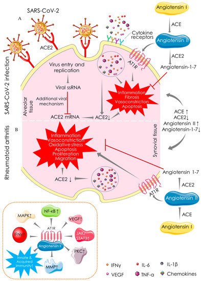
2.1. Angiotensin-Converting Enzyme (ACE)-Dependent Pathway

Immune-inflammatory disorders can be associated with ACE/ACE2 imbalance [42][6]. ACE promotes the conversion of angiotensin I to angiotensin II, while ACE2 catalyzes the conversion of angiotensin II to angiotensin-1–7, which exhibits anti-inflammatory, anti-fibrotic, anti-apoptotic, anti-proliferative, and vasorelaxation effects. ACE2 maintains renin-angiotensin system (Ras) homeostasis to restore normal physiological processes in critical tissues/organs. The role of ACE2 in SARS-CoV-2 infection stands itself as an irony. ACE2 as a receptor serves as a potential cellular target for SARS-CoV-2 to enter the target cells [43][7] (Figure 31). ACE2 binding is essential for the entry of SARS-CoV-2 into the host cells; however, recent evidence revealed a key role of heparan sulfate in facilitating their interaction and thereby potentiating SARS-CoV-2 cell entry and infection [44,45][8][9]. In contrast, ACE2 as an enzyme plays a protective role in SARS-CoV-2 infection [43][7]. The S-protein of SAR-CoV-2 binds to ACE2, resulting in a suppression of ACE2 expression and promoting COVID-19 pathogenesis [46][10]. Inhibition of ACE2/angiotensin-1–7 activates rapidly accelerated fibrosarcoma (Raf)/mitogen-activated protein kinase (MAPK) cascade, which in turn shares identical pathological signalling in both COVID-19 and RA [47][11] (Figure 31). ACE2 activators have been proposed to produce dual benefits in COVID-19 treatment: firstly, by inhibiting the binding of S-protein of SARS-CoV-2 to ACE2 and secondly by offering the protective effect of the ACE2 enzyme [48][12]. ACE2 activation can also be beneficial in RA, which can mitigate inflammation, vasoconstriction, oxidative stress, apoptosis, proliferation, and migration in synovial tissue (Figure 31). ACE activation promotes the accumulation of angiotensin II, which could be pathologically involved in both COVID-19 and RA. In an inflammatory milieu, angiotensin II is known to trigger inflammatory responses and vascular permeability by enhancing the production of prostaglandins and VEGF [49][13]. These inflammatory mediators further endorse nuclear factor kappa-light-chain-enhancer of activated B cells’ (NF-κB) activation, which intensifies the inflammatory responses and promotes infiltration of inflammatory cells into damaged tissues [49][13]. Moreover, angiotensin II can endorse lymphocyte proliferation and activation, as well as the formation of free radicals in leucocytes [50][14]. ACE inhibitors and angiotensin receptor blockers have been regarded to be beneficial in COVID-19, delaying the binding of SARS-CoV-2 by activating ACE2 and increasing the availability of angiotensin-1–7 [48][12]. Pharmacological inhibition of ACE and angiotensin II can reduce the risk of mortality in COVID-19 patients [49][13]. ACE inhibitors limit the production of pro-inflammatory cytokines by suppressing NF-κB activation, and this anti-inflammatory mechanism can be effective against both diseases [51][15]. ACE inhibitors have been proven to improve vascular endothelial function in RA patients [50][14]. Thus, it could be said that both COVID-19 and RA share a common mechanistic pathway of immunopathogenesis mediated through aberrant ACE/ACE2 activities.
Figure 31. Angiotensin-converting enzyme (ACE)-dependent pathway showing the mechanistic similarity between SARS-CoV-2 infection (A) and RA (B). ACE catalyses the conversion of angiotensin I to angiotensin II, which is involved in the pathogenesis of both COVID-19 and RA by promoting inflammation, fibrosis, vasoconstriction, and apoptotic activities. In contrast, ACE2 catalyses the conversion of angiotensin II to angiotensin-1-7 and shares identical protective functions in both COVID-19 and RA. Arrows indicate the downstream cellular events, and red lines indicate inhibition. “↑” indicates upregulation/activation, and “↓” indicates downregulation/suppression. ACE: angiotensin-converting enzyme; ACE2: angiotensin-converting enzyme 2; AT1R: angiotensin II receptor type 1; IFN: interferon; IL: interleukin; JAK: janus tyrosine kinase; MAPK: mitogen-activated protein kinase; MMP: matrix metalloproteinase; NF-κB: nuclear factor kappa-light-chain-enhancer of activated B cells; PKC: protein kinase C; STAT: signal transducer and activator of transcription; TNF: tumor necrosis factor; VEGF: vascular endothelial growth factor.

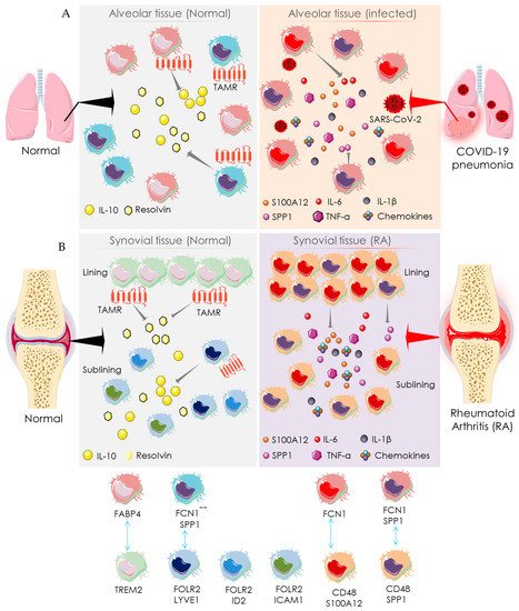
2.2. Macrophage-Mediated Pathway

Macrophages present in bronchial and synovial tissues are heterogeneous (Figure 42). Healthy lungs represent alveolar macrophages expressing fatty-acid-binding protein 4 (FABP4), which help in maintaining gas exchange and compliance. During SARS-CoV-2 infection, the number of FABP4-expressing (FABP4positive) alveolar macrophages is substantially reduced, and thus gas exchange is compromised [52][16] (Figure 42). Bronchoalveolar lavage fluids from COVID-19 patients exhibit more distinct types of macrophages than resident macrophages present in healthy alveoli [53][17]. The alveolar tissue of COVID-19 patients abundantly presents two distinct macrophage clusters expressing ficolin-1 (FCN1), which can be differentiated by their relative expression of secreted phosphoprotein 1/osteopontin (SPP1) [53][17]. These are categorized as only FCN expressing macrophages (FCNpositive) and macrophages that express both FCN and SPP1 (FCNpositiveSPP1positive). FCNpositive macrophages are probably involved in COVID-19 pathogenesis through a strong adaptive immune response mediated through CD8+ T cells. However, their specific pathological role is yet to be established. Similarly, synovial tissue also represents distinct macrophage subsets in RA patients as compared to healthy people [54][18] (Figure 42). In healthy joints, macrophage clusters expressing triggering of receptors expressed on myeloid cells 2 (TREM2), such as TREM2high and TREM2low in association with the macrophages expressing both the folate receptor beta (FOLR2) and lymphatic vessel endothelial hyaluronan receptor 1 (LYVE1), constitute synovial tissue lining. The macrophages expressing FOLR2 in association with LYVE1, inhibitor of DNA binding 2 (ID2), or intercellular adhesion molecule 1 (ICAM1) form the synovial sub-lining. These are categorized as FLOR2positiveLYVE1positive, FLOR2positiveID2positive, and FLOR2positiveICAM1positive macrophage clusters (Figure 42). As compared to healthy joints, synovial tissue of RA patients additionally represents two distinct types of macrophage clusters: one is highly expressing CD48 and S100A12 (S100 calcium-binding protein A12/calgranulin C), while another is expressing both CD48 and SPP1 (Figure 42). Both CD48highS100A12positive and CD48positiveSPP1positive macrophage clusters have been revealed to be associated with RA pathogenesis via producing pro-inflammatory mediators such as IL-1β, IL-6, TNF-α, MMPs, and chemokines and inducing pathogenesis to the adjacent stromal tissue [53][17]. FCNpositive and FCNpositiveSPP1positive macrophages in bronchoalveolar lavage fluids from COVID-19 patients share a transcriptional homology with pathogenic CD48highS100A12positive and CD48positiveSPP1positive macrophage clusters in the synovial tissue of RA patients (Figure 42) [53][17]. In addition, both share similar functional characteristics in the respective tissues [53][17]. Similarly, FABP4positive alveolar macrophages in the bronchoalveolar lavage fluids from healthy individuals share transcriptional and functional homology with TREM2 expressing synovial macrophages in healthy joints (Figure 42) [53][17]. Both TREM2positive and FOLR2positiveLYVE1positive macrophages resolve inflammation by activating anti-inflammatory mediators and repair stromal cells by recruiting Mer receptor tyrosine kinase (MerTK) and its ligand growth arrest-specific protein 6 precursor (GAS6) [53][17]. MerTK, a member of the TAM family with its ligands GAS6 and vitamin K-dependent protein S (PROS1), contributes to an inflammation-alleviating effect. FABP4positive macrophages express Axl receptor tyrosine kinase (Axl) and PROS1, which aids in reducing COVID-19’s severity. Both FABP4positive and TREM2positive macrophages share a similar functional role of a homeostatic brake on inflammation in alveolar and synovial tissues, respectively (Figure 42). Taken together, the alveolar macrophages in healthy individuals share homologies in transcriptomic profiles and regulatory pathways with the macrophages in synovial tissue of healthy individuals. Similarly, macrophages in the alveolar tissue of COVID-19 patients are homologous to that of the synovial macrophages in RA patients. Thus, both SARS-CoV-2 infection and RA share a common mechanistic pathway of immunopathogenesis driven by the activities of analogous macrophage clusters.
Figure 42. Macrophage-mediated pathway showing the mechanistic similarity between SARS-CoV-2 infection and RA. Schematic diagram showing the pro-inflammatory function of distinct macrophage subsets in the normal (left side) and SARS-CoV-2 infected (right side) alveolar tissue (A) and synovial tissue (B) of normal (left side) and RA patients (right side). The identity of distinct participating macrophage subsets is shown at the bottom. Arrows indicate the downstream cellular events; double-headed arrows represent the similarity in transcriptomic homology and regulatory activities. “+” indicates positive expression, and “-“ indicates negative expression. FABP4: fatty acid-binding protein 4; FCN1: ficolin-1; FOLR2: folate receptor beta; ICAM1: intercellular adhesion molecule 1; IFN: interferon; IL: interleukin; LYVE1: lymphatic vessel endothelial hyaluronan receptor 1; S100A12: S100 calcium-binding protein A12/calgranulin C; SPP1: secreted phosphoprotein 1/osteopontin; TAMR: TAM receptor; TREM2: triggering receptor expressed on myeloid cells 2.

3. Recommendation for Anti-Rheumatic Drugs in the COVID-19 Setting

Patients with RA generally represent a compromised immune system, which makes them susceptible to SARS-CoV-2 infection [25][19]. Treatment with immunosuppressant drugs may further increase the risk of acquiring SARS-CoV-2 infection [22][20]. In addition, clinical features of RA flares and SARS-CoV-2 infection frequently overlap [29][21]. Both RA- and COVID-19-positive patients represent some common symptoms like arthralgia, myalgia, and other inflammatory disorders. RA-mediated interstitial lung disease often mimics COVID-19 symptoms. Moreover, RA patients frequently represent increasing evidence of comorbidities [55][22]. Thus, clinical management of RA itself stands as a challenging task in the present COVID-19 setting. Among the possible therapeutic options, the American College of Rheumatology (ACR) and the European League Against Rheumatism (EULAR) recommended several guidelines regarding the use of RA medication in the COVID-19 pandemic [56,57][23][24]. Glucocorticoids have been recommended at the lowest possible dose even in COVID-19-positive cases, and sudden withdrawal has been discouraged. Non-steroidal anti-inflammatory drugs (NSAIDs) have been proposed to be continued unless severe COVID-19 outcomes present to multiple organs. Among disease-modifying antirheumatic drugs (DMARDs), conventional synthetic DMARDs (csDMARDs) have been recommended to be continued; however, leflunomide, methotrexate, and sulfasalazine are suggested to be avoided in suspected or confirmed COVID-19 cases. All biological DMARDs (bDMARDs) except for IL-6 inhibitors and all targeted synthetic DMARDs (tsDMARDs) have been advised to be discontinued in suspected or confirmed cases of COVID-19. Regarding re-initiation of DMARDs, ACR recommended restarting these drugs within 7–14 days of symptom resolution or within 10–17 days of the positive report for symptomatic and asymptomatic patients, respectively [55][22]. However, treatment resumption is required on an individual basis for patients recuperating from a serious illness [56][23]. Thus, adjustments to medication should be done on an individual basis considering disease severity, and specific attention must be given to the recommendations of using antirheumatic drugs in the COVID-19 setting by different professional rheumatology associations, such as the ACR and the EULAR.

References

  1. Kalra, R.S.; Tomar, D.; Meena, A.S.; Kandimalla, R. SARS-CoV-2, ACE2, and Hydroxychloroquine: Cardiovascular Complications, Therapeutics, and Clinical Readouts in the Current Settings. Pathogens 2020, 9, 546.
  2. Schett, G.; Manger, B.; Simon, D.; Caporali, R. COVID-19 revisiting inflammatory pathways of arthritis. Nat. Rev. Rheumatol. 2020, 16, 465–470.
  3. Kalra, R.S.; Dhanjal, J.K.; Meena, A.S.; Kalel, V.C.; Dahiya, S.; Singh, B.; Dewanjee, S.; Kandimalla, R. COVID-19, Neuropathology, and Aging: SARS-CoV-2 Neurological Infection, Mechanism, and Associated Complications. Front. Aging Neurosci. 2021, 13, 662786.
  4. Mukarram, M.S.; Ishaq Ghauri, M.; Sethar, S.; Afsar, N.; Riaz, A.; Ishaq, K. COVID-19: An Emerging Culprit of Inflammatory Arthritis. Case Rep. Rheumatol. 2021, 2021, 6610340.
  5. Dewanjee, S.; Vallamkondu, J.; Kalra, R.S.; Puvvada, N.; Kandimalla, R.; Reddy, P.H. Emerging COVID-19 Neurological Manifestations: Present Outlook and Potential Neurological Challenges in COVID-19 Pandemic. Mol. Neurobiol. 2021, 58, 4694–4715.
  6. Zhu, Z.; Cai, T.; Fan, L.; Lou, K.; Hua, X.; Huang, Z.; Gao, G. The potential role of serum angiotensin-converting enzyme in coronavirus disease 2019. BMC Infect. Dis. 2020, 20, 883.
  7. Ni, W.; Yang, X.; Yang, D.; Bao, J.; Li, R.; Xiao, Y.; Hou, C.; Wang, H.; Liu, J.; Yang, D.; et al. Role of angiotensin-converting enzyme 2 (ACE2) in COVID-19. Crit. Care 2020, 24, 422.
  8. Clausen, T.M.; Sandoval, D.R.; Spliid, C.B.; Pihl, J.; Perrett, H.R.; Painter, C.D.; Narayanan, A.; Majowicz, S.A.; Kwong, E.M.; McVicar, R.N.; et al. SARS-CoV-2 Infection Depends on Cellular Heparan Sulfate and ACE2. Cell 2020, 183, 1043–1057.
  9. Choudhary, S.; Sharma, K.; Silakari, O. The interplay between inflammatory pathways and COVID-19: A critical review on pathogenesis and therapeutic options. Microb. Pathog. 2021, 150, 104673.
  10. Kalra, R.S.; Kandimalla, R. Engaging the spikes: Heparan sulfate facilitates SARS-CoV-2 spike protein binding to ACE2 and potentiates viral infection. Signal. Transduct. Target. Ther. 2021, 6, 39.
  11. Conti, P.; Caraffa, A.; Gallenga, C.E.; Ross, R.; Kritas, S.K.; Frydas, I.; Younes, A.; Ronconi, G. Coronavirus-19 (SARS-CoV-2) induces acute severe lung inflammation via IL-1 causing cytokine storm in COVID-19: A promising inhibitory strategy. J. Biol. Regul. Homeost. Agents 2020, 34, 1971–1975.
  12. Rodríguez-Puertas, R. ACE2 activators for the treatment of COVID 19 patients. J. Med. Virol. 2020, 92, 1701–1702.
  13. Wong, M.C.S.; Wong, S.; Huang, J.; Yan, B. Relating angiotensin-converting enzyme inhibitors or angiotensin receptor blockers with incidence or mortality of COVID-19. ESC Heart Fail. 2020, 7, 3119–3123.
  14. Chang, Y.; Wei, W. Angiotensin II in inflammation, immunity and rheumatoid arthritis. Clin. Exp. Immunol. 2015, 179, 137–145.
  15. Najafi, S.; Rajaei, E.; Moallemian, R.; Nokhostin, F. The potential similarities of COVID-19 and autoimmune disease pathogenesis and therapeutic options: New insights approach. Clin. Rheumatol. 2020, 39, 3223–3235.
  16. Mirastschijski, U.; Dembinski, R.; Maedler, K. Lung Surfactant for Pulmonary Barrier Restoration in Patients with COVID-19 Pneumonia. Front. Med. 2020, 7, 254.
  17. MacDonald, L.; Alivernini, S.; Tolusso, B.; Elmesmari, A.; Somma, D.; Perniola, S.; Paglionico, A.; Petricca, L.; Bosello, S.L.; Carfì, A.; et al. COVID-19 and RA share an SPP1 myeloid pathway that drives PD-L1+ neutrophils and CD14+ monocytes. JCI Insight 2021, 6, e147413.
  18. Alivernini, S.; MacDonald, L.; Elmesmari, A.; Finlay, S.; Tolusso, B.; Gigante, M.R.; Petricca, L.; Di Mario, C.; Bui, L.; Perniola, S.; et al. Distinct synovial tissue macrophage subsets regulate inflammation and remission in rheumatoid arthritis. Nat. Med. 2020, 26, 1295–1306.
  19. Favalli, E.G.; Maioli, G.; Biggioggero, M.; Caporali, R. Clinical management of patients with rheumatoid arthritis during the COVID-19 pandemic. Expert Rev. Clin. Immunol. 2021, 17, 561–571.
  20. Akiyama, S.; Hamdeh, S.; Micic, D.; Sakuraba, A. Prevalence and clinical outcomes of COVID-19 in patients with autoimmune diseases: A systematic review and meta-analysis. Ann. Rheum. Dis. 2020, 80, 384–391.
  21. Roongta, R.; Ghosh, A. Managing rheumatoid arthritis during COVID-19. Clin. Rheumatol. 2020, 39, 3237–3244.
  22. Dougados, M.; Soubrier, M.; Antunez, A.; Balint, P.; Balsa, A.; Buch, M.H.; Casado, G.; Detert, J.; El-Zorkany, B.; Emery, P.; et al. Prevalence of comorbidities in rheumatoid arthritis and evaluation of their monitoring: Results of an international, cross-sectional study (COMORA). Ann. Rheum. Dis. 2014, 73, 62–68.
  23. Mikuls, T.R.; Johnson, S.R.; Fraenkel, L.; Arasaratnam, R.J.; Baden, L.R.; Bermas, B.L.; Chatham, W.; Cohen, S.; Costenbader, K.; Gravallese, E.M.; et al. American College of Rheumatology Guidance for the Management of Rheumatic Disease in Adult Patients During the COVID-19 Pandemic: Version 1. Arthritis Rheumatol. 2020, 72, 1241–1251.
  24. Landewé, R.B.; Machado, P.M.; Kroon, F.; Bijlsma, H.W.; Burmester, G.R.; Carmona, L.; Combe, B.; Galli, M.; Gossec, L.; Iagnocco, A.; et al. EULAR provisional recommendations for the management of rheumatic and musculoskeletal diseases in the context of SARS-CoV-2. Ann. Rheum. Dis. 2020, 79, 851–858.
More