MERTK and AXL in NSCLC: Comparison
Please note this is a comparison between Version 1 by Dan Yan and Version 2 by Lindsay Dong.

MERTK and AXL are members of the TAM family of receptor tyrosine kinases and are abnormally expressed in 69% and 93% of non-small cell lung cancers (NSCLCs), respectively. Expression of MERTK and/or AXL provides a survival advantage for NSCLC cells and correlates with lymph node metastasis, drug resistance, and disease progression in patients with NSCLC. The TAM receptors on host tumor infiltrating cells also play important roles in the immunosuppressive tumor microenvironment. Thus, MERTK and AXL are attractive biologic targets for NSCLC treatment. 

  • MERTK
  • AXL
  • TAM family
  • NSCLC

1. Introduction

Lung cancers are divided into two broad categories: non-small cell lung cancer (NSCLC) and small cell lung cancer (SCLC). Following clinical validation of translational inhibitors targeting two important NSCLC oncogenic drivers, epidermal growth factor receptor (EGFR) [1][2][3][4][15,16,17,18] and anaplastic lymphoma kinase (ALK) [5][6][7][19,20,21], molecular-targeted therapies have been applied to the management of metastatic NSCLC, resulting in remarkably improved prognosis and quality of life relative to patients treated with conventional chemotherapeutics [8][9][10][22,23,24]. Additional mutated oncogenic proteins have been identified in NSCLC, including HER2, BRAF, RET, MET, and ROS1 [6][11][20,25]. Even though patients respond to targeted therapies initially, the majority of patients, if not ultimately all patients, relapse within 1 to 2 years when treated with targeted therapies [12][13][14][15][16][26,27,28,29,30]. Therefore, understanding the mechanisms of primary and secondary resistance to current targeted therapies is critical to enhance patient outcomes. New therapeutic approaches will be required to further enhance outcomes. Both MERTK and AXL, members of the TAM (TYRO3, AXL, and MERTK) family of receptor tyrosine kinases (RTK), are emerging therapeutic targets in NSCLC.

2. Physiologic Roles for MERTK and AXL

The TAM kinases are structurally unique from other RTK subfamilies, possessing two immunoglobulin-like (Ig) repeats and two fibronectin type III (FNIII) domains in their extracellular region and a conserved intracellular kinase domain with an unusual signature sequence, KW(I/L)A(I/L)ES [17][18][31,32] (Figure 1A). Growth Arrest Specific 6 (GAS6) and Protein S (PROS1) are the two best characterized ligands for TAM receptors, although other TAM ligands have been reported including TUBBY [19][33], Tubby-like protein 1 (TULP-1) [19][33], and Galectin-3 (LGALS3) [20][34]. Structurally, both GAS6 and PROS1 contain a N-terminal glutamic acid-rich (GLA) domain, followed by four epidermal growth factor (EGF)-like repeats, and a C-terminal sex hormone binding globulin (SHBG) homology domain comprised of two globular laminin G-like domains (2-LG) [21][35] (Figure 1B). The GLA domain confers the ability of these ligands to bind phosphatidylserine (PtdSer) through their N termini. The complex physiologic role of this signaling system is in part defined by this PtdSer sensing which, for example, recognizes the billions of cells that die by apoptosis in the human body daily. However, GAS6 and PROS1 bind differentially to the TAM receptors. MERTK and TYRO3 are activated by both GAS6 and PROS1, while AXL is only activated by GAS6 [22][23][36,37]. In the absence of apoptotic cells or PtdSer, the affinity of GAS6 for AXL is more than 6-fold higher than for TYRO3 and 70-fold higher than for MERTK [23][24][25][37,38,39] (Figure 1C). GAS6 binding to both MERTK and TYRO3 is enhanced in the presence of PtdSer, while AXL binding is not stimulated.
Figure 1. TAM receptors and their ligands. (A) TAM receptors TYRO3, AXL, and MERTK share a similar structure of two immunoglobulin (Ig)-like domains, two fibronectin type III (FNIII) domains, and an intracellular kinase domain. (B) GAS6 and PROS1 contain a γ-carboxyglutamic acid (Gla) domain, four EGF-like domains, and two lamine G (LG)-like domains. (C) Interaction of TAM receptors with their ligands GAS6 and PROS1. The thickness of the arrows indicates the binding strengths of each ligand to the TAM receptors. “+” indicates the enhanced signal in the presence of phosphatidylserine (PtdSer).
Although TAM receptors are expressed in embryonic tissues [18][26][27][32,40,41], triple knockout mice are viable without obvious developmental defects at birth [28][42], indicating that TAM receptors are not required for embryogenesis. However, knockout of single and/or all 3 TAM receptors is associated with diverse phenotypes in mice, including impaired clearance of apoptotic cells [29][43], enlarged spleen [30][44], increased inflammation [31][45], impairment of tumor cell killing by NK cells [32][46], hyperproliferation of B and T cells [28][42], hyperactivation of antigen-presenting cells [28][42], increased autoantibody production [28][29][33][34][42,43,47,48], auto-immunity [34][48], defects in platelet aggregation [35][49], aborted spermatogenesis and germ cell death [30][44], neurological abnormalities [30][44], multiple organ defects, and blindness in adult triple knockout mice [30][44]. Many of these consequences of TAM receptor loss are related to apoptotic cell clearance and post receptor anti-inflammatory action. This knowledge has spurred research to define the role of TAM RTKs as innate immune checkpoint genes (i.e., guardians against persistent or inappropriate inflammation). Absence of MERTK expression is associated with increased DC activation upon encounter with apoptotic cells, resulting in upregulation of costimulatory molecules and T cell activation [36][50]. Although some of these phenotypes were noted with knockout of single TAM receptor, they are much more pronounced in triple TAM knockout mice, indicating at least some overlap in TAM kinase functions [29][30][35][43,44,49].

3. Oncogenic Roles for MERTK and AXL

3.1. Roles in NSCLC

MERTK and AXL are frequently aberrantly expressed in NSCLC patient samples, but are absent or expressed at low levels in normal human bronchial epithelial cells [37][38][39][40][41][42][43][44][51,52,53,54,55,56,57,58]. High levels of AXL have been described in subsets of both treatment-naïve and relapsed NSCLC [44][45][46][58,59,60]. Increased AXL expression was associated with increased tumor cell invasiveness and tumor grade and predicted poorer survival in patients with NSCLC [42][43][47][48][49][50][51][56,57,61,62,63,64,65]. Inhibition of MERTK in NSCLC cell lines with a small molecule MERTK tyrosine kinase inhibitor (TKI), MERTK-specific blocking monoclonal antibody, or shRNA induced apoptosis and decreased colony formation in vitro and inhibited tumor growth in vivo [52][53][54][55][66,67,68,69]. Treatment with an antibody against active AXL or siRNA/shRNA AXL knockdown also provided anti-tumor activity in NSCLC models [42][50][56][56,64,70].

High levels of MERTK and/or AXL have also been implicated in drug resistance and radioresistance [40][41][46][55][57][58][59][60][61][62][63][64][65][66][67][68][69][70][71][72][54,55,60,69,71,72,73,74,75,76,77,78,79,80,81,82,83,84,85,86]. Increased MERTK or AXL expression in NSCLC correlated with chemotherapy resistance [37][52][56][73][74][75][51,66,70,87,88,89]. Conversely, MERTK or AXL knockdown, treatment with a MERTK monoclonal antibody, or AXL inhibitor R428 or MP-470 promoted apoptosis and increased the sensitivity of NSCLC cells to chemotherapeutic agents [37][52][56][73][74][75][51,66,70,87,88,89]. Upregulated AXL and its interaction with EGFR were associated with resistance to PI3Kα inhibition due to sustained mTOR activation, and addition of AXL inhibitor R428 sensitized tumor cells to PI3Kα [62][76]. In addition, AXL has been implicated in resistance to anti-IGF-1R therapy [76][77][90,91] and resistance to BRAF/MEK inhibitors [78][92].

3.2. Functions in Cancer Cells

MERTK was cloned from a B ALL cell line cDNA library and is ectopically expressed in over 30–50% of childhood acute lymphoblastic leukemia samples and the majority of lymphoid leukemia cell lines, but was not expressed in normal T and B lymphocytes [18][79][32,94]. Similarly, AXL was originally cloned from chronic myelogenous leukemia patient samples but is not expressed in granulocytes or lymphocytes [17][31], suggesting upregulation in the context of hematopoietic malignancy.
Expression and stimulation of a chimeric MERTK protein in NIH3T3 cells was sufficient to confer anchorage-independent colony formation, a hallmark of oncogenic transformation [80][107]. Similarly, expression of MERTK conferred IL-3-independence in Ba/F3 cells [81][82][108,109]. These data suggest a tumor promoting role for MERTK. Similarly, overexpression of AXL induced transformation of NIH3T3 cells and the resulting cells were tumorigenic in nude mice [17][83][31,110]. Although MERTK overexpression in normal lung epithelial cells was not sufficient to drive tumorigenesis in vivo, it promoted expansion of normal lung epithelial cells in culture and enhanced clonogenic potential [41][55].
TAM receptors, particularly AXL, have been associated with epithelial–mesenchymal transition (EMT) [73][84][85][86][87][87,111,112,113,114]. EMT is an important step in the development of metastatic disease in which cell–cell contacts are lost, leading to tumor cell migration, invasion, and metastasis, and has been associated with therapeutic resistance in NSCLC [73][88][89][90][91][92][87,115,116,117,118,119]. Aberrant expression of AXL promotes phenotypes associated with EMT and metastasis, and inhibition of AXL reduces indicators of EMT/metastasis in various cancers [51][85][93][94][95][65,112,120,121,122]. Ectopic overexpression of AXL in NSCLC cell lines caused increased filopodia formation, while silencing of endogenous AXL led to loss of spindle-like morphology [96][123]. NSCLC cells that express high levels of AXL generally expressed abundant vimentin, a transcriptional regulator that contributes to EMT phenotypes [73][87], and downregulation of AXL decreased expression of vimentin in NSCLC [60][73][85][74,87,112]. Further, ectopic expression of AXL increased migration and invasion in NSCLC cells and AXL inhibition reduced the invasive capacity of NSCLC cell lines [47][50][96][61,64,123]. Enhanced metastasis was accompanied by AXL-dependent MMP-9 activation [97][124]. Similarly, overexpression of MERTK promoted migration in both normal lung epithelial and NSCLC cell lines [41][55].

3.3. Signaling in Cancer Cells

Activation of AXL and/or MERTK leads to signaling cascades that are important for tumor progression (Figure 2). MERTK kinase activity is associated with phosphorylation at three tyrosine residues: Y749, Y753, and Y754 on MERTK [98][131] and Y779, Y821, and Y866 on AXL [99][100][132,133]. Both Y779 and Y821 on AXL and two additional phosphorylation sites for MERTK (Y872 and Y929) are docking sites for GRB2 and the p85 regulatory subunit of PI3K, which activate MEK/ERK and PI3K/AKT signaling pathways, respectively [82][99][100][109,132,133]. The MEK/ERK signaling is associated with cell proliferation [101][134], while the PI3K/AKT pathway is preferentially involved in tumor cell survival [102][135]. MERTK-dependent cell migration is mediated by FAK signaling [41][103][55,136], while MERTK induced transformation correlates with activation of STAT-dependent transcription [104][137]. The anti-apoptotic effects of MERTK also correlate with negative regulation of the pro-apoptotic tumor suppressor WW domain-containing oxidoreductase (Wwox) [105][138]. Also, AXL inhibition is reported to mediate apoptosis by reducing the expression of the anti-apoptotic protein MCL1 [106][139]. AXL dimerizes with and phosphorylates EGFR to promote activation of the PLCγ-PKC-mTOR signaling cascade and tumor cell survival [62][76]. Similarly, there is crosstalk between MERTK and EGFR and they are frequently co-expressed on both mtEGFR- and wtEGFR-expressing NSCLC cell lines [55][66][69,80]. In fact, MERTK stabilized the EGFR protein on the cell surface, probably by preventing EGFR internalization and degradation, as EGF-dependent EGFR turnover was reversed by inhibition of lysosomal hydrolase activity [107][140]. Further, inhibition of MERTK expression using siRNA destabilized expression of EGFR protein.

3.4. Immune Regulatory Functions in the Tumor Microenvironment

Figure 2. MERTK and AXL signaling in normal and cancer cells. MERTK and AXL play important physiological roles in phagocytosis, platelet aggregation, and immune suppression. Abnormally expressed MERTK and/AXL on NSCLC and other cancer cells are involved in tumorigenesis, including promoting tumor cell survival and proliferation and tumor cell invasion and metastasis. Besides, cross talk between AXL and EGFR, MERTK and EGFR, and AXL and MERTK have also been implicated in drug resistance in the treatment of NSCLC.

3.4. Immune Regulatory Functions in the Tumor Microenvironment

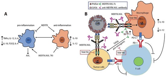
MERTK is upregulated upon monocyte to macrophage differentiation [108][109][110][141,142,143]. Expression of MERTK and AXL on tumor-infiltrating macrophages polarizes them towards a pro-tumor M2-like phenotype [110][111][112][113][143,144,145,146]. M2 macrophages promote an immunosuppressive tumor microenvironment by increasing expression of wound-healing cytokines (IL-10, TGFβ, and IL-4) and decreasing pro-inflammatory cytokines (IL-12, TNFα, and IL-6) [31][110][114][115][116][117][118][45,143,147,148,149,150,151] (Figure 3A). MERTK activation negatively regulates the secretion of pro-inflammatory cytokines, such as TNFα, through suppression of NFκB activation in macrophages [31][119][45,152]. LPS challenge led to over-produced TNFα in  Mertkkd mice, which lack the tyrosine kinase signaling domain, due to hyper-activation of NFκB [31][120][45,153]. Inhibition of MERTK by knockout of Mertk in mice, neutralization of TAM kinase signaling using a recombinant MERTK-Fc protein as a ligand sink or a GAS6 blocking antibody, and knockout of AXL in macrophages also impaired M2-macrophage anti-inflammatory phenotypes, decreased immunosuppressive IL-10 production, and increased pro-inflammatory IL-12 release [72][110][121][122][86,143,154,155]. These cytokine alterations lead to expansion of anti-tumor CD8+ T lymphocytes and inhibition of tumor growth and metastasis (Figure 3B). Indeed, inhibition of MERTK in the tumor microenvironment in  Mertk−/− mice was sufficient to decrease tumor growth and metastasis [121][154]. MERTK-expressing dendritic cells can also regulate T cell activation directly [123][156]. Blocking MERTK on dendritic cells using anti-MERTK antibody promoted T cell proliferation, while treatment with a MERTK-Fc protein to mimic the effect of MERTK expressed on human dendritic cells suppressed naïve CD4+ T cell proliferation [123][156]. The anti-inflammatory effect of MERTK activation in macrophages and apoptotic cell-treated dendritic cells was mediated by inhibition of NFκB activation [36][124][125][50,157,158] or by induction of toll-like receptor (TLR) suppressor of cytokine signaling 1 (SOCS1) and SOCS3 [125][126][127][158,159,160] (Figure 2). Further, the MERTK ligand PROS1 also promotes resolution of inflammation by macrophages and inhibits macrophage M1 polarization to reduce anti-tumor immune response [128][161].
Figure 3. Immune regulatory roles of MERTK and AXL in the TME. (A) MERTK signaling favors macrophage to M2 type to generate immunosuppressive microenvironment through releasing anti-inflammatory cytokine IL-10 and decreasing the release of pro-inflammatory cytokines IL-12 and TNFα. While expression of AXL is promoted by pro-inflammatory M1 macrophage, treatment with MERTK/AXL TKI promotes M2 to M1 macrophage development. (B) MERTK/AXL signaling favors tumor growth in cancer through two independent mechanisms. Enhanced GAS6 secretion by tumor-associated macrophages promote tumor growth through the activation of oncogenic MERTK/AXL signaling in tumor cells. Activation of MERTK in tumor-associated macrophages and in tumor cells promotes PD-L1 expression, resulting in suppression of T cell activation. Besides, the immunosuppressive cytokine environment limits T cell proliferation and effector functions.
More recently, MERTK blockade using anti-MERTK antibody induced a rapid local type I IFN response in tumors [129][162]. The type I IFNs in turn upregulated the TAM receptors through IFNAR-STAT1 signaling and the upregulated TAM system hijacked the IFNAR-STAT1 cassette to induce the cytokine and TLR suppressors SOCS1 and SOC3 [125][130][131][132][133][134][158,163,164,165,166,167] (Figure 3A). AXL knockout in tumor cells also promoted antigen presentation through increased MHCI expression, leading to an enhanced CD8+ T cell response [57][71]. Furthermore, treatment with the pan-TAM kinase inhibitor sitravatinib reduced tumor burden via activated innate and adaptive immune cells [135][168]. Additionally, treatment with sitravatinib converted immunosuppressive M2-type macrophages to immunostimulatory M1-type macrophages [135][168], and this effect was dependent on MERTK expression in bone marrow derived macrophages [125][135][158,168]. These findings support roles for MERTK and AXL as tolerogenic receptors that mediate immunosuppression in the tumor microenvironment [110][118][134][136][143,151,167,169]. Enhanced TAM receptor signaling in response to PtdSer expressed on apoptotic cells resulted in AKT-dependent PD-L1 expression on tumor cells and macrophages, and MERTK inhibition by genetic deletion or treatment with MERTK inhibitor MRX-2843 [137][138][67, 243] led to decreased expression of PD-L1 on tumor cells and innate immune cells [139][170]. In turn, T cell function was indirectly suppressed [24][139][140][38,170,171] (Figure 3B). AXL expression was positively correlated with PD-L1 and CXC chemokine receptor 6 (CXCR6) expression in lung cancer, especially in mtEGFR-expressing NSCLC [141][142][172,173]. Similar to MERTK inhibition, treatment with AXL inhibitor R428 decreased mRNA expression of PD-L1 and CXCR6 in mtEGFR-expressing NSCLC [141][172]. In contrast, increased expression of AXL coincided with reduced overall survival in patients treated with PD-1 blockade [141][142][172,173]. Accordingly, high levels of AXL expression in lung cancer cells correlated with intrinsic resistance to killing by both natural killer cells and cytotoxic T lymphocytes and this phenotype could be reversed by treatment with the AXL inhibitor R428 [143][174]. MERTK plays a role in phagocytosis of apoptotic cells in macrophages, but not in dendritic cells [29][109][144][43,142,175]. In contrast, AXL has a greater role in DCs and a lesser role in apoptotic cell phagocytosis by macrophages [145][176]. Recently, Zhou et al. found that MERTK blockade on tumor-associated macrophages led to accumulation of dying or dead cells in the tumor, resulting in a large increase in extracellular ATP when cells became necrotic [129][146][147][162,177,178]. The increased extracellular ATP in turn opened the ATP-gated P2X7R channel and allowed tumor-derived extracellular cGAMP to reach the cytosol of immune cells to activate the adaptor protein stimulator of interferon genes (STING), which in turn triggered the TANK-binding kinase 1-interferon regulatory factor 3 (TBK1-IRF3)-dependent signaling process, leading to the production of type I IFNs [129][148][149][150][151][152][162,179,180,181,182,183]. Cyclic GAMP-AMP synthase (cGAS)-STING signaling in immune cells is a key determinant for therapeutic efficacy of immune checkpoint inhibitors [151][153][182,184]. Indeed, blockade of MERTK or AXL using a specific antibody or treatment with sitravatinib or R428 synergized with anti-PD-1 or anti-PD-L1 therapy to enhance anti-tumor immune responses [72][129][135][86,162,168]. Immunotherapies, including checkpoint inhibitors, are making an impact as monotherapy and in combination [154][185]. In a clinical trial in patients with advanced NSCLC without activating EGFR or ALK mutation and with PD-L1 expression on greater than 50% of tumor cells, pembrolizumab increased response rate (45% vs. 28%), progression-free survival (PFS, 10.3 vs. 6 months) and overall survival (30 vs. 14.2 months) relative to patients treated with chemotherapy, establishing pembrolizumab as the standard of care for these patients [155][186]. Further work is necessary to explore specific mechanisms of primary and adaptive resistance.
Video Production Service