Campylobacter Biofilms: Comparison
Please note this is a comparison between Version 1 by Bassam Elgamoudi and Version 2 by Jessie Wu.

Microbial biofilms occur naturally in many environmental niches and can be a significant reservoir of infectious microbes in zoonotically transmitted diseases such as that caused by Campylobacter jejuni, the leading cause of acute human bacterial gastroenteritis world-wide. The greatest challenge in reducing the disease caused by this organism is reducing transmission of C. jejuni to humans from poultry via the food chain. Biofilms enhance the stress tolerance and antimicrobial resistance of the microorganisms they harbor and are considered to play a crucial role for Campylobacter spp. survival and transmission to humans. Unconventional approaches to control biofilms and to improve the efficacy of currently used antibiotics are urgently needed. This review summarizes the use plant- and microorganism-derived antimicrobial and antibiofilm compounds such as essential oils, antimicrobial peptides (AMPs), polyphenolic extracts, algae extracts, probiotic-derived factors, d-amino acids (DAs) and glycolipid biosurfactants with potential to control biofilms formed by Campylobacter, and the suggested mechanisms of their action. Further investigation and use of such natural compounds could improve preventative and remedial strategies aimed to limit the transmission of campylobacters and other human pathogens via the food chain.

  • Campylobacter

1. Campylobacter spp. Biofilm Formation and Regulation

The formation of biofilms significantly increases the ability of C. jejuni to survive in extreme conditions [1][2][28,29]. For instant, biofilm encased campylobacter cells survive twice as long under atmospheric conditions, and had been shown to form strong biofilms under aerobic condition [3][4][15,30]. Biofilm formation is also recognized as a potential reservoir for antimicrobial resistance and is known to facilitate exchange of resistance genes between pathogenic and commensal bacteria [5][31]. This is particularly pertinent in case of Campylobacter spp., including C. jejuni and C. coli, which exhibit intrinsic resistance to many antimicrobial agents and are naturally conjugative [6][7][8][32,33,34]. In addition, Campylobacter spp. are becoming increasingly resistant to the most frequently prescribed antibiotics such as erythromycin, tetracycline and fluoroquinolones, and have been listed by WHO as a priority pathogen for the development of new antibiotics [9][10][35,36]. The usage of antibiotics in food animals to control, prevent and treat infections, and to enhance growth, has been implicated in an increased resistance to multiple antibiotics by Campylobacter spp. [11][37]. Majority of C. jejuni and C. coli are now resistant to at least one of the currently used antibiotics, such as penicillin, trimethoprim, sulfamethoxazole, rifampicin and vancomycin [11][37], requiring alternative treatments with either gentamicin or third-generation cephalosporins [12][38].
Several studies have shown that C. jejuni strains are able to attach to, and form mono- or mixed-species biofilms with other bacterial species such as Pseudomonas aeruginosa, Escherichia coli, Staphylococcus simulans, Enterococcus faecalis, Salmonella spp., Flavobacterium spp., and Corynebacterium spp. [13][14][15][6,39,40]. The evidence from these recent publications suggests that the composition of Campylobacter spp. biofilms is similar to that formed by other organisms. While there has been some investigation of the extracellular matrix components of C. jejuni biofilms, the architecture and the composition of these are yet to be fully characterized. C. jejuni NCTC strain 11168 was reported to produce an extracellular fibre-like material as a component of its biofilm, structurally resembling a net-like matrix [16][8]. Such matrices contribute to biofilm-mediated antimicrobial resistance, either by acting as a diffusion barrier or by binding directly to antimicrobial agents and preventing their access to the biofilm-encased cells [17][26]. The extracellular DNA (eDNA) is important for establishment and maintenance of C. jejuni biofilm [18][19][41,42], and appears to be a crucial component of the extracellular matrix of mature biofilms as degradation of eDNA results in reduction of biofilm formation by C. jejuni [18][19][20][41,42,43]. Interestingly, Gaasbeek et al. [21][44] found that a C. jejuni Mu-like prophage-integrated element 1 (CJIE1) containing strain, a non-naturally transformable strain, has a gene encoding an extracellular DNase (eDNase, CJE0256), and eDNase activity could be detected. It is interesting to note that no eDNase activity could be found in naturally transformable C. jejuni strains such as NCTC11168 and 81116.
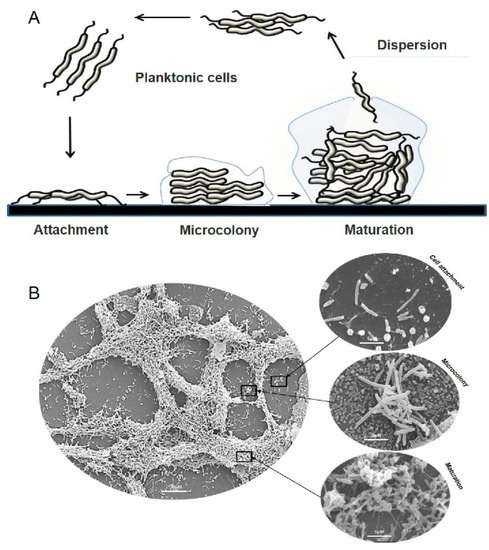
Most of our current knowledge of Campylobacter spp. biofilm architecture is summarised in Figure 1. In the first stage of biofilm formation, planktonic cells attach to the surface via two types of interaction: cell-surface and cell-cell interactions using flagella, fimbriae, amyloid-like fibrils and outer membrane proteins [22][23][24][45,46,47]. This process is critical for bacterial adhesion and is influenced by the properties of both bacterial cells and the surface [25][26][48,49]. Secondly, after initial attachment, the cells start production of extracellular polymeric substance (EPS) consisting of polysaccharides, extracellular DNA (eDNA) [19][42], proteins [27][50], lipids and other glycosylated polymers, in order to initiate micro-colonies and progress to the third stage of a mature biofilm [28][29][51,52]. In a mature biofilm, EPS acts as an adhesive between cells and supports the intricate three-dimensional (3D) structure of the biofilm, protecting the cells from toxic compounds such as antibiotics, but allowing the movement of fluid and nutrients [30][53]. Finally, cell death and autolysis serve as a trigger for the mature biofilm to detach and release cells into the environmental niche in a process called dispersion [31][54]. Biofilm dispersion is believed to be crucial for the propagation and self-renewal of bacterial communities [30][32][53,55] and contributes to bacterial survival, pathogenicity and most importantly, disease transmission [30][33][34][53,56,57].
Figure 1. Cycle of biofilm development. (A) Planktonic cells swim and attach to surfaces (cell-to-surface and cell-to-cell) resulting in the formation of microcolonies. Mature biofilms can return to a planktonic lifestyle through dispersion and released seed cells complete the cycle of biofilm development. (B) Representative scanning electron microscopy (SEM) images of C. jejuni cultured under microaerobic conditions.
The understanding of gene regulation of C. jejuni biofilm formation is still limited. There are a number of genes known to be involved in the biofilm formation process and include those responsible for motility and chemotaxis [35][36][37][58,59,60], lipooligosaccharide biosynthesis [35][36][38][39][58,59,61,62], N-linked protein glycosylation, capsular polysaccharides (CPS) [35][39][40][58,62,63], and stress response proteins. Quorum sensing (QS), which allows the bacteria to regulate population cell density in biofilms was also found to play a role in Campylobacter biofilm formation and to contribute to host colonisation [15][37][41][42][40,60,64,65]. However, an important messenger, the intercellular bis-(3′-5′)-cyclic dimeric guanosine monophosphate (c-di-GMP), which plays an essential role in the transition between sessile and motile lifestyles in many other organisms [43][66], or its homologue, is yet to be found in the C. jejuni genome.

2. Natural Antibiofilm Compounds

Biofilm-disrupting and antimicrobial properties of many naturally occurring compounds against pathogens have been previously explored [44][45][46][67,68,69]. Such compounds (Table 1) include different plant extracts and their components (e.g., containing polyphenols), essential oils (e.g., containing carvacrol) and marine inhabitants (algae extracts), and a number of these have been tested against campylobacters.
Table 1. Antibiofilm activity of natural compounds with their mechanism of action.
Compounds Mechanism of Action Strains MIC * References
    Plant-derived compounds    
Essential oils (EOs)
-
Cinnamaldehyde
-
breakdown of the extracellular matrix
-
inhibit the activity of AI-2 molecules
C. jejuni NCTC 11168

C. coli

C. jejuni S-8

C. jejuni NCTC 81-176

C. jejuni RC039 1.76 mg/L (75.64 mM) [47][48][70,71]
-
Clove oil
0.05–0.4 mg/mL [49][72]
-
Eugenol
2.69 mg/L (60.9 mM) [50][73]
-
Carvacrol
31.25 mg/L (66.56 mM) [51][74]
-
Lavender essential oil
1 mg/mL [52][75]
-
Juniper essential oil
1 mg/mL [51][53][74,76]
-
(-)-α-Pinene
125 mg/L [54][77]
Plant extracts
-
Grapefruit seed extract (GSE)
-
break-down the outer membranes
-
inhibit the activity of AI-2 molecules
C. jejuni NCTC 11168

C. jejuni S-8

C. jejuni F38011

C. jejuni 180ip

C. jejuni 238ip

C. coli 60 mg/L [55][78]
-
Citrus limon peel extract
225 µg/mL [56][79]
-
Ethanol solution extract (EREE)
64–1024 µg/mL [57][80]
-
Green tea (epigallocatechin gallate)
50 μg/mL [58][59][81,82]
-
Polyphenolic extracts
0.15–0.3 mg/L [60][83]
-
Resveratrol
0.1–0.2 mg/mL [61][84]
-
Diallyl sulphide
0.04 mg/mL [62][85]
Antimicrobial peptides (AMPs) Puroindoline A (PinA)
-
quorum sensing-mediated inhibition of EPS production.
C. jejuni 81-176 512 μg/mL [33][63][64][56,86,87]
    Microorganism-derived compounds    
Algae extracts Delisea pulchra extract
-
inhibit the activity of AI-2 molecules
C. jejuni NCTC 11168 230 µg/mL [65][88]
d-amino acids (DAs)
-
d-Methionine
-
d-Tryptophan
-
d-Serine
-
d-Alanine
-
consequence of incorporation of the DAs into the cell.
-
breakdown of the extracellular matrix such as EPS
C. jejuni NCTC 11168 5–100 mM [24][47]
Probiotic-derived factors
-
Bacteriocin
-
Reuterin
-
interfering with DNA synthesis
-
interfering with the membrane integrity of bacterial cells
C. jejuni

C. coli 0.025–32 µg/mL

1.5–5.8 μM
[66]

[67]
[89]

[90]
Glycolipid Biosurfactant Sophorolipid
-
lysis of the cell membrane
C. jejuni subsp. jejuni 33560 0.003% [65][88]
* Minimum inhibitory concentrations (MIC) as determined by the broth microdilution method described in individual references.


2.1. Plant-Derived Compounds

Essential oils (EOs) derived from plants are promising antimicrobial compounds, with over ~300 commercially available EOs. Many EOs (e.g., cinnamon oil, clove oil and lavender essential oil) exhibit antibacterial, antibiofilm and antifungal properties which have a wide range of applications in the food and dietary supplement industry [68][69][70][71][72][91,92,93,94,95]. EOs are also reported to prevent biofilm formation on abiotic surfaces, which has encouraged the development of alternative disinfection strategies, targeting contaminated surfaces and equipment used in food processing [73][74][75][76][96,97,98,99]. Moreover, EOs have been added to animal feed and water as taste enhancers for livestock nutrients and as growth promoters, particularly in poultry and porcine farming [71][77][78][94,100,101]. Here, we describe some compounds that exhibit promising antimicrobial and antibiofilm activities against campylobacters.
Cinnamon oil (Cinnamomum cassia) and clove oil (Eugenia caryophyllus) are reported to have bioactive compounds such as cinnamaldehyde (CA), eugenol (EG) and carvacrol (CR) [69][92]. These compounds act as antimicrobial and antibiofilm agents against many pathogens including P. aeruginosa, Salmonella Typhimurium, Streptococcus mutans and Listeria monocytogenes [79][80][81][82][102,103,104,105]. CA, EG and CR also exhibit an ability to significantly decrease Campylobacter spp. biofilms and remove the biofilms from stainless steel and polystyrene surfaces [48][49][50][51][83][71,72,73,74,106]. Several studies revealed the effectiveness of CR to reduce C. jejuni in vitro and in vivo [84][85][86][87][88][89][107,108,109,110,111,112]. For instance, Wagle et al. [83][106] found that the minimum inhibitory concentration (MIC) of CR (at 0.002%) was able to reduce the C. jejuni adhesion to primary chicken enterocytes (in an in vitro model of chicken intestinal physiology) up to 1.5 log cfu/mL as compared with control. Interestingly, CR downregulated the expression of C. jejuni colonisation factors, critical for persistence in the chicken gut, such as chemotaxis (aspartate chemoreceptor, CcaA), interactions with host cells (aspA) and anaerobic respiration (NapB). Similar to that, šimunović et al. [89][112] demonstrated that CR (MIC 0.0032%), as a pure compound or in synergistic combinations with thymoquinone, and rosmarinic acid, not only has antimicrobial activity against C. jejuni but also can increase the antibiotic susceptibility of C. jejuni by inhibiting the efflux pump activity. Unfortunately, further attempts to determine antibacterial properties of CR against C. jejuni using the broiler chicken model were inconsistent. Arsi et al. [90][113] reported that CR supplemented feed at 0.5–1% could significantly reduce Campylobacter counts in broiler chicks, either alone or in combination with thymol. However, their results could not be replicated in other trials, reportedly due to absorption of those compounds before they reach their target, the small intestine and caeca of chickens, or effects of other enteric microflora [86][109]. To improve the in vivo outcomes, Allaoua et al. [86][109] used a CR-based product, solid galenic CR formulation, designed to delay the CR release to allow it to reach the caeca of broiler chickens in order to control C. jejuni. This new formulation was aimed to preserve the antibacterial efficacy of CR against C. jejuni by allowing CR to reach the caeca and large intestine at an effective concentration (at MIC 0.02 mg/mL), which significantly decreased the C. jejuni caecal load (by 1.5 log). Kelly et al. [85][108] also reported that CR was able to reduce Campylobacter cell adhesion and invasion of chicken intestinal primary cells and also biofilm formation in vitro. They also showed that CR was able to delay colonisation of chicken broilers by inducing changes in gut microflora. Campylobacter spp. was only detected at 35 days of life in the treatment groups compared with the control group where the colonisation occurred at 21 days. Reducing the number of campylobacteria in the chicken intestine is a goal of most studies as quantitative risk assessment models indicate that a reduction of C. jejuni numbers on a broiler carcass by 100-fold (or 2 log units) could result in a significant reduction, by 30 times, in the incidence of campylobacteriosis [91][114]. Even a relatively small reduction in C. jejuni numbers in the chicken cecum by 1 log10 CFU can reduce the public health risk by more than 50% [16][8]. In addition, CR had a significant effect on E. coli numbers in the cecum of the chickens in treatment groups. Similarly, Szott et al. [88][111] found that CR additive could reduce C. jejuni counts in vivo by 1.17 log (up to 28 days of age); however, CR did not successfully reduce Campylobacter caecal colonisation in 33-day-old broilers. Interestingly, addition of CR to the diet decreased feed intake increased feed conversion rates and body weight at all levels of supplementation [92][115]. Similarly, combining basic diet with cinnamon oil (0.3 g of cinnamon oil per kg) could enhance daily weight gain of broiler chickens by 5.1% [93][116]. One more potential advantage of using CR is its effect on probiotic bacteria where the additional proliferation of probiotic bacteria such as Lactobacillus and Bifidobacteria spp. has been proposed to be a potential mechanism of inhibiting avian colonisation by disease-causing organisms such as Campylobacter spp. [68][94][91,117]. The important benefit, all studies agree, is that CR is safe to use as a dietary supplement in the chicken diet and could improve poultry health, feed efficiency, and delay Campylobacter colonisation in chickens.
Lavender essential oil (LEO) has antiviral activity against Herpes simplex virus type 1 [95][118]; antibacterial activity against piperacillin-resistant E. coli J53 R1, chloramphenicol-resistant L. monocytogenes L120, S. aureus MRSA and P. aeruginosa [96][97][98][99][119,120,121,122]; and antifungal activity against Aspergillus niger and Aspergillus tubingensis [100][123]. LEOs also show an antibiofilm activity against C. jejuni with MIC ranged from 0.2 mg/mL to 1 mg/mL [101][124]. LEOs were reported to downregulate a range of genes (i.e., Cj0719c, kpsS, lgt, maf4, waaC and Cj1467), involved in the initial attachment of Campylobacter spp. cells to abiotic and biotic surfaces. Adaszynska et al. [99][122] have evaluated the effect of LEO on chicken production by adding LEO to drinking water given to broiler chickens. The results of the experiments not only showed a significant inhibition of microbial growth, but also a significant increase in the body weight of the chickens in the groups receiving LEO as compared with the control group. Similarly, juniper essential oil (JEO) had shown potent anti-adherent effects against C. jejuni [44][51][53][102][67,74,76,125], where flavonoid-rich fractions from juniper, at 1 mg/mL, were able to inhibit attachment of C. jejuni cells to polystyrene by up to 70–99%, and reduced the invasion of INT407 cells by 76%. α- and β-pinene are another example of essential oil components from Alpinia katsumadai seeds that can have antimicrobial, antimalarial, and antioxidant effects [54][103][104][105][77,126,127,128]. The antimicrobial activities of (-)-α-pinene were reported against Campylobacter spp. in vitro; however, (-)-α-pinene alone showed a low efficacy with MIC50 > 500 mg/L required to inhibit 50% of the strains, but when (-)-α-pinene was combined with antibiotics ciprofloxacin and erythromycin, strong potentiating effects against different Campylobacter strains were observed. The concentrations of antibiotics could be decreased from 1 mg/mL to 0.002 mg/mL for ciprofloxacin, and from 512 mg/mL to <1 mg/mL for erythromycin [106][129]. Possible applications of such natural compounds could be in food packaging to maintain food quality and reduce cross-contamination, or as feed additives to increase weight gain of chickens and by reducing the costs associated with antimicrobial feed additives.
Video Production Service