Metabolism in Retinopathy of Prematurity: Comparison
Please note this is a comparison between Version 1 by Zhongjie Fu and Version 2 by Jessie Wu.

Retinopathy of prematurity is defined as retinal abnormalities that occur during development as a consequence of disturbed oxygen conditions and nutrient supply after preterm birth. Both neuronal maturation and retinal vascularization are impaired, leading to the compensatory but uncontrolled retinal neovessel growth. Current therapeutic interventions target the hypoxia-induced neovessels but negatively impact retinal neurons and normal vessels. Emerging evidence suggests that metabolic disturbance is a significant and underexplored risk factor in the disease pathogenesis. Hyperglycemia and dyslipidemia correlate with the retinal neurovascular dysfunction in infants born prematurely. Nutritional and hormonal supplementation relieve metabolic stress and improve retinal maturation. Here we focus on the mechanisms through which metabolism is involved in preterm-birth-related retinal disorder from clinical and experimental investigations. We will review and discuss potential therapeutic targets through the restoration of metabolic responses to prevent disease development and progression.

  • retinal metabolism
  • retinopathy of prematurity
  • photoreceptor
  • retinal vessel
  • retinal neuron
  • premature infants
  • neovascularization
  • oxygen-induced retinopathy
  • hyperglycemia-associated retinopathy
  • hyperglycemia

1. Introduction

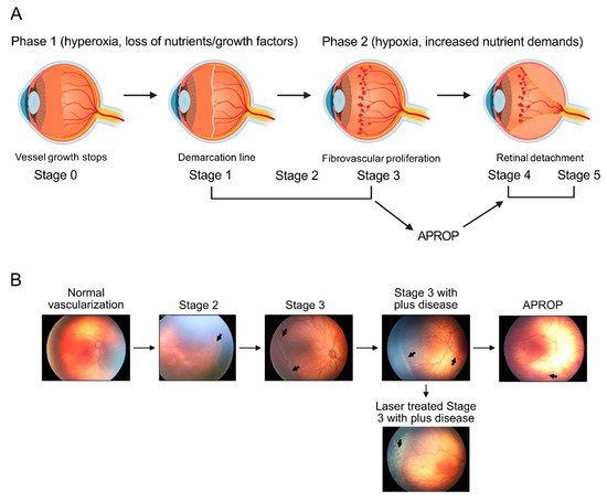
Retinopathy of prematurity (ROP) is a leading cause of blindness in children worldwide, [1] and about 14,000–16,000 infants develop ROP in the US every year. After preterm birth, ROP begins with suppression in the growth of immature retinal vasculature (phase I ROP) (Figure 1A,B), secondary to oxygen supplementation and loss of growth factors normally provided in utero [2]. As the neural retina slowly matures, the increased metabolic demand for nutrients and oxygen is not met in the avascular retinal region. Hypoxia and nutrient deprivation are driving forces to induce retinal vessel growth [3][4][3,4]. However, these newly-formed vessels are uncontrolled and fragile (phase II ROP). Phase II ROP starts at postmenstrual age 30–32 weeks, which coincides with the rapid development of rod photoreceptors [5][6][5,6]. In a rat model of ROP, early photoreceptor dysfunction also predicts subsequent neovascularization [7]. Therefore, modulating retinal metabolic needs may preserve neuronal function and prevent pathologic angiogenesis. Emerging investigations of ROP metabolic changes have been reported with a focus on nutritional interventions such as essential omega-3 and omega-6 long-chain polyunsaturated fatty acids (LCPUFA), insulin-like growth factor 1 (IGF-1), and adiponectin [8][9][10][8,9,10]. Recently, novel blood metabolic biomarkers for ROP have been identified with metabolomics and lipidomics to predict ROP incidence and severity.
Figure 1. ROP progression in premature infants. (A) Schematics of the progression of human retinopathy of prematurity (ROP). Phases 1 and 2 of ROP are associated with different oxygen levels. Loss of essential nutrients and pro-angiogenic growth factors after birth in combination with provision of high supplemental oxygen, leads to hyperoxia that suppresses retinal vascularization (Phase 1). In the second phase of ROP (Phase 2), relative hypoxia and increased nutrient demands of the avascular retina drives fibrovascular proliferation. ROP Phase 2 is defined by anatomic changes, such as the demarcation line (stage 1), ridge (stage 2), extraretinal fibrovascular proliferation (stage 3), partial retinal detachment (stage 4), and total retinal detachment (stage 5). Any stage can develop into aggressive posterior ROP (APROP), which rapidly progresses to tractional retinal detachment (stage 4 or 5). Image made with graphics from ©BioRender (https://biorender.com/ (accessed on 18 October 2021) Agreement number: IA22XF3W0H) (B) Illustration of retinopathy of prematurity (ROP) development, from normal retinal neuro-vascular development, via stage 2 with ridge (arrow), stage 3 with neovascularization and hemorrhage (arrows), stage 3 with plus disease (arrow), APROP with central changes (arrow) and laser treatment (arrow) of stage 3 ROP.

2. Regulation of Retinal Metabolism

2.1. Nutrients

2.1.1. Glucose

Glucose metabolism is one of the most important factors controlling endothelial cell (EC) proliferation, migration, and neovascularization [11][12][13][103,104,105]. Blood-derived glucose penetrates the RPE and the blood–retinal barrier and arrives at the retina facilitated by sodium-independent glucose transporter 1 (Glut1) generating ATP by aerobic glycolysis [14][106]. ECs rely on glycolysis rather than OXPHOS for ATP production and vessel sprouting, and ECs nearly double their glycolytic flux, particularly in tip cells exposed to angiogenic stimuli, such as VEGF [15][107]. Glycolysis in ECs is modulated by the rate-limiting enzyme, 6-phosphofructo-2-kinase/fructose-2,6-biophosphatase 3 (PFKFB3). Pharmacological inhibition of PFKFB3 or EC-specific genetic deletion of Pfkfb3 inhibits pathological retinal neovascularization in mouse OIR [16][17][108,109]. Promotion of glucose uptake during hyperoxia in rat OIR through the inhibition of mitochondrial uncoupling protein 2 (UCP2), a cellular glucose regulator that decreases glucose uptake through Glut1, attenuates the retinal vaso-obliteration and subsequent neovascularization [18][110]. The adenosine A2a receptor (ADORA2A) promotes HIF-1-dependent endothelial cell glycolysis, and the EC-specific Adora2a deletion decreases retinal neovascularization in mouse OIR [19][111]. In addition, under physiological conditions, glycolysis converts glucose to energy, with less than 3% of glucose diverted into the polyol pathway, which reduces glucose to sorbitol and increases oxidative stress through the production of highly toxic advanced glycation end products [20][112]. Aldose reductase is the rate-limiting enzyme in the polyol pathway, and the deletion of the enzyme reduces retinal neovascularization through the attenuation of oxidative stress and protects retinal neurons in mouse OIR [21][22][113,114]. These findings suggest that targeting retinal glucose metabolism is an effective way to control pathological retinal angiogenesis.
Recently, single-cell RNA sequencing reveals that glycolysis gene expression is upregulated in proliferating ECs, but less in tip and immature ECs in a mouse model of choroidal neovascularization [23][115]. Proliferating ECs also upregulated genes involved in one-carbon metabolism, nucleotide synthesis, TCA cycle and OXPHOS [23][115], suggesting the involvement of other metabolic pathways in modulating pathological ocular angiogenesis. Further exploration of their role in ROP is needed.

2.1.2. Amino Acids

Premature infants frequently lack arginine and glutamine because they are unable to maintain the endogenous synthesis of these conditionally essential amino acids [24][25][116,117]. The supplementation of arginine and glutamine (Arg-Gln) suppresses pathological neovascularization in OIR; an in vitro experiment in human RPE cells showed that Arg-Gln decreases VEGF expression [26][63]. ECs have high glutaminase (GLS) activity, which is the enzyme that converts glutamine and glutaminase in the first and rate-limiting step of glutaminolysis, producing energy for proliferation [27][118]. Glutamine is indispensable for vessel sprouting, and the inhibition of GLS1 causes sprouting defects in vitro and in mouse models of developmental angiogenesis and pathological neovascularization in OIR in vivo [28][119].
Serine metabolism via phosphoglycerate dehydrogenase (PHGDH), a key enzyme in the serine synthesis pathway, is important for retinal cell survival, including in EC [29][30][80,120]. Loss of Phgdh in ECs cause defects in retinal angiogenesis and promotes EC apoptosis via heme deficiency, which induces mitochondrial respiration defects and oxidative stress [31][121]. Activation of serine and one carbon metabolism is required for HIF-1 stabilization to protect against hyperoxia-induced retinal vaso-obliteration in mouse OIR [32][78]. Meanwhile, disruption of serine synthesis in the Müller glia also induces mitochondrial dysfunction [33][122] and the Müller glia relies on serine biosynthesis to combat oxidative stress [34][123]. Müller glia is the primary source of VEGF in neovascular retina [35][36][124,125]. Therefore, targeting retinal serine metabolism may protect against retinal neovascularization in ROP.

2.1.3. Fatty Acids

Fatty acids are the other major substrate for energy production in ECs. In vitro, glucose deprivation causes ECs to increase fatty acid oxidation (FAO) flux in an AMP-activated protein kinase (AMPK)-dependent manner [11][103]. Endothelial FAO plays an important role in regulating vessel sprouting [37][126]. As the rate-limiting enzyme of FAO, carnitine palmitoyltransferase 1a (CPT1a) imports FAs into the mitochondria. The endothelial loss of CPT1a causes retinal vascular sprouting defects due to impaired proliferation (not migration) through the inhibition of de novo nucleotide synthesis for DNA replication [37][126]. ECs express fatty acid synthase (FAS), and FAS-mediated de novo lipogenesis is required for vascular sprouting and permeability [38][127]. VEGF enhances the expression of fatty acid uptake and trafficking protein FABP4, which is required for normal EC proliferation [39][128]. Moreover, decreases in both FAO and glycolysis in photoreceptors also induces HIF stabilization and VEGF production, resulting in retinal neovascularization in mice [3][40][3,129]. These findings suggest modulating retinal FAO may also prevent neovascular ROP.

21.2. Hormones

21.2.1. Adiponectin (APN)

APN is an abundant circulating adipokine involved in metabolic modulation [2]. In premature infants, low circulating APN levels correlate with delayed retinal vascularization and ROP progression [9]. In mouse OIR, loss of APN exacerbates and APN administration decreases retinal neovascularization [41][130]. Loss of APN receptor 1 in mice leads to abolished DHA uptake, retention, conservation, elongation in photoreceptors, and eventual photoreceptor degeneration [42][43][131,132]. In mouse HAR, pharmacologic activation of the APN pathway by recombinant APN or APN receptor agonist exerts protective effects on retinal vessel growth and neuronal development [44][99]. These studies suggest that increasing circulating APN levels might benefit the preterm infants and decrease the risk for ROP incidence and progression.
Omega-3 LCPUFA increases circulating APN, which mediates omega-3 LCPUFA’s inhibitory effects on neovascularization in OIR mice [9], as well as in other mouse models with proliferative retinopathy [45][133]. In premature infants, circulating APN is positively correlated with DHA [9]. The increase in circulating APN by dietary omega-3 LCPUFA has also been demonstrated in various studies [46][47][48][49][134,135,136,137]. These reports suggest that omega-3 LCPUFA supplementation is essential in maintaining circulating APN levels to prevent ROP.
In addition, APN levels could be modulated by fibroblast growth factor 21 (FGF21) [50][138], which is expressed in many tissues but mainly in the liver under physiologic conditions [51][139]. FGF21 plays an essential role in modulating lipid and glucose use [52][53][54][140,141,142]. FGF21 is also a key regulator of browning of white adipose tissue and increases energy expenditure [55][143]. FGF21 via APN inhibits choroidal and retinal neovascularization in mice [56][144]. FGF21 also increases APN secretion in obese mice [50][138] and protects diabetes-induced retinal neuronal dysfunction [57][145]. Furthermore, FGF21 preserves retinal neuronal responses in mice with inherited retinal degeneration [58][146]. In preterm infants, circulating FGF21 levels are very low, and the postnatal increase in FGF21 observed in full-term infants seems absent in preterm infants [59][60][61][147,148,149]. Taken together, these reports suggest that circulating FGF21 levels may be correlated with increase in APN levels and ROP progression in preterm infants. Further clinic investigations are needed to validate this hypothesis.

21.2.2. Insulin-Growth Factor 1 (IGF-1)

IGF-1 is an important liver-derived growth factor and a key regulator of body growth and development [62][63][150,151]. In premature infants, persistent low circulating IGF-1 levels strongly correlate with ROP development [64][65][66][67][68][69][94,152,153,154,155,156]. IGF-1 is critical for normal retinal vascularization as a lack of IGF-1 in mice prevents retinal vessel growth [65][152]. IGF-1 also supports VEGF activation of endothelial cell proliferation [65][70][152,157]. Therefore, early restoration of IGF-1 may prevent ROP. Mice with early supplementation of IGF-1 before exposure to hyperoxia have less vessel loss and neovascularization in the OIR model [71][158]. In premature infants with postnatal hyperglycemia in the first month, there are also lower plasma IGF-1 levels [64][94]. In mouse OIR model combined with the HAR model, decreased liver IGF-1 expression is observed before the induction of hyperglycemia; IGF-1 treatment reduces retinal neovascularization and improves retinal revascularization [64][94]. These findings suggest that early supplementation of IGF-1 may improve retinal vascularization and decrease ROP risk. The phase 2 randomized controlled trial (ClinicalTrials.gov Identifier: NCT01096784) shows that rhIGF-1/rhIGFBP-3 decreases the occurrence of severe bronchopulmonary dysplasia, but the dose needs to be further optimized for ROP prevention [72][159]. Increasing the number of patients in the study would also help evaluate the effects of IGF-1 on ROP with completion of the current phase 2b clinical trial using SHP607 (recombinant protein complex of IGF-1/IGFBP3) in preterm infants (ClinicalTrials.gov Identifier: NCT03253263). Moreover, recent investigations have also demonstrated that low circulating IGF-1 levels are correlated with low weekly platelet counts [69][156], which is associated with ROP progression in premature infants [73][74][160,161]. Platelet transfusions inhibit retinal neovascularization in OIR mice [73][160], suggesting that normalizing platelet levels and platelet-derived growth factors (IGF-1, VEGFA, PDGFBB [69][156]) might prevent ROP in premature infants.