Distribution of Sulfate-Reducing Bacteria in the Environment: Comparison
Please note this is a comparison between Version 2 by Beatrix Zheng and Version 1 by Ivan Kushkevych.

Sulfate-reducing bacteria (SRB) are a heterogeneous group of anaerobic microorganisms that play an important role in producing hydrogen sulfide not only in the natural environment, but also in the gastrointestinal tract and oral cavity of animals and humans.

  • anaerobic microorganisms
  • sulfate-reducing bacteria
  • hydrogen sulfide
  • toxicity

1. Introduction

Sulfate-reducing bacteria (SRB) are microorganisms that occur in different ecosystems globally [1,2,3,4,5][1][2][3][4][5]. They can also be isolated from the gastrointestinal tract and the oral cavity of humans and animals [6,7,8,9,10,11][6][7][8][9][10][11]. The cultivation of SRB is sometimes fastidious, as they require anaerobic conditions, strict temperature regulations and precise pH requirements [12]. Consequently, research on SRB is uncommon, and a method of long-term cryopreservation has not been thoroughly developed. Therefore, in this work we review methods for cryopreservation and their application for preservation of SRB [13,14][13][14].

For cryopreservation, it is necessary to choose the right laboratory equipment in which long-term storage can be performed [15]. Another important step in cryopreservation is to select the right cryoprotectant to maximize the viability of the microorganism after freezing [13]. Although the type and concentration of the chosen cryoprotectant is critical, the possibility to combine different preservation compounds to achieve successful cryopreservation may be equally important. The viability of microorganisms is influenced by a number of factors [16]. Colony age, amount of inoculum, cell size or rate of cooling may impact the survival of the culture. Furthermore, viability can also vary between individual species within the same genus [13].

2. Sulfate-Reducing Bacteria in Various Biotopes

In terms of physiology, ecology and function, SRB can be isolated from various biotopes (water, mud, river sediment, sea sediment, human and animal intestinal tracts, etc.) [17]. SRB are mostly classified in the class Deltaproteobacteria and differ from other classes in their characteristic type of metabolism [18]. SRB use sulfate reduction respiration to obtain necessary energy [19,20,21,22][19][20][21][22]. Another descriptive feature of SRB is their cellular shape. Their cells can be spherical, oval, spiral or vibroid [1,18][1][18]. The positive occurrence of SRB is characterized by a strong odor of hydrogen sulfide emission [23,24][23][24]. At the moment, the classification of sulfate-reducing microorganisms has been validly revised. However, according to Bergey’s Manual of Determinative Bacteriology (1994), SRB were divided on the basis of 16S rRNA into the following groups [17,23,24][17][23][24]:
  • Gram-negative mesophilic SRB; these do not form spores and are one of the most widespread SRB in nature (genera Desulfovibrio, Desulfobotulus, Desulfobulbus, Desulfohalobium and Desulfomicrobium);
  • Gram-positive spore-forming bacteria; these are a typical representative (genus Desulfotomaculum) that can be identified from soil samples (according to the updated classification, these microorganisms are represented and included in order Clostridiales);
  • Gram-negative thermophilic sulfate-reducing microorganisms (genus Thermodesulfobacterium);
  • Gram-negative thermophilic archaeal sulfate-reducing microorganisms; these include members of the genus Archaeoglobus that can only be found in anaerobic, underwater, hydrothermal environments because they require salt and high temperature for their growth.
The majority of these groups use sulfate as a terminal electron acceptor during anaerobic respiration. The presence of SRB with high metabolic activity can be identified by the blackening of water and sediments [23].
SRB are important hydrogen-utilizing organisms that, despite their occurrence in other ecosystems, colonize the digestive tract of mammals [11,25,26,27,28,29][11][25][26][27][28][29]. Previous studies have indicated that SRB play an important role in the development of intestinal bowel disease. SRB are also an important factor with regards to food biotechnology, and they can also play a role in part methylation of mercury. Certainly, the presence of different microorganisms in the gut, and the application of probiotics, can influence the eco-physiology of SRB in the intestinal environment [25]. Moreover, SRB have successfully adapted to almost all ecosystems on Earth [2].

2.1. Water Environment

SRB are often found in aquatic polluted environments [1,12][1][12]. Pollution can be of anthropogenic or of natural origin. The presence of sulfate can lead to a number of microbial processes and sulfide formation. Large microbial pollution due to the growth of SRB was recorded in canals and ports such as Venice or the city of Bruges in Belgium. SRB can also be found in the aqueous phases of oil and gasoline storage tanks [12].
In marine waters, SRB can be found more in the upper layers of sediment, where low redox conditions are encountered. Their known competitors, methanogenic archaea, are commonly found in the lower parts of sediment [30]. When SRB and methanogenic archaea occur together in marine sediments, they do not compete against each other, but rather complement each other in the degradation of organic matter. For instance, in marine sediment, SRB and methanogenic archaea are often present together, but methanogens degrade non-competitive substrates and produce methane [25]. Samples of Desulfovibrio spp., Desulfotalea and Desulfuromonas have been found in the upper part of the marine sediment (100 cm) [1,31][1][31]. At the same time, Desulfosporosinus and Desulfovibrio have been most often isolated from the deeper layers. However, a study conducted by Barton & Hamilton (2007) [1] reported that the amount of SRB was low in comparison to the total microbial population inhabiting saltwater environments. It was confirmed that SRB from deep-sea habitats are much more barotolerant than species from near-surface environments [1].
A relatively high population density of SRB has been observed in wastewater biofilms. The composition of the microbial community in wastewater depends on the ability of the organisms to adhere to the surface of the biofilm [32]. Six major genera of SRB have been found in wastewater biofilms: Desulfomicrobium, Desulfovibrio, Desulfonema, Desulforegula, Desulfobacterium, and Desulfobulbus [1]. The authors found that Desulfobulbus spp. generated the highest population density of about 108–109 cells per cm−3 from SRB [32]. High sulfur reduction was found in a narrow anaerobic zone, which was located 150–300 µm below the biofilm surface. As a result, the biofilm formed in the wastewater facilitated the growth of anaerobic SRB under aerobic conditions [32].

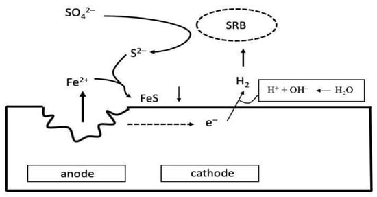
2.2. Surfaces of Corrosive Metals

The colonization of surfaces by SRB is one of the issues of the oil and gas industry, as hydrogen sulfide produced by SRB can cause corrosion and contamination of hydrocarbon products [33]. Corrosion of iron and ferrous alloys occurs not only in aquatic but also in terrestrial environments, regardless of nutrient content, temperature, pressure and pH [1]. Microbial corrosion (MIC) is a biological process that damages the surfaces of corrosive materials due to the action of not only SRB, but also other microorganisms such as aerobic and autotrophic bacteria [24,34][24][34]. A scheme of iron metal corrosion by SRB is shown in Figure 21.
Figure 21. Scheme of iron metal corrosion by SRB (modified from Barton and Fauque, 2009 [2]).
SRB consume H2, and as a result, depolarize the cathode. When Fe2+ is released from the anode, a depression is formed in the metal and insoluble FeS is created. H+ from ionizing water is combined with electrons to form H2 for SRB [2].

2.3. Corrosion of Concrete, Stone Elements and Masonry

Concrete pipes can also be subject to microbial corrosion [2]. The main cause of corrosion of concrete pipes is the metabolic process of SRB. Bacteria grow in water sediment at the bottom of the pipes and hydrogen sulfide is formed there. Once the hydrogen sulfide is produced by SRB, an aerobic zone occurs, and the sulfate-oxidizing bacteria begin to form sulfuric acid, which gradually dissolves the stone surfaces [32,34][32][34].
MIC is the result of a chemical interaction between a metal material and the environment in which the metal is located [2]. The result is a loss of material. Most often, it is an electrochemical process in which electrons from a metal are transported through several redox reactions to a final electron acceptor that is close to the metal surface [32]. There are several mechanisms by which SRB affect corrosion [2], including biofilm formation and attachment on the anode side. In this process, a set of natural bacteria, including SRB, accumulates on the metal surface; it is assumed that the effect of the so-called “quorum sensing” tunes the oxidation, localizes the bacteria on the metal material and creates a depression in this place [2]. Another mechanism by which SRB accelerate corrosion is depolarization at the cathode, which occurs because SRB consume H2 facilitated by hydrogenases. Corrosion can be prevented by the use of protective materials [34]. Plastic pipes with an uneven inner surface or pipes that are highly alkaline on their walls [2].

2.4. Gastrointestinal Tract

The large intestine is a complex microbial ecosystem inhabited by a number of different microbial species [1]. The abundance and composition of organisms plays an important role in human metabolism, and also in the health, disease or physiology of the human body. There are about 1011–1012 microbial cells in 1 g of intestinal contents. 143 stool samples were examined for the abundance of SRB, and it was found that 83% of the specimens contained SRB at a concentration of 102–1011 per 1 g of feces [35]. It was also shown that the incidence of SRB influence the number of methanogenic archaea [36,37][36][37]. It is well-known that SRB and methanogenic archaea compete for nutrients in the gastrointestinal tract (GIT). A study revealed a negative correlation between the concentration of methane in the breath to the number of SRB in fecal samples [36].
In the intestinal microbial composition, hydrogen-utilizing microorganisms play an important role in the metabolism of molecular hydrogen (H2) and sulfur [19]. Due to the fact that SRB use H2 as an electron donor, they facilitate fermentation processes [38,39][38][39].
Anaerobic bacteria represent an integral component of the human microbiome. While many of them are associated with maintaining optimal health, others are involved in a variety of pathological processes, both in immune-competent and immunocompromised individuals [40,41,42][40][41][42]. The most common SRB species that occur in humans and animals are: Desulfovibrio (64–81%), Desulfobacter (9–16%), Desulfobulbus (5–8%), Desulfomonas (3–10%) and Desulfotomaculum (2%). The genus Desulfovibrio is the most common genus of SRB [43,44][43][44]. Desulfovibrio is the most isolated genus of SRB and is found in samples in which inflammatory bowel disease has been confirmed [4,5,45,46,47][4][5][45][46][47]. Desulfotomaculum ruminis and D. acetoxidans originate from intestines [12].
SRB most commonly occur along with Actinobacteria, Firmicutes, and Proteobacteria [11]. SRB significantly affect the pH in the gastrointestinal tract, since they form hydrogen sulfide and acetic acid, and these substances lower pH [5]. The growth conditions of intestinal SRB in the GIT are greatly influenced by the concentration of sulfates, which varies among individuals. However, this depends on the type of diet [48].

3. Conditions Determining the Viability of Sulfate-Reducing Bacteria

Representatives of SRB occur at all sites that meet anaerobic conditions [12]. SRB are able to tolerate temperatures from –5 °C to 75 °C. They are able to tolerate a large pH range (5–9.5) and a large osmotic pressure range [1,12][1][12].
The presence of sulfate and lactate in the human gut contributes significantly to the growth support of SRB [4,8][4][8]. This is also related to the subsequent accumulation of their metabolites, acetate and hydrogen sulfide, in the gastrointestinal environment. In a mixed culture, the growth of SRB was supported by increased sulfur availability [1]. Sulfated polysaccharides, such as mucin and chondroitin sulfate, could be used by SRB as electron acceptors. It has also been shown that when sulfate concentrations increased, the growth of Desulfovibrio desulfuricans also increased [44].

3.1. Physical Conditions

Temperature. SRB strains, which are classified as mesophilic, are known to have a temperature optimum of about 30 °C, but can also tolerate up to 45–48 °C [12]. However, such a large temperature range is more connected with environmental SRB [12,49][12][49]. Intestinal SRB species are grown at 37 °C. This temperature corresponds to warm-blooded animals and humans. Most thermophilic SRB were found in geothermal environments and in oil field waters [23]. The optimum growth temperature of thermophilic SRB (Thermodesulfobacterium) is from 54 °C to 70 °C, and the maximum temperature at which the bacteria are still able to grow is 85 °C [1].
pH. As mentioned above, SRB are able to tolerate a range of pH from 5 to 9.5, but this depends on the environment from which they originally isolated [12]. Although the pH level in the large intestine of humans or animals is limited and depends on a number of different factors (composition and enzymatic activity of intestinal microorganisms, digestion process and consumed food), the pH in the human digestive tract is most often reported to be around 7.6 to 8. However, the pH in the colon is at least one unit lower and lies between 5.7 in the caecum and 6.7 in the rectum [50]. The pH measured in feces is 7 [51]. Microbial growth is said to reach a maximum when the medium has a pH between 7 to 8 [5]. When the medium reached a pH of less than 6, a 26% decrease in microbial growth was recorded when compared to the medium at a pH of 7 to 8 [49]. It was concluded that adults and elderly people (64 to 83 years) supported a higher number of SRB than young people aged around 15 to 20 years [52].

3.2. Competition and Coexistence with Other Intestinal Microorganisms

Coexistence between hydrogenotrophic microbes (SRB and methanogenic archaea) and hydrogenogenic microbes (Clostridium, Bacteroides and Escherichia) is essential to maintain fermentative processes in the gut [11]. Desulfovibrio, which uses H2 produced by Clostridium and Bacteroides, can serve as an example [27]. Methanobrevibacter competes with Desulfovibrio for H2. The inhibition of SRB by methanogenic archaea (increased methanogenesis) results in the accumulation of short chain fatty acids with succinate and lactate [37]. A concentration of 200 mg L−1 of hydrogen sulfide is regarded as an upper limit and can cause inhibition of methanogenesis [53]. The increased availability of sulfate in the gut can lead to the inhibition of methanogenesis [37].
When sulfate is present in the large intestine, the occurrence of SRB is promoted [54]. However, when sulfate levels are reduced, methanogenic archaea predominate in the large intestine that can strongly compete with SRB for the availability of important metabolites of the intestinal microbiota [11,54][11][54]. Sulfate present in the digestive tract can come from both exogenous and endogenous environments [55]. Exogenous sources most often include drinking water and diet. Specific sulfate concentrations have been measured in more than 200 commonly available foods and beverages [48]. Foods high in sulfate (>10 µmol g−1 or up to 1 mg g−1) include certain types of bread, soy flour, dried fruit and sausages. Beverages which commonly contain sulfates (>2.5 µmol mL−1 or 0.25 mg mL−1) include some beers, ciders and wines. About 95% of the sulfate is absorbed in the gastrointestinal tract, and the remaining 5% can be found in the feces. Sources of sulfates of endogenous origin include sulfate mucins, sulfate-conjugated bile, and also, for example, chondroitin sulfate [55]. Sulfate ions in organic compounds need to lose the sulfate group, and in that case, sulfate becomes available. Sulfate does not only serve to support SRB growth and suppress growth of methanogenic archaea, but during sulfate dissimilation reduction, sulfate is used as the final electron acceptor [56,57,58,59][56][57][58][59].
If sulfate is present, Desulfovibrio and Desulfobulbus intestinal species are able to use H2 in the intestinal environment. The coexistence of SRB and methanogenic archaea found in the same ecosystem is possible if both groups of microorganisms use different electron donors [23]. Lactate is one of the main electron donors that occurs within the large intestine [49]. Lactate can be produced by lactic acid-producing bacteria such as Lactobacillus, Streptococcus, Bifidobacterium and others. Lactic acid as a final product of metabolism is then used by SRB [46].

3.3. Biochemical Characteristics of Sulfate-Reducing Bacteria

For intestinal bacteria, polysaccharides, starch and cellulose serve as the main sources of energy and carbon [1,12][1][12]. They can also use a certain amount of oligosaccharides and proteins. The main products of intestinal metabolism are acetate, short chain fatty acids, propionate, butyrate, H2 and CO2. The most common electron donors of SRB are H2, lactate and acetate [11,12][11][12]. Frequent removal of H2 from the lumen by SRB is essential for maintaining a healthy gut [11]. In contrast, sulfate or sulfite serve as electron acceptors (similar to thiosulfate and sulfur, in some cases) for the formation of hydrogen sulfide. Other possible electron donors for SRB growth are fatty acids, glutamate, serine, alanine, ethanol, and a variety of other organic acids such as succinate and pyruvate [36]. The most frequently used electron donors of intestinal SRB are lactate, pyruvate, acetate and ethanol (Figure 32).
Figure 32. Different types of electron donors used by intestinal SRB which were isolated from human fecal samples (data from Gibson et al., 1988 [36]).
Gibson et al., 1988, found that SRB differ with regards to their substrate utilization sprectrum; Desulfovibrio spp. use lactate and H2, Desulfobacter spp. utilize acetate, Desulfobulbus spp. use propionate and H2, Desulfomonas spp. utilize lactate, and lastly, acetate and butyrate are used by Desulfotomaculum spp. [37].
Inhibitors. It is clear that, for SRB, one of the main inhibitors is molecular oxygen (O2) [12,24][12][24]. However, it is mentioned that SRB are capable of some adaptation to the environment where O2 is present [60]. For example, SRB colonizing a drinking water biofilm have been able to survive up to 72 h of exposure to aeration. Of the SRB, the genus Desulfovibrio is the most tolerant to an environment with a certain amount of O2 [61].
High concentrations of various metals are reported to be inhibitors of SRB growth. The toxicity of individual metals depends on the experimental conditions (amount of inoculum, number of cells, pH, temperature). Molybdates have been found to inhibit SRB growth by inhibiting sulfate reduction, thereby reducing the possibility of sulfate transport into the bacterial cell and, thus, reducing possible energy production [33].
The following organisms were used to test the degree of toxicity of molybdates: Desulfotomaculum ruminis, Desulfovibrio vulgaris, and Desulfovibrio desulfuricans (two species). Postgate C medium was used in the experiment. Bacterial growth in the medium was inhibited at a concentration ranging from 40–200 µmol L−1 of the molybdate. Simultaneously, selenium inhibition of SRB growth was tested. Selenium inhibited SRB growth at a concentration between 160 and 320 µmol L−1. The presence of 50 µmol L−1 thiosulfate completely suppressed the effect of selenium. A comparison of the effect of selenium and molybdates shows that molybdates have more mechanisms of inhibition than selenium, since the addition of thiosulfate to the molybdate resulted in only partial inhibition [33].
Hydrogen sulfide. Hydrogen sulfide formation is dependent on SRB growth, which is strongly influenced by environmental pH [45,46,62,63,64][45][46][62][63][64]. This compound is highly toxic. If hydrogen sulfide is not effectively removed from the gut, its accumulation can lead to damage of the colon’s epithelial cells. It has been reported that increased levels of hydrogen sulfide in the gut may be associated with inflammatory bowel disease [44,55][44][55]. Hydrogen sulfide can be removed in the gut by detoxification with the help of intestinal epithelial cells or, due to ongoing bacterial growth, can be incorporated into cellular material [37,44][37][44]. The accumulation of hydrogen sulfide in the intestinal environment can be the result of sulfate metabolism inhibition in mammals and increased SRB activity [45]. Hydrogen sulfide toxicity can even cause DNA damage [62], leading to the formation of an unstable genome, the accumulation of mutations and, in extreme cases, the outbreak of colorectal cancer [65].
The extent that hydrogen sulfide influences the human gut is still not fully understood [44,55][44][55]. Further knowledge of this could be useful particularly in the medical sector, as hydrogen sulfide production can lead to inflammatory bowel diseases [46,47][46][47]. The construction industry is likewise affected by SRB. Here, SRB are responsible for corroding various surfaces, and thereby the structural integrity of these materials is weakened. It is therefore desirable to find suitable protection to combat these issues [2]. Furthermore, SRB are involved in water pollution [12]. Water pollution caused by SRB occurs mostly in canals.

References

  1. Cambridge University Press. Sulphate-Reducing Bacteria: Environmental and Engineered Systems; Barton, L., Hamilton, W.A., Eds.; Cambridge University Press: Cambridge, NY, USA, 2007; ISBN 978-0-521-85485-6.
  2. Barton, L.L.; Fauque, G.D. Chapter 2 Biochemistry, Physiology and Biotechnology of Sulfate-Reducing Bacteria. In Advances in Applied Microbiology; Elsevier: Amsterdam, The Netherlands, 2009; Volume 68, pp. 41–98. ISBN 978-0-12-374803-4.
  3. Kushkevych, I. Isolation and Purification of Sulfate-Reducing Bacteria. In Microorganisms; Blumenberg, M., Shaaban, M., Elgaml, A., Eds.; IntechOpen: London, UK, 2020; ISBN 978-1-83880-187-8.
  4. Kushkevych, I.; Dordević, D.; Kollár, P. Analysis of Physiological Parameters of Desulfovibrio Strains from Individuals with Colitis. Open Life Sci. 2019, 13, 481–488.
  5. Kushkevych, I.; Dordević, D.; Vítězová, M. Analysis of PH Dose-Dependent Growth of Sulfate-Reducing Bacteria. Open Med. 2019, 14, 66–74.
  6. Kushkevych, I.; Kollar, P.; Suchy, P.; Parak, T.; Pauk, K.; Imramovsky, A. Activity of Selected Salicylamides against Intestinal Sulfate-Reducing Bacteria. Neuro Endocrinol. Lett. 2015, 36 (Suppl. 1), 106–113.
  7. Kushkevych, I.V. Kinetic Properties of Pyruvate Ferredoxin Oxidoreductase of Intestinal Sulfate-Reducing Bacteria Desulfovibrio Piger Vib-7 and Desulfomicrobium Sp. Rod-9. Pol. J. Microbiol. 2015, 64, 107–114.
  8. Kushkevych, I.; Fafula, R.; Parák, T.; Bartoš, M. Activity of Na+/K+-Activated Mg2+-Dependent ATP-Hydrolase in the Cell-Free Extracts of the Sulfate-Reducing Bacteria Desulfovibrio Piger Vib-7 and Desulfomicrobium Sp. Rod-9. Acta Vet. Brno 2015, 84, 3–12.
  9. Kushkevych, I.V. Activity and Kinetic Properties of Phosphotransacetylase from Intestinal Sulfate-Reducing Bacteria. Acta Biochim. Pol. 2015, 62, 103–108.
  10. Kushkevych, I.; Coufalová, M.; Vítězová, M.; Rittmann, S.K.-M.R. Sulfate-Reducing Bacteria of the Oral Cavity and Their Relation with Periodontitis—Recent Advances. JCM 2020, 9, 2347.
  11. Ran, S.; Mu, C.; Zhu, W. Diversity and Community Pattern of Sulfate-Reducing Bacteria in Piglet Gut. J. Animal Sci. Biotechnol. 2019, 10, 40.
  12. Postgate, J. The Suphate-Reducing Bacteria, 2nd ed.; Cambridge University: Cambridge, NY, USA, 1984.
  13. Hubálek, Z. Cryopreservation of Microorganisms at Ultra-Low Temperatures; Elsevier: Amsterdam, The Netherlands, 1996.
  14. Trsic-Milanovic, N.; Kodzic, A.; Baras, J.; Dimitrijevic-Brankovic, S. The Influence of a Cryoprotective Medium Containing Glycerol on the Lyophilization of Lactic Acid Bacteria. J. Serb. Chem. Soc. 2001, 66, 435–441.
  15. Butterfield, W.; Jong, S.C.; Alexander, M.T. Polypropylene Vials for Preserving Fungi in Liquid Nitrogen. Mycologia 1978, 70, 1122–1124.
  16. Grout, B.; Morris, J.; Mclellan, M. Cryopreservation and the Maintenance of Cell Lines. Trends Biotechnol. 1990, 8, 293–297.
  17. Castro, H.F.; Williams, N.H.; Ogram, A. Phylogeny of Sulfate-Reducing Bacteria1. FEMS Microbiol. Ecol. 2000, 31, 1–9.
  18. Brenner, D.J.; Krieg, N.R.; Staley, J.T.; Garrity, G.M. The Proteobacteria, Part C: The Alpha-, Beta-, Delta-, and Epsilonproteobacteria. In Bergey’s Manual of Systematic Bacteriology; Springer: Boston, MA, USA, 2005; p. 1388.
  19. Kushkevych, I.; Vítězová, M.; Kos, J.; Kollár, P.; Jampílek, J. Effect of Selected 8-Hydroxyquinoline-2-Carboxanilides on Viability and Sulfate Metabolism of Desulfovibrio Piger. J. Appl. Biomed. 2018, 16, 241–246.
  20. Kushkevych, I.; Cejnar, J.; Treml, J.; Dordević, D.; Kollar, P.; Vítězová, M. Recent Advances in Metabolic Pathways of Sulfate Reduction in Intestinal Bacteria. Cells 2020, 9, 698.
  21. Kushkevych, I.; Kováč, J.; Vítězová, M.; Vítěz, T.; Bartoš, M. The Diversity of Sulfate-Reducing Bacteria in the Seven Bioreactors. Arch. Microbiol. 2018, 200, 945–950.
  22. Abdulina, D.; Kováč, J.; Iutynska, G.; Kushkevych, I. ATP Sulfurylase Activity of Sulfate-Reducing Bacteria from Various Ecotopes. Biotech 2020, 10, 55.
  23. Fauque, G.D. Ecology of Sulfate-Reducing Bacteria. In Sulfate-Reducing Bacteria; Barton, L.L., Ed.; Springer: Boston, MA, USA, 1995; pp. 217–241. ISBN 978-1-4899-1584-9.
  24. Hao, O.J.; Chen, J.M.; Huang, L.; Buglass, R.L. Sulfate-reducing Bacteria. Crit. Rev. Environ. Sci. Technol. 1996, 26, 155–187.
  25. Dordević, D.; Jančíková, S.; Vítězová, M.; Kushkevych, I. Hydrogen Sulfide Toxicity in the Gut Environment: Meta-Analysis of Sulfate-Reducing and Lactic Acid Bacteria in Inflammatory Processes. J. Adv. Res. 2020, 27, 55–69.
  26. Kushkevych, I.; Dordević, D.; Vítězová, M. Possible Synergy Effect of Hydrogen Sulfide and Acetate Produced by Sulfate-Reducing Bacteria on Inflammatory Bowel Disease Development. J. Adv. Res. 2020, 21, 71–78.
  27. Černý, M.; Vítězová, M.; Vítěz, T.; Bartoš, M.; Kushkevych, I. Variation in the Distribution of Hydrogen Producers from the Clostridiales Order in Biogas Reactors Depending on Different Input Substrates. Energies 2018, 11, 3270.
  28. Kováč, J.; Vítězová, M.; Kushkevych, I. Metabolic Activity of Sulfate-Reducing Bacteria from Rodents with Colitis. Open Med. 2018, 13, 344–349.
  29. Kushkevych, I.; Vítězová, M.; Fedrová, P.; Vochyanová, Z.; Paráková, L.; Hošek, J. Kinetic Properties of Growth of Intestinal Sulphate-Reducing Bacteria Isolated from Healthy Mice and Mice with Ulcerative Colitis. Acta Vet. Brno 2017, 86, 405–411.
  30. Plugge, C.M.; Zhang, W.; Scholten, J.C.M.; Stams, A.J.M. Metabolic Flexibility of Sulfate-Reducing Bacteria. Front. Microbio. 2011, 2, 81.
  31. Köpke, B.; Wilms, R.; Engelen, B.; Cypionka, H.; Sass, H. Microbial Diversity in Coastal Subsurface Sediments: A Cultivation Approach Using Various Electron Acceptors and Substrate Gradients. AEM 2005, 71, 7819–7830.
  32. Okabe, S.; Itoh, T.; Satoh, H.; Watanabe, Y. Analyses of Spatial Distributions of Sulfate-Reducing Bacteria and Their Activity in Aerobic Wastewater Biofilms. Appl. Environ. Microbiol. 1999, 65, 5107–5116.
  33. Newport, P.J.; Nedwell, D.B. The Mechanisms of Inhibition of Desulfovibrio and Desulfotomaculum Species by Selenate and Molybdate. J. Appl. Bacteriol. 1988, 65, 419–423.
  34. Islander, R.L.; Devinny, J.S.; Mansfeld, F.; Postyn, A.; Shih, H. Microbial Ecology of Crown Corrosion in Sewers. J. Environ. Eng. 1991, 117, 751–770.
  35. Leclerc, H.; Oger, C.; Beerens, H.; Mossel, D.A.A. Occurrence of Sulphate Reducing Bacteria in the Human Intestinal Flora and in the Aquatic Environment. Water Res. 1980, 14, 253–256.
  36. Gibson, G.R.; Macfarlane, G.T.; Cummings, J.H. Occurrence of Sulphate-Reducing Bacteria in Human Faeces and the Relationship of Dissimilatory Sulphate Reduction to Methanogenesis in the Large Gut. J. Appl. Bacteriol. 1988, 65, 103–111.
  37. Gibson, G.R.; Macfarlane, S.; Macfarlane, G.T. Metabolic Interactions Involving Sulphate-Reducing and Methanogenic Bacteria in the Human Large Intestine. FEMS Microbiol. Ecol. 1993, 12, 117–125.
  38. Kushkevych, I.; Vítězová, M.; Vítěz, T.; Bartoš, M. Production of Biogas: Relationship between Methanogenic and Sulfate-Reducing Microorganisms. Open Life Sci. 2017, 12, 82–91.
  39. Kushkevych, I.; Vítězová, M.; Vítěz, T.; Kováč, J.; Kaucká, P.; Jesionek, W.; Bartoš, M.; Barton, L. A New Combination of Substrates: Biogas Production and Diversity of the Methanogenic Microorganisms. Open Life Sci. 2018, 13, 119–128.
  40. Gajdács, M.; Spengler, G.; Urbán, E. Identification and Antimicrobial Susceptibility Testing of Anaerobic Bacteria: Rubik’s Cube of Clinical Microbiology? Antibiotics 2017, 6, 25.
  41. Gajdács, M.; Urbán, E. Relevance of Anaerobic Bacteremia in Adult Patients: A Never-Ending Story? Eur. J. Microbiol. Immunol. 2020, 10, 64–75.
  42. Gajdács, M.; Ábrók, M.; Lázár, A.; Terhes, G.; Urbán, E. Anaerobic Blood Culture Positivity at a University Hospital in Hungary: A 5-Year Comparative Retrospective Study. Anaerobe 2020, 63, 102200.
  43. Gibson, G.R.; Macfarlane, G.T. Chemostat Enrichment of Sulphate-Reducing Bacteria from the Large Gut. Lett. Appl. Microbiol. 1988, 7, 127–133.
  44. Gibson, G.R.; Cummings, J.H.; Macfarlane, G.T. Growth and Activities of Sulphate-Reducing Bacteria in Gut Contents of Healthy Subjects and Patients with Ulcerative Colitis. FEMS Microbiol. Lett. 1991, 86, 103–112.
  45. Kushkevych, I.; Dordević, D.; Kollar, P.; Vítězová, M.; Drago, L. Hydrogen Sulfide as a Toxic Product in the Small-Large Intestine Axis and Its Role in IBD Development. JCM 2019, 8, 1054.
  46. Kushkevych, I.; Kotrsová, V.; Dordević, D.; Buňková, L.; Vítězová, M.; Amedei, A. Hydrogen Sulfide Effects on the Survival of Lactobacilli with Emphasis on the Development of Inflammatory Bowel Diseases. Biomolecules 2019, 9, 752.
  47. Kushkevych, I.; Castro Sangrador, J.; Dordević, D.; Rozehnalová, M.; Černý, M.; Fafula, R.; Vítězová, M.; Rittmann, S.K.-M.R. Evaluation of Physiological Parameters of Intestinal Sulfate-Reducing Bacteria Isolated from Patients Suffering from IBD and Healthy People. JCM 2020, 9, 1920.
  48. Florin, T.H.J.; Neale, G.; Goretski, S.; Cummings, J.H. The Sulfate Content of Foods and Beverages. J. Food Compos. Anal. 1993, 6, 140–151.
  49. Kováč, J.; Kushkevych, I. New Modification of Cultivation Medium for Isolation and Growth of Intestinal Sulfate-Reducing Bacteria. In Proceedings of the 24th International Ph.D. Students Conference, Brno, Czech Republic, 8–9 November 2017; Volume 2017, pp. 702–707.
  50. Fallingborg, J.; Christensen, L.A.; Ingeman-Nielsen, M.; Jacobsen, B.A.; Abildgaard, K.; Rasmussen, H.H. PH-Profile and Regional Transit Times of the Normal Gut Measured by a Radiotelemetry Device. Aliment. Pharmacol. Ther. 2007, 3, 605–614.
  51. Beerens, H.; Romond, C. Sulfate-Reducing Anaerobic Bacteria in Human Feces. Am. J. Clin. Nutr. 1977, 30, 1770–1776.
  52. Fite, A. Identification and Quantitation of Mucosal and Faecal Desulfovibrios Using Real Time Polymerase Chain Reaction. Gut 2004, 53, 523–529.
  53. Hilton, B.L.; Oleszkiewicz, J.A. Sulfide-Induced Inhibition of Anaerobic Digestion. J. Environ. Eng. 1988, 114, 1377–1391.
  54. Christl, S.U.; Gibson, G.R.; Cummings, J.H. Role of Dietary Sulphate in the Regulation of Methanogenesis in the Human Large Intestine. Gut 1992, 33, 1234–1238.
  55. Deplancke, B.; Hristova, K.R.; Oakley, H.A.; McCracken, V.J.; Aminov, R.; Mackie, R.I.; Gaskins, H.R. Molecular Ecological Analysis of the Succession and Diversity of Sulfate-Reducing Bacteria in the Mouse Gastrointestinal Tract. Appl. Environ. Microbiol. 2000, 66, 2166–2174.
  56. Kushkevych, I.; Dordević, D.; Vítězová, M.; Kollár, P. Cross-Correlation Analysis of the Desulfovibrio Growth Parameters of Intestinal Species Isolated from People with Colitis. Biologia 2018, 73, 1137–1143.
  57. Postgate, J.R. On the Nutrition of Desulphovibrio Desulphuricans. J. Gen. Microbiol. 1951, 5, 714–724.
  58. Postgate, J.R. Sulphate Reduction by Bacteria. Annu. Rev. Microbiol. 1959, 13, 505–520.
  59. Postgate, J.R.; Campbell, L.L. Classification of Desulfovibrio Species, the Nonsporulating Sulfate-Reducing Bacteria. Bacteriol. Rev. 1966, 30, 732–738.
  60. Bade, K.; Manz, W.; Szewzyk, U. Behavior of Sulfate Reducing Bacteria under Oligotrophic Conditions and Oxygen Stress in Particle-Free Systems Related to Drinking Water. FEMS Microbiol. Ecol. 2000, 32, 215–223.
  61. Krekeler, D.; Sigalevich, P.; Teske, A.; Cypionka, H.; Cohen, Y. A Sulfate-Reducing Bacterium from the Oxic Layer of a Microbial Mat from Solar Lake (Sinai), Desulfovibrio oxyclinae Sp. Nov. Arch. Microbiol. 1997, 167, 369–375.
  62. Attene-Ramos, M.S.; Wagner, E.D.; Plewa, M.J.; Gaskins, H.R. Evidence That Hydrogen Sulfide Is a Genotoxic Agent. Mol. Cancer Res. 2006, 4, 9–14.
  63. Kushkevych, I.; Leščanová, O.; Dordević, D.; Jančíková, S.; Hošek, J.; Vítězová, M.; Buňková, L.; Drago, L. The Sulfate-Reducing Microbial Communities and Meta-Analysis of Their Occurrence during Diseases of Small–Large Intestine Axis. JCM 2019, 8, 1656.
  64. Kushkevych, I.; Dordević, D.; Vítězová, M. Toxicity of Hydrogen Sulfide toward Sulfate-Reducing Bacteria Desulfovibrio Piger Vib-7. Arch. Microbiol. 2019, 201, 389–397.
  65. Pitcher, M.C.; Cummings, J.H. Hydrogen Sulphide: A Bacterial Toxin in Ulcerative Colitis? Gut 1996, 39, 1–4.
More