Your browser does not fully support modern features. Please upgrade for a smoother experience.
Please note this is a comparison between Version 2 by Catherine Yang and Version 1 by Masutaka Furue.
The aryl hydrocarbon receptor (AHR)/AHR-nuclear translocator (ARNT) system is a sensitive sensor for small molecular, xenobiotic chemicals of exogenous and endogenous origin, including dioxins, phytochemicals, microbial bioproducts, and tryptophan photoproducts. AHR/ARNT are abundantly expressed in the skin. Once activated, the AHR/ARNT axis strengthens skin barrier functions and accelerates epidermal terminal differentiation by upregulating filaggrin expression. In addition, AHR activation induces oxidative stress. However, some AHR ligands simultaneously activate the nuclear factor-erythroid 2-related factor-2 (NRF2) transcription factor, which is a master switch of antioxidative enzymes that neutralizes oxidative stress.
- aryl hydrocarbon receptor (AHR)
- atopic dermatitis
- psoriasis
1. Introduction
The skin is the outermost surface of the body and is vulnerable to a myriad of external chemicals and internal substances. To maintain homeostasis, skin cells, including keratinocytes, sebocytes, fibroblasts, dendritic cells, and other immune cells, express several chemical sensors, such as aryl hydrocarbon receptor (AHR), pregnane X receptor, constitutive androstane receptor, and peroxisome proliferator-activated receptors [1,2,3,4][1][2][3][4]. Among these chemical receptors, AHR has gained special attention because it plays a crucial role in photoaging, epidermal differentiation, and immunomodulation [2,3,5,6,7][2][3][5][6][7].
AHR, also called dioxin receptor, binds to environmental polyaromatic hydrocarbons and dioxins with high affinity and induces oxidative stress by generating abundant reactive oxygen species (ROS) [5,6,7][5][6][7]. Additionally, AHR is a promiscuous receptor and is activated by a plethora of exogenous and endogenous ligands, such as photo-induced chromophores, phytochemicals, and microbial bioproducts [8,9,10,11,12][8][9][10][11][12]. Many AHR ligands exert antioxidative activity by activating antioxidative transcription factor nuclear factor-erythroid 2-related factor-2 (NRF2) [10,13][10][13]. Medicinal coal tar and soybean tar Glyteer activate both AHR and NRF2 and have been used to treat inflammatory skin diseases, such as atopic dermatitis (AD) and psoriasis [14,15][14][15].
AD and psoriasis are common inflammatory skin diseases. An excellent therapeutic response to biologics indicates a pivotal pathogenic role of interleukin (IL)-4/IL-13 signaling in AD [16,17][16][17] and the tumor necrosis factor (TNF)-α/IL-23/IL-17A axis in psoriasis [18,19][18][19]. Although distinct signaling pathways operate in developing full-blown AD and psoriasis, 81% of dysregulated genes in AD are shared with those in psoriasis in skin lesions [20]. Notably, recent phase II, randomized dose-finding studies have demonstrated that topical application of the natural AHR agonist tapinarof is efficacious and well tolerated in patients with AD and psoriasis [21,22][21][22].
2. AHR and Atopic Dermatitis
AD is a common and heterogenous eczematous skin disorder characterized by Th2-deviated skin inflammation, barrier disruption, and chronic pruritus [17,82,83][17][23][24]. Frequent relapse with intense pruritus deteriorates quality of life and decreases treatment satisfaction of the afflicted patients [84,85,86,87,88][25][26][27][28][29]. The lifetime incidence of AD is as high as 20% in the general population [89][30]. Skin barrier dysfunction is associated with the reduced production of terminal differentiation molecules such as filaggrin [15,51][15][31]. Abnormal skin barrier integrity also causes an increased colonization of microbes such as Staphylococcus aureus, which further exacerbate Th2-deviated skin inflammation [90,91][32][33]. In addition, some autoimmune diseases are comorbid with AD [92][34].
Investigation on AHR gene polymorphism reveals that AHR rs10249788 and rs2066853 polymorphisms are found in patients with AD, psoriasis, and healthy controls, but no significant differences were detected in genotype or allele frequencies between the three groups [93][35]. However, the AHR rs2066853 (AG + AA) or rs10249788 (CT + TT) genotypes are a risk factor for severe dry skin phenotype and the combined rs10249788 (CT + TT) and rs2066853 (AG + AA) genotypes lead to a higher risk for severe dry skin in Chinese patients with AD [93][35]. rs10249788 exists in the AHR promoter region where nuclear factor 1C (NF1C) binds and suppresses the transcription and protein expression of AHR [94][36]. Notably, NF1C prefers to associate with the C allele compared to the T allele at rs10249788. Thus, subjects with the rs10249788 (CC) allele express less AHR than those with the rs10249788 (TT) allele [94][36]. In fact, AHR mRNA levels for the TT genotype are 1.7-fold higher than those for the CC genotype [95][37]. No significant differences were obtained in AHR production between the CC and CT genotypes [95][37]. In parallel with increased levels of AHR, cells with the TT genotype express significantly higher levels of CYP1A1, IL-24, and IL-1β [95][37]. It is intriguing that IL-24 downregulates the filaggrin expression via STAT3 activation [67][38].
Immunohistological and real time PCR studies for AHR have been reported in AD [96,97][39][40]. Hong et al. showed an increased expression of both AHR and ARNT without CYP1A1 induction in the lesioned skin of AD compared with normal control skin [96][39]. Alternatively, Kim et al. demonstrated an increased expression of ARNT and CYP1A1 but not AHR in the lesional skin of AD [97][40]. As the Th2-deviated milieu potently reduces filaggrin and other barrier-related molecules, the upregulation of AHR/ARNT may be compensatory to attenuate the Th2-mediated filaggrin reduction. A recent study by Yu et al. demonstrated the possibility that the Th2-deviated milieu decreases the production of endogenous AHR ligand such as indole-3-aldehyde by commensal skin microbiota [98][41]. These findings collectively suggest that most AHR likely lack physiological ligands in the Th2-prone milieu in AD. Therefore, rapid-metabolizing AHR ligands, such as FICZ and indole-3-aldehyde, appropriately activate the AHR/ARNT/FLG axis and may be beneficial in treating AD [58,98][42][41]. However, vigorous and long-lasting activation of the AHR/ARNT/FLG axis by slow-metabolizing dioxins and environmental pollutants may exacerbate barrier dysfunction and aggravate AD [96,99][39][43].
Although the pathogenic implication of AHR and its gene polymorphism in AD remain elusive, recent clinical trials using topical AHR ligand tapinarof have reported its efficacy for AD [100,101,102][44][45][46]. Tapinarof (5-[(E)-2-phenylethenyl]-2-[propan-2-yl] benzene-1, 3-diol, WBI-1001, GSK2894512 or bentivimod) is a naturally derived (but is now a fully synthetic) hydroxylated stilbene produced by bacterial symbionts of entomopathogenic nematodes [100,101,102,103][44][45][46][47]. Tapinarof is a high affinity AHR ligand with antioxidative activity via NRF2 activation and a ROS-scavenging structure [102][46] (Figure 1). Tapinarof has gained increased attention because its topical application is efficacious for patients with AD in clinical trials [21,100,104][21][44][48]. Tapinarof activates the AHR/CYP1A1 axis and augments the expression of filaggrin and involucrin [102][46]. Even in barrier-disrupted AD patients, systemic absorption of topical tapinarof is limited and likely decreases during the treatment course in parallel with treatment success that restores the barrier dysfunction [104][48]. In general, topical tapinarof is tolerable but frequent adverse events include headaches and folliculitis [104][48].
In an early clinical trial, patients with AD affecting 3–20% of their body surface area (BSA) and with an Investigator’s Global Assessment (IGA; 0: clear, 1: almost clear, 2: mild, 3: moderate, 4: severe, 5: very severe) of 2–4 were randomized (1:1:1) to receive a placebo (n = 51), topical tapinarof 0.5% (n = 50) or 1% (n = 47) in a cream formulation applied twice daily for six weeks [100][44]. There was a decrease of 1.3 (43%; p < 0.001; 95% confidence interval (CI) −1.2 to −0.5) and 1.8 (56.3%; p < 0.001; 95% CI −1.6 to −0.9) in IGA at day 42 in the topical tapinarof 0.5% and 1% groups, respectively, compared with a decrease of 0.5 (14.7%) in the placebo group. At day 42, improvement in Eczema Area and Severity Index (EASI) score was 68.9% (p < 0.001) and 76.3% (p < 0.001) for tapinarof 0.5% and 1%, respectively, compared with 23.3% for placebo. Improvement in pruritus severity score at day 42 was 29.8% (p < 0.001) and 66.9% (p < 0.001) for tapinarof 0.5% and 1%, respectively, compared with 9.5% for placebo [100][44]. Adverse events included headaches (placebo: 0%; 0.5% tapinarof: 8%; 1% tapinarof: 14%), migraines (placebo: 0%; 0.5% tapinarof: 4%; 1% tapinarof: 3%), folliculitis (placebo: 0%; 0.5% tapinarof: 6%; 1% tapinarof: 8%), and contact dermatitis (placebo: 0%; 0.5% tapinarof: 3%: 1% tapinarof: 5%) [100][44].
A phase II, double-blind, vehicle-controlled, randomized, six-arm trial (1:1:1:1:1:1) in patients aged 12 to 65 years, with BSA involvement of at least 5% to 35% and an IGA score of 3 or higher (moderate to severe) at baseline was performed. Primary end points included an IGA score of clear or almost clear (0 or 1) and a minimum two-grade improvement (treatment success) at week 12 [21]. The rates of treatment success with topical tapinarof cream at week 12 were 53% (1% twice daily, n = 40), 46% (1% once daily, n = 41), 37% (0.5% twice daily, n = 43), 34% (0.5% once daily, n = 41), 24% (vehicle twice daily, n = 42), and 28% (vehicle once daily, n = 40). The rate with tapinarof 1% twice daily (53%) was statistically significantly higher than the rate with vehicle twice daily (24%). Notably, treatment success was maintained for four weeks after the end of tapinarof treatment. The proportion of patients achieving EASI75 (75% or greater improvement in EASI) score reduction at week 12 was significantly higher in the groups treated with 1% tapinarof (60% and 51% for twice daily and once daily, respectively) than with vehicle (26% and 25% in the groups receiving vehicle twice daily and once daily, respectively) [21]. Headaches (e.g., 10% (1% twice daily), 2% (0.5% twice daily), and 0% (0.5% twice daily)) and folliculitis (e.g., 10% (1% twice daily), 7% (0.5% twice daily), and 0% (0.5% twice daily)) were again frequent adverse events [21].
In a murine dermatitis model, topically applied FICZ activated AHR and significantly reduced the dermatitis score and histological inflammation with a decrease of Il22 gene expression in chronic mite antigen-induced dermatitis [58][42]. In addition, topical FICZ restored the dermatitis-induced filaggrin downregulation [58][42]. CCL17 and CCL22 are crucial chemokines to recruit Th2 cells [68][49]. IL-4/IL-13 stimulates dendritic cells to produce CCL17 and CCL22 via STAT6 activation and contributes to the recruitment of Th2 cells in the lesional skin of AD [68][49]. Soybean tar Glyteer inhibits the IL-4/IL-13-mediated STAT6 activation and subsequent production of CCL17 and CCL22 in dendritic cells [68][49]. In addition, pruritogenic Th2 cytokine IL-31 synergistically upregulates the IL-4/IL-13-mediated CCL17 and CCL22 production in dendritic cells because IL-4/IL-13 increase IL-31 receptor A (IL31RA) expression [105][50]. Glyteer again attenuates the IL-4/IL-13-mediated IL31RA upregulation and subsequent CCL17 and CCL22 production by inhibiting STAT6 activation [105][50]. It is known that coal tar inhibits STAT6 activation via the NRF2-antioxidative pathway [15]. Ligation of AHR by FICZ also reduces the expression of type 1 IgE Fc receptor in Langerhans cells [106][51].
Although antioxidative AHR ligands are therapeutic for dermatitis, exaggerated activation of AHR by genetic manipulation in transgenic mice or by dioxin treatment induces itchy dermatitis most likely due to an abnormally accelerated keratinization process, epidermal acanthosis, elongation of nerve fibers, and production of pruritogenic artemin [47,99,107][52][43][53]. Therefore, extreme activation of AHR is deleterious for skin. In parallel, ovalbumin-induced delayed hypersensitivity is enhanced by topical benzopyrene with upregulation of IL-5, IL-13, and IL-17 expression in lymph node cells [96][39].
Since FICZ is an endogenous UVB photoproduct [8], the barrier-protecting effects of FICZ may explain, at least in part, why UVB phototherapy is efficacious for the treatment of AD and psoriasis [108,109][54][55].
3. AHR and Psoriasis
Psoriasis is an (auto)immune-mediated disease that manifests as widespread desquamative erythema [110,111][56][57]. Males are twice as likely to be affected than females [112,113][58][59]. The cosmetic disfigurement associated with psoriasis profoundly impairs the patients’ quality of life, treatment satisfaction and adherence, and socioeconomic stability [114,115][60][61]. The autoimmune nature of psoriasis is exemplified by its high comorbidity with psoriatic arthritis [110,116,117,118][56][62][63][64] and other autoimmune diseases including autoimmune bullous diseases [119,120,121,122,123,124][65][66][67][68][69][70]. Psoriasis is also comorbid with cardiovascular diseases, metabolic diseases, and renal disorders, which represent a condition called inflammatory skin march [111,125,126,127,128,129][57][71][72][73][74][75]. The excellent therapeutic efficacy of anti-TNF-α/IL-23/IL-17A biologics for psoriasis point to the central role of the TNF-α/IL-23/IL-17A axis in its pathogenesis [18,19,130,131,132,133,134][18][19][76][77][78][79][80] Additionally, genetic and environmental factors are known to be involved in its pathogenesis [135,136][81][82].
As AHR predominantly regulates the immune balance of Th17/22 and Treg cells [28[83][84][85][86],29,69,70], AHR is expected to play a significant role in psoriasis [102][46]. In an imiquimod-induced psoriasis model, AhR deficiency exacerbates skin inflammation with upregulated gene expression of Il22, Il17a, and Il23 [137][87]. The intensity of delayed type-hypersensitivity is also enhanced in Ahr-deficient mice [137][87]. However, further experiments demonstrated that Ahr-deficiency in nonhematopoietic cells, including keratinocytes, but not in hematopoietic cells, was likely responsible for the exacerbation of inflammation [137][87]. Notably, intraperitoneal injection of FICZ ameliorated the imiquimod-induced psoriasis-like inflammation. Tapinarof and FICZ also reduced the imiquimod-induced psoriasiform skin inflammation by inhibiting Il17a, Il17f, Il19, Il22, Il23a, and Il1b gene expression [102][46]. The therapeutic action of tapinarof and FICZ was AHR-dependent because it was not observed in Ahr-deficient mice [102][46]. In an ex vivo activation assay of skin-resident immunocompetent cells using normal human skin, tapinarof inhibited the expression of IL17A message approximately 50% but increased the IL22 expression [102,138][46][88] (Figure 1). In mice, IL-22 is produced from Th17, γδT, ILC3, and CD4−CD8−TCRβ+ cells [139][89]. AHR was required for IL-22 production by Th17, but not by the three other cell types, in the imiquimod-treated ears [139][89]. Although imiquimod-induced skin inflammation is popular as a psoriasis model, attention should be paid because imiquimod is degraded by CYP1A1 so the efficacy of AHR agonists may partly rely on this effect in the imiquimod model [140][90].
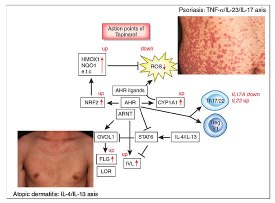
Figure 1. Aryl hydrocarbon receptor (AHR) signal and action points of tapinarof (red words and arrows). AHR is a promiscuous chemical sensor that is activated by various oxidative and antioxidative ligands. Once activated, cytoplasmic AHR translocates into the nucleus where it heterodimerizes with an AHR-nuclear translocator (ARNT) and then induces the transcription of AHR-responsive genes such as cytochrome P450 1A1 (CYP1A1). CYP1A1 degrades AHR ligands. Some ligands such as dioxins are chemically stable and long-lived. Therefore, CYP1A1 generates high amounts of reactive oxygen species (ROS) after sustained efforts to degrade them. Some antioxidative AHR ligands activate nuclear factor-erythroid 2-related factor-2 (NRF2) transcription factor, which upregulates gene expression of various antioxidative enzymes, such as heme oxygenase 1 (HMOX1), NAD(P)H dehydrogenase, and quinone 1 (NQO1), and these antioxidative enzymes neutralize ROS. AHR/ARNT signaling also activates OVO-like 1 (OVOL1) transcription factor and upregulates the expression of filaggrin (FLG) and loricrin (LOR). AHR upregulates the expression of involucrin (IVL) in an OVOL1-independent manner. Therefore, AHR/ARNT signaling accelerates epidermal terminal differentiation and enhances the repair of barrier disruption. Interleukin (IL)-4 and IL-13 activate signal transducer and activator of transcription 6 (STAT6) and inhibit the OVOL1/FLG, OVOL1/LOR, and AHR/IVL axes. However, suitable AHR activation can inhibit the IL-4/IL-13-mediated STAT6 activation and restore the expression of FLG, LOR, and IVL. Regarding immune response, AHR signaling affects T-helper (Th17) differentiation and is essential for IL-22 production. AHR ligation (especially by high concentrations of ligands) induces the differentiation of regulatory cell populations, Treg and Tr1 cells. Tapinarof is an antioxidative AHR ligand and upregulates CYP1A1 expression. Topical tapinarof is efficacious in psoriasis and atopic dermatitis. Current studies demonstrate that tapinarof activates NRF2/antioxidative signaling and reduces oxidative stress. Tapinarof also upregulates FLG and IVL expression. Tapinarof downregulates IL-17A production and increases IL-22 production.
Immunohistological and real time PCR studies have demonstrated that the expression of AHR and ARNT is upregulated in the lesional skin of psoriasis, whereas CYP1A1 expression was significantly decreased compared to normal controls [97][40]. In contrast, serum levels of both AHR and CYP1A1 are elevated in patients with psoriasis compared to normal controls [141][91]. Further studies are warranted to investigate these controversial data.
References
- Furue, K.; Mitoma, C.; Tsuji, G.; Furue, M. Protective role of peroxisome proliferator-activated receptor α agonists in skin barrier and inflammation. Immunobiology 2018, 223, 327–330.
- Furue, M.; Fuyuno, Y.; Mitoma, C.; Uchi, H.; Tsuji, G. Therapeutic agents with AHR inhibiting and NRF2 activating activity for managing chloracne. Antioxidants (Basel) 2018, 7, 90.
- Furue, M.; Hashimoto-Hachiya, A.; Tsuji, G. Antioxidative phytochemicals accelerate epidermal terminal differentiation via the AHR-OVOL1 pathway: Implications for atopic dermatitis. Acta Derm. Venereol. 2018, 98, 918–923.
- Omiecinski, C.J.; Vanden Heuvel, J.P.; Perdew, G.H.; Peters, J.M. Xenobiotic metabolism, disposition, and regulation by receptors: From biochemical phenomenon to predictors of major toxicities. Toxicol. Sci. 2011, 120, S49–S75.
- Esser, C.; Bargen, I.; Weighardt, H.; Haarmann-Stemmann, T.; Krutmann, J. Functions of the aryl 1002 hydrocarbon receptor in the skin. Semin. Immunopathol. 2013, 35, 677–691.
- Furue, M.; Takahara, M.; Nakahara, T.; Uchi, H. Role of AhR/ARNT system in skin homeostasis. Arch. Dermatol. Res. 2014, 306, 769–779.
- Mimura, J.; Fujii-Kuriyama, Y. Functional role of AhR in the expression of toxic effects by TCDD. Biochim. Biophys. Acta 2003, 1619, 263–268.
- Fritsche, E.; Schäfer, C.; Calles, C.; Bernsmann, T.; Bernshausen, T.; Wurm, M.; Hübenthal, U.; Cline, J.E.; Hajimiragha, H.; Schroeder, P.; et al. Lightening up the UV response by identification of the aryl hydrocarbon receptor as a cytoplasmatic target for ultraviolet B radiation. Proc. Natl. Acad. Sci. USA 2007, 104, 8851–8856.
- Rannug, A.; Rannug, U.; Rosenkranz, H.S.; Winqvist, L.; Westerholm, R.; Agurell, E.; Grafström, A.K. Certain photooxidized derivatives of tryptophan bind with very high affinity to the Ah receptor and are likely to be endogenous signal substances. J. Biol. Chem. 1987, 262, 15422–15427.
- Furue, M.; Uchi, H.; Mitoma, C.; Hashimoto-Hachiya, A.; Chiba, T.; Ito, T.; Nakahara, T.; Tsuji, G. Antioxidants for healthy skin: The emerging role of aryl hydrocarbon receptors and nuclear factor-erythroid 2-related factor-2. Nutrients 2017, 9, 223.
- Magiatis, P.; Pappas, P.; Gaitanis, G.; Mexia, N.; Melliou, E.; Galanou, M.; Vlachos, C.; Stathopoulou, K.; Skaltsounis, A.L.; Marselos, M.; et al. Malassezia yeasts produce a collection of exceptionally potent activators of the Ah (dioxin) receptor detected in diseased human skin. J. Investig. Dermatol. 2013, 133, 2023–2030.
- Takei, K.; Mitoma, C.; Hashimoto-Hachiya, A.; Takahara, M.; Tsuji, G.; Nakahara, T.; Furue, M. Galactomyces fermentation filtrate prevents T helper 2-mediated reduction of filaggrin in an aryl hydrocarbon receptor-dependent manner. Clin. Exp. Dermatol. 2015, 40, 786–793.
- Takei, K.; Hashimoto-Hachiya, A.; Takahara, M.; Tsuji, G.; Nakahara, T.; Furue, M. Cynaropicrin attenuates UVB-induced oxidative stress via the AhR-Nrf2-Nqo1 pathway. Toxicol. Lett. 2015, 234, 74–80.
- Takei, K.; Mitoma, C.; Hashimoto-Hachiya, A.; Uchi, H.; Takahara, M.; Tsuji, G.; Kido-Nakahara, M.; Nakahara, T.; Furue, M. Antioxidant soybean tar Glyteer rescues T-helper-mediated downregulation of filaggrin expression via aryl hydrocarbon receptor. J. Dermatol. 2015, 42, 171–180.
- Van den Bogaard, E.H.; Bergboer, J.G.; Vonk-Bergers, M.; van Vlijmen-Willems, I.M.; Hato, S.V.; van der Valk, P.G.; Schröder, J.M.; Joosten, I.; Zeeuwen, P.L.; Schalkwijk, J. Coal tar induces AHR-dependent skin barrier repair in atopic dermatitis. J. Clin. Investig. 2013, 123, 917–927.
- Simpson, E.L.; Bieber, T.; Guttman-Yassky, E.; Beck, L.A.; Blauvelt, A.; Cork, M.J.; Silverberg, J.I.; Deleuran, M.; Kataoka, Y.; Lacour, J.P.; et al. Two Phase 3 Trials of dupilumab versus placebo in atopic dermatitis. N. Engl. J. Med. 2016, 375, 2335–2348.
- Furue, M.; Ulzii, D.; Vu, Y.H.; Tsuji, G.; Kido-Nakahara, M.; Nakahara, T. Pathogenesis of atopic dermatitis: Current paradigm. Iran. J. Immunol. 2019, 16, 97–107.
- Furue, K.; Ito, T.; Furue, M. Differential efficacy of biologic treatments targeting the TNF-α/IL-23/IL-17 axis in psoriasis and psoriatic arthritis. Cytokine 2018, 111, 182–188.
- Furue, K.; Ito, T.; Tsuji, G.; Kadono, T.; Furue, M. Psoriasis and the TNF/IL23/IL17 axis. G. Ital. Dermatol. Venereol. 2019, 154, 418–424.
- Tsoi, L.C.; Rodriguez, E.; Degenhardt, F.; Baurecht, H.; Wehkamp, U.; Volks, N.; Szymczak, S.; Swindell, W.R.; Sarkar, M.K.; Raja, K.; et al. Atopic dermatitis is an IL-13-dominant disease with greater molecular heterogeneity compared to psoriasis. J. Investig. Dermatol. 2019, 139, 1480–1489.
- Peppers, J.; Paller, A.S.; Maeda-Chubachi, T.; Wu, S.; Robbins, K.; Gallagher, K.; Kraus, J.E. A phase 2, randomized dose-finding study of tapinarof (GSK2894512 cream) for the treatment of atopic dermatitis. J. Am. Acad. Dermatol. 2019, 80, 89–98.
- Robbins, K.; Bissonnette, R.; Maeda-Chubachi, T.; Ye, L.; Peppers, J.; Gallagher, K.; Kraus, J.E. Phase 2, randomized dose-finding study of tapinarof (GSK2894512 cream) for the treatment of plaque psoriasis. J. Am. Acad. Dermatol. 2019, 80, 714–721.
- Furue, M.; Chiba, T.; Tsuji, G.; Ulzii, D.; Kido-Nakahara, M.; Nakahara, T.; Kadono, T. Atopic dermatitis: Immune deviation, barrier dysfunction, IgE autoreactivity and new therapies. Allergol. Int. 2017, 66, 398–403.
- Seo, E.; Yoon, J.; Jung, S.; Lee, J.; Lee, B.H.; Yu, J. Phenotypes of atopic dermatitis identified by cluster analysis in early childhood. J. Dermatol. 2019, 46, 117–123.
- Arima, K.; Gupta, S.; Gadkari, A.; Hiragun, T.; Kono, T.; Katayama, I.; Demiya, S.; Eckert, L. Burden of atopic dermatitis in Japanese adults: Analysis of data from the 2013 National Health and Wellness Survey. J. Dermatol. 2018, 45, 390–396.
- Igarashi, A.; Fujita, H.; Arima, K.; Inoue, T.; Dorey, J.; Fukushima, A.; Taguchi, Y. Health-care resource use and current treatment of adult atopic dermatitis patients in Japan: A retrospective claims database analysis. J. Dermatol. 2019, 46, 652–661.
- Jung, H.J.; Bae, J.Y.; Kim, J.E.; Na, C.H.; Park, G.H.; Bae, Y.I.; Shin, M.K.; Lee, Y.B.; Lee, U.H.; Jang, Y.H.; et al. Survey of disease awareness, treatment behavior and treatment satisfaction in patients with atopic dermatitis in Korea: A multicenter study. J. Dermatol. 2018, 45, 1172–1180.
- Komura, Y.; Kogure, T.; Kawahara, K.; Yokozeki, H. Economic assessment of actual prescription of drugs for treatment of atopic dermatitis: Differences between dermatology and pediatrics in large-scale receipt data. J. Dermatol. 2018, 45, 165–174.
- Takeuchi, S.; Oba, J.; Esaki, H.; Furue, M. Non-corticosteroid adherence and itch severity influence perception of itch in atopic dermatitis. J. Dermatol. 2018, 45, 158–164.
- Williams, H.; Stewart, A.; von Mutius, E.; Cookson, W.; Anderson, H.R. Is eczema really on the increase worldwide? J. Allergy Clin. Immunol. 2008, 121, 947–954.
- Geng, S.; Mezentsev, A.; Kalachikov, S.; Raith, K.; Roop, D.R.; Panteleyev, A.A. Targeted ablation of Arnt in mouse epidermis results in profound defects in desquamation and epidermal barrier function. J. Cell Sci. 2006, 119, 4901–4912.
- Furue, M.; Iida, K.; Imaji, M.; Nakahara, T. Microbiome analysis of forehead skin in patients with atopic dermatitis and healthy subjects: Implication of Staphylococcus and Corynebacterium. J. Dermatol. 2018, 45, 876–877.
- Iwamoto, K.; Moriwaki, M.; Miyake, R.; Hide, M. Staphylococcus aureus in atopic dermatitis: Strain-specific cell wall proteins and skin immunity. Allergol. Int. 2019, 68, 309–315.
- Furue, M.; Kadono, T. “Inflammatory skin march” in atopic dermatitis and psoriasis. Inflamm. Res. 2017, 66, 833–842.
- Li, Z.Z.; Zhong, W.L.; Hu, H.; Chen, X.F.; Zhang, W.; Huang, H.Y.; Yu, B.; Dou, X. Aryl hydrocarbon receptor polymorphisms are associated with dry skin phenotypes in Chinese patients with atopic dermatitis. Clin. Exp. Dermatol. 2019, 44, 613–619.
- Li, D.; Takao, T.; Tsunematsu, R.; Morokuma, S.; Fukushima, K.; Kobayashi, H.; Saito, T.; Furue, M.; Wake, N.; Asanoma, K. Inhibition of AHR transcription by NF1C is affected by a single-nucleotide polymorphism, and is involved in suppression of human uterine endometrial cancer. Oncogene 2013, 32, 4950–4959.
- Liu, G.; Asanoma, K.; Takao, T.; Tsukimori, K.; Uchi, H.; Furue, M.; Kato, K.; Wake, N. Aryl hydrocarbon receptor SNP -130 C/T associates with dioxins susceptibility through regulating its receptor activity and downstream effectors including interleukin. Toxicol. Lett. 2015, 232, 384–392.
- Mitamura, Y.; Nunomura, S.; Nanri, Y.; Ogawa, M.; Yoshihara, T.; Masuoka, M.; Tsuji, G.; Nakahara, T.; Hashimoto-Hachiya, A.; Conway, S.J.; et al. The IL-13/periostin/IL-24 pathway causes epidermal barrier dysfunction in allergic skin inflammation. Allergy 2018, 73, 1881–1891.
- Hong, C.H.; Lee, C.H.; Yu, H.S.; Huang, S.K. Benzopyrene, a major polyaromatic hydrocarbon in smoke fume, mobilizes Langerhans cells and polarizes Th2/17 responses in epicutaneous protein sensitization through the aryl hydrocarbon receptor. Int. Immunopharmacol. 2016, 36, 111–117.
- Kim, H.O.; Kim, J.H.; Chung, B.Y.; Choi, M.G.; Park, C.W. Increased expression of the aryl hydrocarbon receptor in patients with chronic inflammatory skin diseases. Exp. Dermatol. 2014, 23, 278–281.
- Yu, J.; Luo, Y.; Zhu, Z.; Zhou, Y.; Sun, L.; Gao, J.; Sun, J.; Wang, G.; Yao, X.; Li, W. A tryptophan metabolite of the skin microbiota attenuates inflammation in patients with atopic dermatitis through the aryl hydrocarbon receptor. J. Allergy Clin. Immunol. 2019, 143, 2108–2119.
- Kiyomatsu-Oda, M.; Uchi, H.; Morino-Koga, S.; Furue, M. Protective role of 6-formylindolocarbazole (FICZ), an endogenous ligand for arylhydrocarbon receptor, in chronic mite-induced dermatitis. J. Dermatol. Sci. 2018, 90, 284–294.
- Hidaka, T.; Ogawa, E.; Kobayashi, E.H.; Suzuki, T.; Funayama, R.; Nagashima, T.; Fujimura, T.; Aiba, S.; Nakayama, K.; Okuyama, R.; et al. The aryl hydrocarbon receptor AhR links atopic dermatitis and air pollution via induction of the neurotrophic factor artemin. Nat. Immunol. 2017, 18, 64–73.
- Bissonnette, R.; Poulin, Y.; Zhou, Y.; Tan, J.; Hong, H.C.; Webster, J.; Ip, W.; Tang, L.; Lyle, M. Efficacy and safety of topical WBI-1001 in patients with mild to severe atopic dermatitis: Results from a 12-week, multicentre, randomized, placebo-controlled double-blind trial. Br. J. Dermatol. 2012, 166, 853–860.
- Richardson, W.H.; Schmidt, T.M.; Nealson, K.H. Identification of an anthraquinone pigment and a hydroxystilbene antibiotic from Xenorhabdus luminescens. Appl. Environ. Microbiol. 1988, 54, 1602–16055.
- Smith, S.H.; Jayawickreme, C.; Rickard, D.J.; Nicodeme, E.; Bui, T.; Simmons, C.; Coquery, C.M.; Neil, J.; Pryor, W.M.; Mayhew, D.; et al. Tapinarof is a natural AhR agonist that resolves skin inflammation in mice and humans. J. Investig. Dermatol. 2017, 137, 2110–2119.
- Zang, Y.N.; Jiang, D.L.; Cai, L.; Chen, X.; Wang, Q.; Xie, Z.W.; Liu, Y.; Zhang, C.Y.; Jing, S.; Chen, G.H.; et al. Use of a dose-response model to guide future clinical trial of Benvitimod cream to treat mild and moderate psoriasis. Int. J. Clin. Pharmacol. Ther. 2016, 54, 87–95.
- Bissonnette, R.; Vasist, L.S.; Bullman, J.N.; Collingwood, T.; Chen, G.; Maeda-Chubachi, T. Systemic pharmacokinetics, safety, and preliminary efficacy of topical AhR agonist Tapinarof: Results of a phase 1 study. Clin. Pharmacol. Drug Dev. 2018, 7, 524–531.
- Takemura, M.; Nakahara, T.; Hashimoto-Hachiya, A.; Furue, M.; Tsuji, G. Glyteer, soybean tar, impairs IL-4/Stat6 signaling in murine bone marrow-derived dendritic cells: The basis of its therapeutic effect on atopic dermatitis. Int. J. Mol. Sci. 2018, 19, 1169.
- Miake, S.; Tsuji, G.; Takemura, M.; Hashimoto-Hachiya, A.; Vu, Y.H.; Furue, M.; Nakahara, T. IL-4 augments IL-31/IL-31 receptor alpha interaction leading to enhanced Ccl17 and Ccl22 production in dendritic cells: Implications for atopic dermatitis. Int. J. Mol. Sci. 2019, 20, 4053.
- Koch, S.; Stroisch, T.J.; Vorac, J.; Herrmann, N.; Leib, N.; Schnautz, S.; Kirins, H.; Forster, I.; Weighardt, H.; Bieber, T. AhR mediates an anti-inflammatory feedback mechanism in human Langerhans cells involving FcεRI and IDO. Allergy 2017, 72, 1686–1693.
- Kennedy, L.H.; Sutter, C.H.; Leon Carrion, S.; Tran, Q.T.; Bodreddigari, S.; Kensicki, E.; Mohney, R.P.; Sutter, T.R. 2,3,7,8-Tetrachlorodibenzo-p-dioxin-mediated production of reactive oxygen species is an essential step in the mechanism of action to accelerate human keratinocyte differentiation. Toxicol. Sci. 2013, 132, 235–249.
- Edamitsu, T.; Taguchi, K.; Kobayashi, E.H.; Okuyama, R.; Yamamoto, M. Aryl hydrocarbon receptor directly regulates artemin gene expression. Mol. Cell Biol. 2019, 39.
- Morita, A. Current developments in phototherapy for psoriasis. J. Dermatol. 2018, 45, 287–292.
- Ortiz-Salvador, J.M.; Pérez-Ferriols, A. Phototherapy in Atopic Dermatitis. Adv. Exp. Med. Biol. 2017, 996, 279–286.
- Boehncke, W.H.; Schön, M.P. Psoriasis. Lancet 2015, 386, 983–994.
- Furue, M.; Kadono, T. The contribution of IL-17 to the development of autoimmunity in psoriasis. Innate Immun. 2019, 25, 337–343.
- Ogawa, E.; Okuyama, R.; Seki, T.; Kobayashi, A.; Oiso, N.; Muto, M.; Nakagawa, H.; Kawada, A. Epidemiological survey of patients with psoriasis in Matsumoto city, Nagano Prefecture, Japan. J. Dermatol. 2018, 45, 314–317.
- Ito, T.; Takahashi, H.; Kawada, A.; Iizuka, H.; Nakagawa, H. Epidemiological survey from 2009 to 2012 of psoriatic patients in Japanese Society for Psoriasis Research. J. Dermatol. 2018, 45, 293–301.
- Ichiyama, S.; Ito, M.; Funasaka, Y.; Abe, M.; Nishida, E.; Muramatsu, S.; Nishihara, H.; Kato, H.; Morita, A.; Imafuku, S.; et al. Assessment of medication adherence and treatment satisfaction in Japanese patients with psoriasis of various severities. J. Dermatol. 2018, 45, 727–731.
- Takahashi, H.; Satoh, K.; Takagi, A.; Iizuka, H. Cost-efficacy and pharmacoeconomics of psoriatic patients in Japan: Analysis from a single outpatient clinic. J. Dermatol. 2019, 46, 478–481.
- Komatsu-Fujii, T.; Honda, T.; Otsuka, A.; Kabashima, K. Improvement of nail lesions in a patient with psoriatic arthritis by switching the treatment from an anti-interleukin-17A antibody to an anti-tumor necrosis factor-α antibody. J. Dermatol. 2019, 46, e158–e160.
- Tsuruta, N.; Narisawa, Y.; Imafuku, S.; Ito, K.; Yamaguchi, K.; Miyagi, T.; Takahashi, K.; Fukamatsu, H.; Morizane, S.; Koketsu, H.; et al. Cross-sectional multicenter observational study of psoriatic arthritis in Japanese patients: Relationship between skin and joint symptoms and results of treatment with tumor necrosis factor-α inhibitors. J. Dermatol. 2019, 46, 193–198.
- Yamamoto, T.; Ohtsuki, M.; Sano, S.; Morita, A.; Igarashi, A.; Okuyama, R.; Kawada, A. Late-onset psoriatic arthritis in Japanese patients. J. Dermatol. 2019, 46, 169–170.
- Chujo, S.; Asahina, A.; Itoh, Y.; Kobayashi, K.; Sueki, H.; Ishiji, T.; Umezawa, Y.; Nakagawa, H. New onset of psoriasis during nivolumab treatment for lung cancer. J. Dermatol. 2018, 45, e55–e56.
- Furue, K.; Ito, T.; Tsuji, G.; Kadono, T.; Nakahara, T.; Furue, M. Autoimmunity and autoimmune co-morbidities in psoriasis. Immunology 2018, 154, 21–27.
- Ho, Y.H.; Hu, H.Y.; Chang, Y.T.; Li, C.P.; Wu, C.Y. Psoriasis is associated with increased risk of bullous pemphigoid: A nationwide population-based cohort study in Taiwan. J. Dermatol. 2019, 46, 604–609.
- Ichiyama, S.; Hoashi, T.; Kanda, N.; Hashimoto, H.; Matsushita, M.; Nozawa, K.; Ueno, T.; Saeki, H. Psoriasis vulgaris associated with systemic lupus erythematosus successfully treated with apremilast. J. Dermatol. 2019, 46, e219–e221.
- Kamata, M.; Asano, Y.; Shida, R.; Maeda, N.; Yoshizaki, A.; Miyagaki, T.; Kawashima, T.; Tada, Y.; Sato, S. Secukinumab decreased circulating anti-BP180-NC16a autoantibodies in a patient with coexisting psoriasis vulgaris and bullous pemphigoid. J. Dermatol. 2019, 46, e216–e217.
- Masaki, S.; Bayaraa, B.; Imafuku, S. Prevalence of inflammatory bowel disease in Japanese psoriatic patients. J. Dermatol. 2019, 46, 590–594.
- Chiu, H.Y.; Chang, W.L.; Shiu, M.N.; Huang, W.F.; Tsai, T.F. Psoriasis is associated with a greater risk for cardiovascular procedure and surgery in patients with hypertension: A nationwide cohort study. J. Dermatol. 2018, 45, 1381–1388.
- Momose, M.; Asahina, A.; Fukuda, T.; Sakuma, T.; Umezawa, Y.; Nakagawa, H. Evaluation of epicardial adipose tissue volume and coronary artery calcification in Japanese patients with psoriasis vulgaris. J. Dermatol. 2018, 45, 1349–1352.
- Takamura, S.; Takahashi, A.; Inoue, Y.; Teraki, Y. Effects of tumor necrosis factor-α, interleukin-23 and interleukin-17A inhibitors on bodyweight and body mass index in patients with psoriasis. J. Dermatol. 2018, 45, 1130–1134.
- Han, J.H.; Lee, J.H.; Han, K.D.; Kim, H.N.; Bang, C.H.; Park, Y.M.; Lee, J.Y.; Kim, T.Y. Increased risk of psoriasis in subjects with abdominal obesity: A nationwide population-based study. J. Dermatol. 2019, 46, 695–701.
- Yamazaki, F.; Takehana, K.; Tamashima, M.; Okamoto, H. Improvement in abnormal coronary arteries estimated by coronary computed tomography angiography after secukinumab treatment in a Japanese psoriatic patient. J. Dermatol. 2019, 46, e51–e52.
- Bayaraa, B.; Imafuku, S. Sustainability and switching of biologics for psoriasis and psoriatic arthritis at Fukuoka University Psoriasis Registry. J. Dermatol. 2019, 46, 389–398.
- Nakajima, K.; Sano, S. Mouse models of psoriasis and their relevance. J. Dermatol. 2018, 45, 252–263.
- Ogawa, E.; Sato, Y.; Minagawa, A.; Okuyama, R. Pathogenesis of psoriasis and development of treatment. J. Dermatol. 2018, 45, 264–272.
- Kamata, M.; Tada, Y. Safety of biologics in psoriasis. J. Dermatol. 2018, 45, 279–286.
- Tada, Y.; Ishii, K.; Kimura, J.; Hanada, K.; Kawaguchi, I. Patient preference for biologic treatments of psoriasis in Japan. J. Dermatol. 2019, 46, 466–477.
- Bayaraa, B.; Imafuku, S. Relationship between environmental factors, age of onset and familial history in Japanese patients with psoriasis. J. Dermatol. 2018, 45, 715–718.
- Elder, J.T. Expanded genome-wide association study meta-analysis of psoriasis expands the catalog of common psoriasis-associated variants. J. Investig. Dermatol. Symp. Proc. 2018, 19, S77–S78.
- Esser, C. The aryl hydrocarbon receptor in immunity: Tools and potential. Methods Mol. Biol. 2016, 1371, 239–257.
- Stockinger, B.; Di Meglio, P.; Gialitakis, M.; Duarte, J.H. The aryl hydrocarbon receptor: Multitasking in the immune system. Annu. Rev. Immunol. 2014, 32, 403–432.
- Schiering, C.; Vonk, A.; Das, S.; Stockinger, B.; Wincent, E. Cytochrome P4501-inhibiting chemicals amplify aryl hydrocarbon receptor activation and IL-22 production in T helper 17 cells. Biochem. Pharmacol. 2018, 151, 47–58.
- Ye, J.; Qiu, J.; Bostick, J.W.; Ueda, A.; Schjerven, H.; Li, S.; Jobin, C.; Chen, Z.E.; Zhou, L. The aryl hydrocarbon receptor preferentially marks and promotes gut regulatory T cells. Cell Rep. 2017, 21, 2277–2290.
- Di Meglio, P.; Duarte, J.H.; Ahlfors, H.; Owens, N.D.; Li, Y.; Villanova, F.; Tosi, I.; Hirota, K.; Nestle, F.O.; Mrowietz, U.; et al. Activation of the aryl hydrocarbon receptor dampens the severity of inflammatory skin conditions. Immunity 2014, 40, 989–1001.
- Smith, S.H.; Peredo, C.E.; Takeda, Y.; Bui, T.; Neil, J.; Rickard, D.; Millerman, E.; Therrien, J.P.; Nicodeme, E.; Brusq, J.M.; et al. Development of a topical treatment for psoriasis targeting RORγ: From bench to skin. PLoS ONE 2016, 11, e0147979.
- Cochez, P.M.; Michiels, C.; Hendrickx, E.; Van Belle, A.B.; Lemaire, M.M.; Dauguet, N.; Warnier, G.; de Heusch, M.; Togbe, D.; Ryffel, B.; et al. AhR modulates the IL-22-producing cell proliferation/recruitment in imiquimod-induced psoriasis mouse model. Eur. J. Immunol. 2016, 46, 1449–1459.
- Mescher, M.; Tigges, J.; Rolfes, K.M.; Shen, A.L.; Yee, J.S.; Vogeley, C.; Krutmann, J.; Bradfield, C.A.; Lang, D.; Haarmann-Stemmann, T. The Toll-like receptor agonist imiquimod is metabolized by aryl hydrocarbon receptor-regulated cytochrome P450 enzymes in human keratinocytes and mouse liver. Arch. Toxicol. 2019, 93, 1917–1926.
- Beranek, M.; Fiala, Z.; Kremlacek, J.; Andrys, C.; Krejsek, J.; Hamakova, K.; Palicka, V.; Borska, L. Serum levels of aryl hydrocarbon receptor, cytochromes P450 1A1 and 1B1 in patients with exacerbated psoriasis vulgaris. Folia Biol. (Praha) 2018, 64, 97–102.
More