GSL-Enriched Microdomains in Immune Functions: Comparison
Please note this is a comparison between Version 2 by Peter Tang and Version 1 by Hitoshi Nakayama.

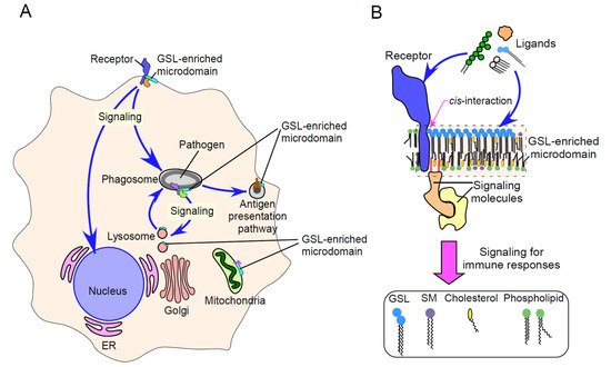
Glycosphingolipids (GSLs), together with cholesterol, sphingomyelin (SM), and glycosylphosphatidylinositol (GPI)-anchored and membrane-associated signal transduction molecules, form GSL-enriched microdomains. These specialized microdomains interact in a cis manner with various immune receptors, affecting immune receptor-mediated signaling.

  • glycosphingolipids
  • microdomain
  • immune signaling

1. Introduction

Membrane microdomains, also called lipid rafts, consist of glycosphingolipids (GSLs), cholesterol, sphingomyelin (SM), glycosylphosphatidylinositol (GPI)-anchored proteins and membrane-associated signal transduction molecules [1,2][1][2]. GSLs, a predominant component of microdomains, are characterized structurally by their hydrophobic ceramide and hydrophilic glycan moieties. The ceramide moiety contains fatty acid chains that vary widely in length [3]. More than 400 types of GSLs have been identified based on differences in their glycan structures [4]. Cell surface receptors bind ligands expressed on other cells (in trans) to communicate with neighboring cells, whereas a large number of cell surface receptors interact with ligands expressed on the same cell (in cis) [5]. The hydrophobic ceramide moiety enables GSLs to interact with the steroid ring system of cholesterol via van der Waals forces and hydrogen bonds [6,7][6][7]. In addition, the hydrophilic glycan moieties of GSLs interact in cis with each other, promoting the lateral interaction of GSLs with other components of cellular membranes. These interactions result in the phase separation of GSL-enriched membrane microdomains. GSL-enriched microdomains in the outer surfaces of membranes are able to associate with membrane proteins and lipid-anchored signal transduction molecules, which are localized in the inner surfaces of membranes [8,9,10][8][9][10]. These supramolecular complexes provide signaling platforms for cellular functions. The types of GSLs and their metabolism are not only cell type specific but also depend on whether the cells are proliferating or differentiating [4,11][4][11]. GSL-enriched microdomains are therefore thought to be involved in a large number of biological functions, including immunological functions (Figure 1 and Figure 2).
Figure 1. Schematic pathway of GSL biosynthesis. GSLs and the related molecules referred to in this review are shown.
Figure 2. Multiplicity of GSL-enriched microdomain-driven immune signaling. (A) Schematic image showing multiplicity of GSL-enriched microdomain-driven signaling in immune cells. In both innate and adaptive immunity, GSL-enriched microdomains affect immune signaling by themselves or by cis-interaction with various immune receptors. This results in various immune responses, such as cytokine production, phagocytosis/phagosome maturation, antigen presentation and apoptosis. GSL-enriched microdomains are present not only in plasma membranes but also in membranes of intracellular organelles, such as lysosomes and phagosomes. LacCer forms membrane microdomains on intracellular granules, including lysosomes, in human neutrophils, and these LacCer-enriched microdomains on phagosomes act as a platform of intracellular signaling required for phagosome maturation (fusion of lysosomes to pathogen-containing phagosomes). GD3 forms membrane microdomains on mitochondria-associated membranes (MAMs) and contributes to autophagosome assembly. ER, endoplasmic reticulum. Golgi, Golgi apparatus. (B) Schematic image showing cis-interactions between GSL-enriched microdomains and immune receptors. The transfer of signals induced by ligand binding into cells involves the direct binding of these ligands to GSL-enriched microdomains in plasma membranes, followed by the transduction of the signals through membrane-associated signal transduction molecules. In addition, signaling molecule-associated GSL-enriched microdomains interact in cis with various receptor proteins, leading to a variety of immune responses. Thus, GSL-enriched microdomains provide signaling platforms for ligand binding to the plasma membranes of immune cells. Intracellular GSL-enriched microdomains may provide platforms for cross-talk among several types of proteins, such as membrane-associated and signaling proteins and sphingolipid metabolites.

2. Physicochemical Properties of GSL-Enriched Microdomains

A biological membrane consisting of a lipid bilayer is often compared to a sea of phospholipids and cholesterol with floating sphingolipids and membrane proteins. Depending on their physicochemical properties, membrane components are distributed in a non-homogeneous manner throughout the cell membrane, leading to the formation of membrane microdomains that differ in molecular composition. These microdomains form supramolecular structures, which are stabilized by lateral intermolecular interactions. The properties of membrane components provide physical boundaries between the outside and the inside of cells. GSLs are specifically expressed in the outer layer of cell membranes [2]. GSLs can form clusters on cell membranes by lateral interactions based on their physicochemical properties, including hydrogen bonds from hydroxyl groups, the acetamide structure of the ceramide moiety and van der Waals interactions between hydrocarbon chains [11]. The physicochemical properties of GSLs suggest that they form defined clusters and that certain proteins cannot undergo free and continuous lateral diffusion in the membrane but rather are transiently confined to microdomains [4,12,13][4][12][13]. Because there are many difficulties evaluating GSL-enriched membrane microdomains in their original state, the actual state of GSL-enriched membrane microdomains remains unclear. Techniques to determine the structural and molecular arrangements of GSL-enriched membrane microdomains include single-molecule fluorescence tracking and electron microscopy. Although several GSL analogues have been generated by attaching a fluorescent label to the carbohydrate or lipid portion, it is unclear whether these analogues behave identically to natural unlabeled molecules [14]. Recently, hydrophilic fluorophore-conjugated analogues of gangliosides (sialylated GSLs), such as GM3, GM2 and GM1, were shown to be useful for the assessment of these microdomains, because these analogues retain binding specificity to their ligands [15,16][15][16]. Fluorescent-labeled SM analogues have been developed, consisting of hydrophilic fluorophores conjugated to the choline headgroup of SM via a hydrophilic nonaethylene glycol linker, which retain their positive charge [17]. These analogues have been shown to behave similarly to native SM in artificial liquid ordered (Lo)–disordered (Ld) phase-separated giant unilamellar vesicles (GUVs) and detergent-resistant membranes (DRMs) from cells.
The sizes of lipid microdomains range from 10 to 200 nm in diameter [1]. In comparison, GSL-enriched microdomains, consisting of GSLs that assemble laterally due to their physicochemical properties, have been reported to be 50–100 nm in diameter [18,19][18][19]. Immunoelectron microscopy showed that the neutral GSL lactosylceramide (LacCer, CDw17) forms microdomains with a diameter of about 43 nm in the plasma membranes of resting human neutrophils [18]. The phagocytosis of microorganisms by neutrophils induced the formation of larger LacCer-enriched microdomains, about 60 nm in diameter [20], on phagosomal membranes, suggesting that the reorganized LacCer-enriched microdomains provide platforms for transducing phagosome–lysosome fusion through molecular interactions, such as protein–lipid interactions. Single-molecule imaging with fluorescently labeled GPI-anchored proteins biophysically determined that these microdomains are dynamic domains with a diameter of about 10 nm [21,22][21][22]. Thus, the sizes of microdomains depend on the types of observed molecules. These differences may depend not only on the experimental methods used for observation but also on the physical properties of the observed molecules. Unlike GPI-anchored proteins, carbohydrates in GSLs are almost the same as ceramide, and the melting point of neutral GSLs is above 65 °C [23]. Therefore, the lateral interactions among GSLs appear to be very strong, enabling GSLs to pack together in plasma membranes. Acidic GSLs, such as gangliosides, have a much lower melting point than neutral GSLs. Moreover, the phase transition temperature of reconstructed ganglioside-enriched microdomains is around 40 °C, indicating that these microdomains can exist as distinct domains on phospholipid temperatures at body temperature [24]. Indeed, GSLs with different types of glycan structures have been shown to constitute distinct microdomains on the same cells and to have distinct functions [19,25][19][25]. Moreover, the interdigitation of long acyl chain SM in the outer leaflet and phosphatidylserine (PS) in the inner leaflet of the plasma membrane has been reported [26,27][26][27]. Interestingly, GM1 containing C16:0 but not C16:1 acyl chains was reported to form nanoclusters without the ligand cholera toxin B subunit (CTxB) [28]. Moreover, CTxB was found to induce the co-clustering of GM1 containing C16:0 with the GPI-anchored protein CD59, whereas such co-clustering did not occur with GM1 containing C16:1. These results suggest that the acyl chain structures of the ceramide moiety in GSLs also affect the properties of microdomains.

3. GSL-Enriched Microdomains as Regulators of Immune Receptor Signaling
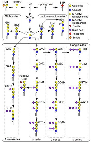
Following the binding of ligands to membrane receptors, the receptors undergo conformational changes, transferring signaling into the cells. During this process, GSL-enriched microdomains interact with various receptor proteins in cis, inducing physiological and immune system activities (Figure 1). This section describes the mechanisms by which receptors are regulated by GSL-enriched microdomains. The best known mechanisms are ganglioside–receptor protein interactions. Lateral interactions between GM3-enriched microdomains and insulin receptors (IRs) through a basic lysine residue (K944) lead to the inhibition of insulin-induced signaling in adipocytes [29]. Therefore, an increase in GM3 expression in cells may result in insulin resistance. Moreover, in the absence of epidermal growth factor (EGF), autophosphorylation of the EGF receptor (EGFR) is suppressed by its cis-interaction with GM3 through a lysine residue (K642) [30]. Adipogenic stimulation of adipose progenitor cells induces the accumulation of GM3- and caveolin 1-positive microdomains at the base of the primary cilia [31]. This accumulation, however, is reduced by the loss of trichoplein, resulting in the inhibition of IR/insulin-like growth factor-1 (IGF-1) receptor (IGF1R)–Akt signaling and protection from obesity and metabolic syndrome. Molecular dynamics (MD) simulations have shown that GM3 binds to the extracellular domain of the glucagon receptor, a class B1 G-protein-coupled receptor (GPCR), and modulates the dynamics of the extracellular domain, suggesting that GM3 plays a role in regulating the insulin/glucagon signaling ratio [32]. Thus, GM3-enriched microdomains appear to be critical for metabolic regulation. An evaluation of these GM3-receptor interactions suggested that an increase in GM3 content involved the glucose-induced inhibition of IGF1R–Rac1 signaling, affecting keratinocyte motility [33]. This finding suggests possible therapeutic approaches for treating wounds in patients with diabetes.
In addition to GM3, GM1, which is most abundant in neurons, interacts in cis with neurotrophin receptors [34] and modulates laminin-1-induced neurite outgrowth via TrkA and β1 integrin [35]. A study using tritium-labeled GM1 photoactivable derivatives suggested mechanisms by which GM1 interact in cis with receptors, such as TrkA [36]. The formation of the GM1–TrkA complex through those oligosaccharide interactions was found to promote neuroblastoma cell differentiation. In addition, GM1 was found to interact in cis with the GPCR serotonin-1A receptor [37]. Most (>90%) gangliosides in adult mammalian brains are composed of GM1, GD1a, GD1b and GT1b, which differ in the number and position of sialic acids linked to a common tetrasaccharide core [38]. GT1b is particularly recognized by botulinum neurotoxin type B (BoNT/B) [39], with a GT1b–synaptotagmin (SYT) cis-interactive molecular complex constituting a high-affinity BoNT/B receptor [40]. Similar to the cis-interactions of GM3 with IR and EGFR, a lysine residue (K52) on the SYT-juxtamembrane was found to be critical for the SYT–GT1b cis-interactions required for BoNT/B binding.
GSL-enriched microdomains are also associated with receptor-mediated immune signaling (Table 1). In innate immune responses, various pattern-recognition receptors (PRRs), including TLRs and integrins, are crucial for the detection of invading pathogens. Immune signaling is subsequently activated in neutrophils, macrophages and dendritic cells, resulting in pathogen removal. This elimination pathway is initiated by the binding of pathogen-derived molecules, called pathogen-associated molecular patterns (PAMPs), to PRRs expressed on phagocytes, leading to the formation of nascent phagosomes containing pathogens and their subsequent fusion to lysosomes. In particular, macrophages and dendritic cells are responsible for the antigen presentation of pathogen-derived molecules via major histocompatibility complex (MHC)-mediated pathways, resulting in the induction of acquired immune responses. TLRs are the best-characterized germline-encoded PRR proteins, and they transmit signals through several adaptor molecules [41]. TLR4 was the first member of the TLR family to be identified functionally [42]. TLR4 binds to lipopolysaccharide (LPS) with the support of GPI-anchored protein CD14, suggesting that TLR4 is activated at the sites of membrane microdomains [43]. The interaction of receptor molecules with membrane microdomains containing GM1 during LPS-induced cellular activation [44] was evaluated by fluorescence resonance energy transfer (FRET), which showed that TLR4 is associated with GM1-positive membrane microdomains after stimulation with LPS. A cholesterol-binding motif of TLR4 is regarded as critical for its translocation into membrane microdomains [45], suggesting that sphingolipids may not be involved in TLR4 signaling [46]. Recent atomistic molecular dynamics (MD) simulations regarding the TLR4 dimer complex showed that glucosylceramide (GlcCer) enhanced electrostatic interactions of the TLR4 extracellular domain with membranes [47]. This result suggests that the effects of GlcCer on LPS/TLR4 orientation affects LPS/TLR4 signaling through MyD88 adapter-like (Mal), also termed TIRAP. Thus, additional investigations are required to elucidate the mechanisms underlying the cis-interactions of TLR4 with membrane microdomain components. Cross-talk between neurotrophic receptors and TLR4 is also thought to be involved in neuroprotection mechanisms [48]. An extract from inflamed rabbit skin inoculated with vaccinia virus (Neurotropin®, NTP) was shown to control nerve growth factor (NGF) receptor TrkA-mediated TLR4-associated signaling through clusters of newly formed membrane microdomains in PC12 cells. GSL-enriched microdomains involve not only TLR4-mediated immune functions but also functions mediated by other members of the TLR family. For example, the binding of bacterial flagellin to asialoGM1 and TLR5 expressed on human lung epithelial cells was found to induce the autocrine release of ATP [49]. This released ATP binds to and activates ATP receptors in plasma membranes, leading to Ca2+ mobilization and Erk1/2 phosphorylation. GD1a on human monocytes binds to the subunit of type IIb Escherichia coli enterotoxin, promoting its interaction with the TLR2/TLR1 signaling complex and activating NF-κB [50].
Table 1. GSLs and receptor-mediated immune signaling.

GSLs

Co-Receptors

Cell Type

Immune Signaling

Ref. No.

GlcCer

TLR4

Macrophages

Impact on LPS/TLR4 orientation and Mal-associated signaling

[47]

GA1

TLR5

Lung epithelial cells NCIH292

Flagellin-mediated autocrine release of ATP

[49]

GD1a

TLR2/TLR1

Monocytes

LT-IIb-B5-mediated NFκB activation

[50]

LacCer

CD11b/CD18

Neutrophils

Lyn and Akt activations, and the resulting phagocytosis of zymosan and mycobacteria

[20,51]

[20][51]

Gb3Cer

CD59

Lung epithelial cells H1299

PIP3 and flotillin-associated uptake of P. aeruginosa

[52]

Neolacto-series GSLs

MHC class I

HAP1 cells

Interference of the accessibility of MHC class I molecules for immune cell receptors and the resulting suppression of CD8+ T-cell activation

[53]

GM1, GM3

CD4, LFA-1

T-cell line

PI3K and p56lck-associated T-cell responses

[54]

a-Series gangliosides

CD4, TCR

T cells

Helper T-cell activation

[55]

Asialo-series

gangliosides

CD8, TCR

T cells

Killer T-cell activation

[55]

GM1

IgM-BCR

Immature B cells

Removal of autoreactive immature B cells (apoptosis)

[56]

GM3

CD95/Fas

T cells

Formation of death-inducing signaling complex upon CD95/Fas engagement (apoptosis)

[57]

4. GSL-Enriched Microdomains in Immune Functions

Several properties of neutrophils, including adhesion, migration and phagocytosis, are modulated by the CD11b/CD18 integrin, also called Mac-1, CR3 or αMβ2 integrin [58]. Despite CD11b/CD18 cytoplasmic regions lacking catalytic activity, CD11b/CD18 can transduce signals inside the cells [59]. These results suggest that CD11b/CD18-mediated outside-in signaling requires partner molecules as a signaling platform. Microdomains enriched in the neutral GSL LacCer can not only bind microbial ligands but can act as a signaling platform. That is, LacCer-enriched microdomains are able to act as a PRR for pathogens. Indeed, LacCer was found to bind to various pathogens and their PAMPs, including Candida albicans-derived β-glucan (CSBG) and mycobacterial lipoarabinomannan (LAM) [20[20][60],60], and to form membrane microdomains coupled to various signaling molecules, including the Src family kinase Lyn, through their very long C24 fatty acid chains, transducing ligand-binding signals to the inside of the cells [18]. In addition, Lyn-coupled LacCer-enriched microdomains serve as a platform for CD11b/CD18-dependent outside-in and phagocytic signaling in human neutrophils [20,51,61][20][51][61]. LacCer-enriched microdomains are likely to interact in cis with the extracellular juxtamembrane region of CD18 [51]. In lung epithelial cells, the interaction of the Pseudomonas aeruginosa-derived virulence factor LecA with globotriaosylceramide (Gb3Cer) in the outer leaflet of plasma membranes was found to induce the formation of membrane domains enriched in saturated long fatty acyl chain-containing Gb3Cer species, the GPI-anchored protein CD59, phosphatidylinositol (3,4,5)-trisphosphate (PIP3) and flotillin, thereby promoting the efficient uptake of P. aeruginosa [52]. Mechanisms were suggested by which Gb3Cer and its associated molecules mediate signal transduction from extracellular to intracellular sites through transbilayer coupling. Thus, by providing a signaling platform, several types of GSL-enriched microdomains can regulate the function of receptors that lack catalytic moieties, such as CD11b/CD18. Moreover, the structures of GSLs, especially their fatty acid chains, may be a key component of GSL-enriched microdomains that act as signaling platforms.
GSLs may also be involved in MHC-mediated antigen presentation pathways. For example, MHC class II molecules have been reported to contain possible Gb3Cer binding sites [62]. Results suggested that Gb3Cer modulates MHC class II-mediated antigen presentation from B cells to helper T cells, although the molecular mechanisms of Gb3Cer-MHC class II binding are still unknown. Tumors, however, may limit MHC class I-mediated antigen presentation [63]. A recent study investigating the roles of GSLs and related enzymes in MHC class I pathways using genome-wide haploid genetic screening and CRISPR/Cas9 systems [53] found that, in the absence of signal peptide peptidase-like 3 (SPPL3) protease, high amounts of negatively charged neolacto-series GSLs interfere with the accessibility of MHC class I molecules for immune cell receptors, suppressing the activation of CD8+ T cells. In this pathway, SPPL3 catabolizes the glycosyltransferase B3GNT5, which generates neolacto-series GSLs, and controls the ability of MHC class I molecules to access their receptors. Thus, neolacto-series GSLs may affect antigen presentation and help tumor cells escape from immune surveillance [63]. In addition, the sialic acid residues on GSLs were found to be critical for MHC class I shielding [53]. The molecular basis of the interactions between GSL-enriched microdomains and antigen presentation-related molecules may provide critical information that can help in the treatment of immune disorders.
In addition to their roles in innate immune signaling, GSL-enriched microdomains are also essential in acquired immune signaling. During T-cell responses, lymphocyte function-associated antigen-1 (LFA-1) moves into membrane microdomains upon CD4 ligation, becoming associated mainly with GM3 [54]. At this time, phosphoinositide 3-kinase (PI3K) is mainly associated with GM1, and its association with p56lck was increased. During the reaction, LFA-1 becomes primarily associated with GM1. However, the mechanisms by which these gangliosides interact (directly or indirectly) with protein molecules remain unknown. a-Series gangliosides and asialo-series gangliosides have been implicated in the function and stimulation of T-cell receptors (TCRs) on CD4-positive (CD4+) and CD8-positive (CD8+) T cells, respectively [55], indicating the possibility that CD4 and CD8 interact with a-series and asialo-series gangliosides, respectively, through their common glycan structures [55,64][55][64]. Similarly, the ceramide structures of gangliosides may participate in these interactions. Although future studies are needed to address these possibilities, individual gangliosides may be involved in the movement of CD4 and CD8 to specific and correct locations in cell membranes [55,64][55][64].
Notch signaling is critical for T-cell development in the thymus [65]. Notch ligands, such as Dll1, may interact with GSLs through their GSL-binding motif [66], suggesting that the molecular interactions of protein receptors and ligands with GSLs may be associated with the regulation of T-cell functions. T cells play important roles in the pathogenesis of various autoimmune diseases, including systemic lupus erythematosus (SLE) [67]. GSL expression is dysregulated in CD4+ T cells from patients with SLE [68], and T cells from these patients show alternations in GSL recycling and turnover. Thus, GSL-enriched microdomains may be implicated in the pathogenesis of autoimmune diseases. GSL-enriched microdomains interact with B-cell receptors (BCRs) as well as participating in TCR-mediated functions. Indeed, GM1-enriched microdomains associated with BCR signaling may be critical for ganglioside-related B lymphocyte functions [56,69,70,71][56][69][70][71]. BCRs are indispensable for the B-cell clonal selection process and their differentiation into plasma cells. GM1-enriched microdomains may be involved in the compartmentalization of different types of BCRs, such as IgM and IgD types, expressed on mature B cells [56,70][56][70], although the mechanisms by which GM1-enriched microdomains modulate BCR signaling remain to be determined.

References

  1. Pike, L.J. Rafts defined: A report on the Keystone Symposium on Lipid Rafts and Cell Function. J. Lipid Res. 2006, 47, 1597–1598.
  2. Murate, M.; Abe, M.; Kasahara, K.; Iwabuchi, K.; Umeda, M.; Kobayashi, T. Transbilayer distribution of lipids at nano scale. J. Cell Sci. 2015, 128, 1627–1638.
  3. Kaga, N.; Kazuno, S.; Taka, H.; Iwabuchi, K.; Murayama, K. Isolation and mass spectrometry characterization of molecular species of lactosylceramides using liquid chromatography-electrospray ion trap mass spectrometry. Anal. Biochem. 2005, 337, 316–324.
  4. Hakomori, S. Structure, organization, and function of glycosphingolipids in membrane. Curr. Opin. Hematol. 2003, 10, 16–24.
  5. Held, W.; Mariuzza, R.A. Cis-trans interactions of cell surface receptors: Biological roles and structural basis. Cell Mol. Life Sci. 2011, 68, 3469–3478.
  6. Hakomori, S. Glycosphingolipids in cellular interaction, differentiation, and oncogenesis. Annu. Rev. Biochem. 1981, 50, 733–764.
  7. Mukherjee, S.; Maxfield, F.R. Membrane domains. Annu. Rev. Cell Dev. Biol. 2004, 20, 839–866.
  8. Brown, D.A.; London, E. Structure of detergent-resistant membrane domains: Does phase separation occur in biological membranes? Biochem. Biophys. Res. Commun. 1997, 240, 1–7.
  9. Iwabuchi, K.; Handa, K.; Hakomori, S. Separation of “glycosphingolipid signaling domain” from caveolin-containing membrane fraction in mouse melanoma B16 cells and its role in cell adhesion coupled with signaling. J. Biol Chem. 1998, 273, 33766–33773.
  10. Iwabuchi, K.; Yamamura, S.; Prinetti, A.; Handa, K.; Hakomori, S. GM3-enriched microdomain involved in cell adhesion and signal transduction through carbohydrate-carbohydrate interaction in mouse melanoma B16 cells. J. Biol. Chem. 1998, 273, 9130–9138.
  11. Sonnino, S.; Prinetti, A.; Mauri, L.; Chigorno, V.; Tettamanti, G. Dynamic and structural properties of sphingolipids as driving forces for the formation of membrane domains. Chem. Rev. 2006, 106, 2111–2125.
  12. Jacobson, K.; Sheets, E.D.; Simson, R. Revisiting the fluid mosaic model of membranes. Science 1995, 268, 1441–1442.
  13. Hakomori, S.; Handa, K.; Iwabuchi, K.; Yamamura, S.; Prinetti, A. New insights in glycosphingolipid function: “glycosignaling domain”, a cell surface assembly of glycosphingolipids with signal transducer molecules, involved in cell adhesion coupled with signaling. Glycobiology 1998, 8, xi–xix.
  14. Singh, R.D.; Puri, V.; Valiyaveettil, J.T.; Marks, D.L.; Bittman, R.; Pagano, R.E. Selective caveolin-1-dependent endocytosis of glycosphingolipids. Mol. Biol. Cell 2003, 14, 3254–3265.
  15. Komura, N.; Suzuki, K.G.; Ando, H.; Konishi, M.; Koikeda, M.; Imamura, A.; Chadda, R.; Fujiwara, T.K.; Tsuboi, H.; Sheng, R.; et al. Raft-based interactions of gangliosides with a GPI-anchored receptor. Nat. Chem. Biol. 2016, 12, 402–410.
  16. Kusumi, A.; Fujiwara, T.K.; Tsunoyama, T.A.; Kasai, R.S.; Liu, A.A.; Hirosawa, K.M.; Kinoshita, M.; Matsumori, N.; Komura, N.; Ando, H.; et al. Defining raft domains in the plasma membrane. Traffic 2020, 21, 106–137.
  17. Kinoshita, M.; Suzuki, K.G.; Matsumori, N.; Takada, M.; Ano, H.; Morigaki, K.; Abe, M.; Makino, A.; Kobayashi, T.; Hirosawa, K.M.; et al. Raft-based sphingomyelin interactions revealed by new fluorescent sphingomyelin analogs. J. Cell Biol. 2017, 216, 1183–1204.
  18. Iwabuchi, K.; Prinetti, A.; Sonnino, S.; Mauri, L.; Kobayashi, T.; Ishii, K.; Kaga, N.; Murayama, K.; Kurihara, H.; Nakayama, H.; et al. Involvement of very long fatty acid-containing lactosylceramide in lactosylceramide-mediated superoxide generation and migration in neutrophils. Glycoconj. J. 2008, 25, 357–374.
  19. Fujita, A.; Cheng, J.; Fujimoto, T. Segregation of GM1 and GM3 clusters in the cell membrane depends on the intact actin cytoskeleton. Biochim. Biophys. Acta 2009, 1791, 388–396.
  20. Nakayama, H.; Kurihara, H.; Morita, Y.S.; Kinoshita, T.; Mauri, L.; Prinetti, A.; Sonnino, S.; Yokoyama, N.; Ogawa, H.; Takamori, K.; et al. Lipoarabinomannan binding to lactosylceramide in lipid rafts is essential for the phagocytosis of mycobacteria by human neutrophils. Sci. Signal. 2016, 9, ra101.
  21. Pralle, A.; Keller, P.; Florin, E.L.; Simons, K.; Horber, J.K. Sphingolipid-cholesterol rafts diffuse as small entities in the plasma membrane of mammalian cells. J. Cell Biol. 2000, 148, 997–1008.
  22. Suzuki, K.G.; Fujiwara, T.K.; Sanematsu, F.; Iino, R.; Edidin, M.; Kusumi, A. GPI-anchored receptor clusters transiently recruit Lyn and G alpha for temporary cluster immobilization and Lyn activation: Single-molecule tracking study 1. J. Cell Biol. 2007, 177, 717–730.
  23. Saxena, K.; Zimmermann, P.; Schmidt, R.R.; Shipley, G.G. Bilayer properties of totally synthetic C16:0-lactosyl-ceramide. Biophys. J. 2000, 78, 306–312.
  24. Ferraretto, A.; Pitto, M.; Palestini, P.; Masserini, M. Lipid domains in the membrane: Thermotropic properties of sphingomyelin vesicles containing GM1 ganglioside and cholesterol. Biochemistry 1997, 36, 9232–9236.
  25. Iwabuchi, K.; Nagaoka, I. Lactosylceramide-enriched glycosphingolipid signaling domain mediates superoxide generation from human neutrophils. Blood 2002, 100, 1454–1464.
  26. Róg, T.; Orłowski, A.; Llorente, A.; Skotland, T.; Sylvänne, T.; Kauhanen, D.; Ekroos, K.; Sandvig, K.; Vattulainen, I. Interdigitation of long-chain sphingomyelin induces coupling of membrane leaflets in a cholesterol dependent manner. Biochim. Biophys. Acta 2016, 1858, 281–288.
  27. Skotland, T.; Sandvig, K. The role of PS 18:0/18:1 in membrane function. Nat. Commun. 2019, 10, 2752.
  28. Arumugam, S.; Schmieder, S.; Pezeshkian, W.; Becken, U.; Wunder, C.; Chinnapen, D.; Ipsen, J.H.; Kenworthy, A.K.; Lencer, W.; Mayor, S.; et al. Ceramide structure dictates glycosphingolipid nanodomain assembly and function. Nat. Commun. 2021, 12, 3675.
  29. Kabayama, K.; Sato, T.; Saito, K.; Loberto, N.; Prinetti, A.; Sonnino, S.; Kinjo, M.; Igarashi, Y.; Inokuchi, J. Dissociation of the insulin receptor and caveolin-1 complex by ganglioside GM3 in the state of insulin resistance. Proc. Natl. Acad. Sci. USA 2007, 104, 13678–13683.
  30. Coskun, U.; Grzybek, M.; Drechsel, D.; Simons, K. Regulation of human EGF receptor by lipids. Proc. Natl. Acad. Sci. USA 2011, 108, 9044–9048.
  31. Yamakawa, D.; Katoh, D.; Kasahara, K.; Shiromizu, T.; Matsuyama, M.; Matsuda, C.; Maeno, Y.; Watanabe, M.; Nishimura, Y.; Inagaki, M. Primary cilia-dependent lipid raft/caveolin dynamics regulate adipogenesis. Cell Rep. 2021, 34, 108817.
  32. Ansell, T.B.; Song, W.; Sansom, M.S.P. The Glycosphingolipid GM3 Modulates Conformational Dynamics of the Glucagon Receptor. Biophys. J. 2020, 119, 300–313.
  33. Dam, D.H.M.; Wang, X.Q.; Sheu, S.; Vijay, M.; Shipp, D.; Miller, L.; Paller, A.S. Ganglioside GM3 Mediates Glucose-Induced Suppression of IGF-1 Receptor-Rac1 Activation to Inhibit Keratinocyte Motility. J. Investig. Dermatol. 2017, 137, 440–448.
  34. Mutoh, T.; Tokuda, A.; Miyadai, T.; Hamaguchi, M.; Fujiki, N. Ganglioside GM1 binds to the Trk protein and regulates receptor function. Proc. Natl. Acad. Sci. USA 1995, 92, 5087–5091.
  35. Ichikawa, N.; Iwabuchi, K.; Kurihara, H.; Ishii, K.; Kobayashi, T.; Sasaki, T.; Hattori, N.; Mizuno, Y.; Hozumi, K.; Yamada, Y.; et al. Binding of laminin-1 to monosialoganglioside GM1 in lipid rafts is crucial for neurite outgrowth. J. Cell Sci. 2009, 122, 289–299.
  36. Chiricozzi, E.; Biase, E.D.; Maggioni, M.; Lunghi, G.; Fazzari, M.; Pome, D.Y.; Casellato, R.; Loberto, N.; Mauri, L.; Sonnino, S. GM1 promotes TrkA-mediated neuroblastoma cell differentiation by occupying a plasma membrane domain different from TrkA. J. Neurochem. 2019, 149, 231–241.
  37. Prasanna, X.; Jafurulla, M.; Sengupta, D.; Chattopadhyay, A. The ganglioside GM1 interacts with the serotonin1A receptor via the sphingolipid binding domain. Biochim. Biophys. Acta. 2016, 1858, 2818–2826.
  38. Schnaar, R.L. Gangliosides of the Vertebrate Nervous System. J. Mol. Biol. 2016, 428, 3325–3336.
  39. Chai, Q.; Arndt, J.W.; Dong, M.; Tepp, W.H.; Johnson, E.A.; Chapman, E.R.; Stevens, R.C. Structural basis of cell surface receptor recognition by botulinum neurotoxin B. Nature 2006, 444, 1096–1100.
  40. Flores, A.; Ramirez-Franco, J.; Desplantes, R.; Debreux, K.; Ferracci, G.; Wernert, F.; Blanchard, M.P.; Maulet, Y.; Youssouf, F.; Sangiardi, M.; et al. Gangliosides interact with synaptotagmin to form the high-affinity receptor complex for botulinum neurotoxin B. Proc. Natl. Acad. Sci. USA 2019, 116, 18098–18108.
  41. Brubaker, S.W.; Bonham, K.S.; Zanoni, I.; Kagan, J.C. Innate immune pattern recognition: A cell biological perspective. Annu. Rev. Immunol. 2015, 33, 257–290.
  42. Medzhitov, R.; Preston-Hurlburt, P.; Janeway, C.A., Jr. A human homologue of the Drosophila Toll protein signals activation of adaptive immunity. Nature 1997, 388, 394–397.
  43. Ciesielska, A.; Matyjek, M.; Kwiatkowska, K. TLR4 and CD14 trafficking and its influence on LPS-induced pro-inflammatory signaling. Cell. Mol. Life Sci. 2021, 78, 1233–1261.
  44. Triantafilou, M.; Miyake, K.; Golenbock, D.T.; Triantafilou, K. Mediators of innate immune recognition of bacteria concentrate in lipid rafts and facilitate lipopolysaccharide-induced cell activation. J. Cell Sci. 2002, 115, 2603–2611.
  45. Ruysschaert, J.M.; Lonez, C. Role of lipid microdomains in TLR-mediated signalling. Biochim. Biophys. Acta 2015, 1848, 1860–1867.
  46. Carroll, R.G.; Zaslona, Z.; Galvan-Pena, S.; Koppe, E.L.; Sevin, D.C.; Angiari, S.; Triantafilou, M.; Triantafilou, K.; Modis, L.K.; O’Neill, L.A. An unexpected link between fatty acid synthase and cholesterol synthesis in proinflammatory macrophage activation. J. Biol. Chem. 2018, 293, 5509–5521.
  47. Mobarak, E.; Haversen, L.; Manna, M.; Rutberg, M.; Levin, M.; Perkins, R.; Rog, T.; Vattulainen, I.; Boren, J. Glucosylceramide modifies the LPS-induced inflammatory response in macrophages and the orientation of the LPS/TLR4 complex in silico. Sci. Rep. 2018, 8, 13600.
  48. Fukuda, Y.; Nakajima, K.; Mutoh, T. Neuroprotection by Neurotropin through Crosstalk of Neurotrophic and Innate Immune Receptors in PC12 Cells. Int. J. Mol. Sci. 2020, 21, 6456.
  49. McNamara, N.; Gallup, M.; Sucher, A.; Maltseva, I.; McKemy, D.; Basbaum, C. AsialoGM1 and TLR5 cooperate in flagellin-induced nucleotide signaling to activate Erk1/2. Am. J. Respir. Cell Mol. Biol. 2006, 34, 653–660.
  50. Liang, S.; Wang, M.; Tapping, R.I.; Stepensky, V.; Nawar, H.F.; Triantafilou, M.; Triantafilou, K.; Connell, T.D.; Hajishengallis, G. Ganglioside GD1a is an essential coreceptor for Toll-like receptor 2 signaling in response to the B subunit of type IIb enterotoxin. J. Biol. Chem. 2007, 282, 7532–7542.
  51. Nakayama, H.; Yoshizaki, F.; Prinetti, A.; Sonnino, S.; Mauri, L.; Takamori, K.; Ogawa, H.; Iwabuchi, K. Lyn-coupled LacCer-enriched lipid rafts are required for CD11b/CD18-mediated neutrophil phagocytosis of nonopsonized microorganisms. J. Leukoc. Biol. 2008, 83, 728–741.
  52. Brandel, A.; Aigal, S.; Lagies, S.; Schlimpert, M.; Melendez, A.V.; Xu, M.; Lehmann, A.; Hummel, D.; Fisch, D.; Madl, J.; et al. The Gb3-enriched CD59/flotillin plasma membrane domain regulates host cell invasion by Pseudomonas aeruginosa. Cell. Mol. Life Sci. 2021, 78, 3637–3656.
  53. Jongsma, M.L.M.; de Waard, A.A.; Raaben, M.; Zhang, T.; Cabukusta, B.; Platzer, R.; Blomen, V.A.; Xagara, A.; Verkerk, T.; Bliss, S.; et al. The SPPL3-Defined Glycosphingolipid Repertoire Orchestrates HLA Class I-Mediated Immune Responses. Immunity 2021, 54, 132–150.e139.
  54. Barbat, C.; Trucy, M.; Sorice, M.; Garofalo, T.; Manganelli, V.; Fischer, A.; Mazerolles, F. p56lck, LFA-1 and PI3K but not SHP-2 interact with GM1- or GM3-enriched microdomains in a CD4-p56lck association-dependent manner. Biochem. J. 2007, 402, 471–481.
  55. Nagafuku, M.; Okuyama, K.; Onimaru, Y.; Suzuki, A.; Odagiri, Y.; Yamashita, T.; Iwasaki, K.; Fujiwara, M.; Takayanagi, M.; Ohno, I.; et al. CD4 and CD8 T cells require different membrane gangliosides for activation. Proc. Natl. Acad. Sci. USA 2012, 109, E336–E342.
  56. Minguet, S.; Klasener, K.; Schaffer, A.M.; Fiala, G.J.; Osteso-Ibanez, T.; Raute, K.; Navarro-Lerida, I.; Hartl, F.A.; Seidl, M.; Reth, M.; et al. Caveolin-1-dependent nanoscale organization of the BCR regulates B cell tolerance. Nat. Immunol. 2017, 10, 1150–1159.
  57. Garofalo, T.; Misasi, R.; Mattei, V.; Giammarioli, A.M.; Malorni, W.; Pontieri, G.M.; Pavan, A.; Sorice, M. Association of the death-inducing signaling complex with microdomains after triggering through CD95/Fas. Evidence for caspase-8-ganglioside interaction in T cells. J. Biol. Chem. 2003, 278, 8309–8315.
  58. Lamers, C.; Pluss, C.J.; Ricklin, D. The Promiscuous Profile of Complement Receptor 3 in Ligand Binding, Immune Modulation, and Pathophysiology. Front. Immunol. 2021, 12, 662164.
  59. Rabb, H.; Michishita, M.; Sharma, C.P.; Brown, D.; Arnaout, M.A. Cytoplasmic tails of human complement receptor type 3 (CR3, CD11b/CD18) regulate ligand avidity and the internalization of occupied receptors. J. Immunol. 1993, 151, 990–1002.
  60. Sato, T.; Iwabuchi, K.; Nagaoka, I.; Adachi, Y.; Ohno, N.; Tamura, H.; Seyama, K.; Fukuchi, Y.; Nakayama, H.; Yoshizaki, F.; et al. Induction of human neutrophil chemotaxis by Candida albicans-derived beta-1,6-long glycoside side-chain-branched beta-glucan. J. Leukoc. Biol. 2006, 80, 204–211.
  61. Chiricozzi, E.; Ciampa, M.G.; Brasile, G.; Compostella, F.; Prinetti, A.; Nakayama, H.; Ekyalongo, R.C.; Iwabuchi, K.; Sonnino, S.; Mauri, L. Direct interaction, instrumental for signaling processes, between LacCer and Lyn in the lipid rafts of neutrophil-like cells. J. Lipid Res. 2015, 56, 129–141.
  62. George, T.; Boyd, B.; Price, M.; Lingwood, C.; Maloney, M. MHC class II proteins contain a potential binding site for the verotoxin receptor glycolipid CD77. Cell. Mol. Biol. 2001, 47, 1179–1185.
  63. Jongsma, M.L.M.; Neefjes, J.; Spaapen, R.M. Playing hide and seek: Tumor cells in control of MHC class I antigen presentation. Mol. Immunol. 2021, 136, 36–44.
  64. Nakayama, H.; Nagafuku, M.; Suzuki, A.; Iwabuchi, K.; Inokuchi, J.I. The regulatory roles of glycosphingolipid-enriched lipid rafts in immune systems. FEBS Lett. 2018, 592, 3921–3942.
  65. Ciofani, M.; Zuniga-Pflucker, J.C. The thymus as an inductive site for T lymphopoiesis. Annu. Rev. Cell Dev. Biol. 2007, 23, 463–493.
  66. Heuss, S.F.; Tarantino, N.; Fantini, J.; Ndiaye-Lobry, D.; Moretti, J.; Israel, A.; Logeat, F. A glycosphingolipid binding domain controls trafficking and activity of the mammalian notch ligand delta-like 1. PLoS ONE 2013, 8, e74392.
  67. Sharabi, A.; Tsokos, G.C. T cell metabolism: New insights in systemic lupus erythematosus pathogenesis and therapy. Nat. Rev. Rheumatol. 2020, 16, 100–112.
  68. McDonald, G.; Deepak, S.; Miguel, L.; Hall, C.J.; Isenberg, D.A.; Magee, A.I.; Butters, T.; Jury, E.C. Normalizing glycosphingolipids restores function in CD4+ T cells from lupus patients. J. Clin. Investig. 2014, 124, 712–724.
  69. Kimata, H. GM1, a ganglioside that specifically enhances immunoglobulin production and proliferation in human plasma cells. Eur. J. Immunol. 1994, 24, 2910–2913.
  70. Klasener, K.; Maity, P.C.; Hobeika, E.; Yang, J.; Reth, M. B cell activation involves nanoscale receptor reorganizations and inside-out signaling by Syk. eLife 2014, 3, e02069.
  71. Shrestha, D.; Exley, M.A.; Vereb, G.; Szollosi, J.; Jenei, A. CD1d favors MHC neighborhood, GM1 ganglioside proximity and low detergent sensitive membrane regions on the surface of B lymphocytes. Biochim. Biophys. Acta. 2014, 1840, 667–680.
More