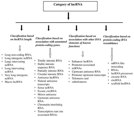
Long noncoding RNAs (lncRNAs) are the largest groups of ribonucleic acids, but, despite the increasing amount of literature data, the least understood. Given the involvement of lncRNA in basic cellular processes, especially in the regulation of transcription, the role of these noncoding molecules seems to be of great importance for the proper functioning of the organism. Studies have shown a relationship between disturbed lncRNA expression and the pathogenesis of many diseases, including cancer.
- tumors
- breast cancer
- lncRNA
- expression
1. lncRNA—History
2. lncRNA—Characteristics

3. lncRNA—Functions
lncRNA affects the transcription process directly, acting as an enhancer, by “stopping” transcription factors, and by affecting chromatin looping and gene methylation (using epigenetic complexes such as PCR2) [21][9].
4. lncRNA and Malignant Tumors
The characteristics of long noncoded RNA described above—such as tissue or cellular specificity and the regulation of gene expression at the transcriptional and post-transcriptional levels—indicate that lncRNAs may be important in the formation of malignant tumors. Studies have shown that lncRNAs affect the pathways of division, growth, and cell differentiation, and are also involved in cellular death processes [21,22][9][11]. Modifications to these processes may lead to carcinogenesis [22][11]. Moreover, some lncRNAs are regulated by oncogene products or cancer transformation suppressors, which means that they are believed to indirectly perform tumorigenic functions (Table 1) [22][11].
| lncRNA | Genomic Location | Expression in Patients | Function in Tumorigenesis |
|---|---|---|---|
| PCGEM1 | 2q32.2 | Increased in prostate cancer | oncogene |
| MALAT1 | 11q13.1 | Increased in colon, lung, and liver cancers | oncogene |
| MEG3 | 14q32.2 | Down-regulated in multiple cancers | tumor suppressor |
| HOTAIR | 12q13.13 | Increased in primary breast tumors and metastases, GIST, and pancreatic cancers |
oncogene |
The first observed transcripts of altered expression in the tissue of a malignant tumor—prostate cancer—were PCA3 and PCGEM1. PCA3 currently functions as a cancer marker [22][11]. The previously mentioned MALAT1 was also discovered in cancerous tissue as one of the first lncRNAs. Of prognostic significance, its altered expression was discovered in the tissues of lung cancer [24][12]. It is now known that altered expression of this transcript occurs in many cancers, which may indicate its importance in the process of cellular proliferation [25][13].
DNA damage, a specific accumulation of which is observed in cancer cells, causes activation of the transcription factor p53. This factor, depending on the degree of DNA damage, induces apoptosis or halts the cell cycle for the duration of repair. One element of the proapoptotic pathway or the suspension of cell division is an increase in lncRNA transcription through the p53 factor. The resulting transcripts are involved in the regulation of these pathways and thus modulate responses to cellular stress [22,26,27][11][14][15]. On the other hand, some lncRNAs affect p53 function by interacting with the gene enhancers of this protein [28][16]. Finally, the MEG3 transcript—a reduced expression of which has been found in many malignancies [22][11]—activates factor p53 itself [26,29][14][17]. There are also many lncRNA genes in the region that, like the MYC oncogene, are translated more intensively [22][11]. Some of these lncRNAs regulate expression of the MYC gene in a cis manner [22][11]. On the other hand, expression of lncRNAs from the described region, involved in repression of the genes regulating the cell cycle according to MYC, is modified by the aforementioned proto-oncogene [22,30,31][11][18][19]. Studies have shown that the expression of oncogene and tumor transformation suppressors in malignant tumor tissues is increased and decreased, respectively [32][20]. This suggests that the interaction between lncRNAs and oncogenes or tumor suppressors is an important mechanism that contributes to the initiation of carcinogenesis. Another important mechanism seems to be the effect of lncRNAs on chromatin-modulating complexes through epigenetic changes, as an accumulation of epigenetic DNA modifications is very common in malignant tumors [33][21]. Long noncoding RNA can also affect cell homeostasis by post-transcriptional regulation; mRNA splicing, processing, and translation; or post-translational protein modification [22][11]. In addition, it has an impact on mRNA by binding microRNAs, reducing the amount of free short RNA and thereby reducing their impact on encoding transcripts [22][11]. The proper functioning of the cell is conditioned by the balance of metabolic processes. Studies show that lncRNAs are involved in the basic pathways of cellular metabolism, including in cancer cells, e.g., in the production of ATP under hypoxic conditions via HIF-1alpha factor or the Warburg effect [26][14]. A characteristic feature of malignant tumors is the ability to bypass immune control. Reports indicate that lncRNAs are involved in regulating the immune response by modifying the activity of immunocompetent cells [38][22]. For this reason, lncRNAs appear likely to be involved in the formation of immunomicelles of malignant tumors, even improving their defenses against the immune system [26,39][14][23]. It was mentioned above that overexpression of the HOTAIR transcript, which connects to the PCR2 complex, contributes to the formation of metastasis. Long noncoding RNAs also support the metastatic process by participating in epithelial–mesenchymal transition (EMT), as well as in signaling pathways associated with the activation of cancer stem cells [26][14].65. lncRNA—T-UCRs
Of those lncRNAs whose expression is altered in the tissues of malignant tumors, including breast cancer, some transcripts arise from so-called ultraconservative regions (UCRs). UCRs are genomic sequences that have survived evolution and are 100% compatible between the orthologous regions of humans, mice, and rats [3,52][3][24]. Of the 481 UCRs discovered, 111 coincide with sequences of genes encoding a human protein (exonic UCRs), 256 bear no resemblance to either the coding sequence or the resulting mRNA (nonexonic UCRs), and, for the remaining 114, insufficient evidence has been obtained to determine whether they are transcribed (possibly exonic UCRs) [52][24]. Of all known UCRs, 39% are intergenically located, 43% are found in intron sequences (including one hundred nonexonic UCRs), and 15% are in exon sequences [3]. Nonexonic UCRs, both intronic and intergenic, often form clusters near the genes of transcription factors and developmental proteins, but are also often found in so-called “gene deserts”, i.e., long, noncoding sections of DNA [53][25].76. T-UCRs and Malignant Tumors
The importance of T-UCRs in the process of carcinogenesis also remains unknown, although they are associated with oncogenes and suppressors of tumor transformation; participate in the apoptosis, proliferation, and migration of cancer cells; and also seem to regulate microRNA functions [43,54][26][27]. Moreover, studies have shown that transcription of certain T-UCRs is induced by hypoxia [55][28]. Despite the lack of relevant literature, there is evidence of a changed expression of T-UCRs in malignant tumor tissues. One of the first studies confirming the presence of a changed number, compared with healthy tissues, of numerous T-UCRs in cancerous tissues—including cancer cells of chronic lymphocytic leukemia and tissues of colorectal and hepatocellular carcinoma—was that of Calin et al., published in 2007 [53,55][25][28]. In 2010, a changed expression of T-UCRs was also discovered in neuroblastoma tissues [56][29].97. Conclusive Summary
References
- Lander, E.S.; Linton, L.M.; Birren, B.; Nusbaum, C.; Zody, M.C.; Baldwin, J.; Devon, K.; Dewar, K.; Doyle, M.; FitzHugh, W.; et al. Inter-national human genome sequencing consortium. Initial sequencing and analysis of the human ge-nome. Nature 2001, 409, 860–921.
- Venter, J.C.; Adams, M.D.; Myers, E.W.; Li, P.W.; Mural, R.J.; Sutton, G.G.; Smith, H.O.; Yandell, M.; Evans, C.A.; Holt, R.A.; et al. The sequence of the human genome. Science 2001, 291, 1304–1351.
- Jarroux, J.; Morillon, A.; Pinskaya, M. Discovery, and Classification of lncRNAs. Adv. Exp. Med. Biol. 2017, 1008, 1–46.
- Brannan, C.I.; Dees, E.C.; Ingram, R.S.; Tilghman, S.M. The product of the H19 gene may function as an RNA. Mol. Cell. Biol. 1990, 10, 28–36.
- XIST Gene—GeneCards|XIST RNA Gene. Weizmann Institute of Science. Available online: https://www.genecards.org/cgi-bin/carddisp.pl?gene=XIST&keywords=xist (accessed on 4 August 2021).
- Lyon, M.F. Gene action in the X-chromosome of the mouse (Mus musculus L.). Nature 1961, 190, 372–373.
- Ayupe, A.C.; Tahira, A.C.; Camargo, L.; Beckedorff, F.C.; Verjovski-Almeida, S.; Reis, E.M. Global analysis of biogenesis, stability and sub-cellular localization of lncRNAs mapping to intragenic re-gions of the human genome. RNA Biol. 2015, 12, 877–892.
- Giannakakis, A.; Zhang, J.; Jenjaroenpun, P.; Nama, S.; Zainolabidin, N.; Aau, M.Y.; Yarmishyn, A.A.; Vaz, C.; Ivshina, A.V.; Grinchuk, O.V.; et al. Contrasting expression patterns of coding and noncoding parts of the human genome upon oxidative stress. Sci. Rep. 2015, 5, 9737.
- Schmitt, A.M.; Chang, H.Y. Long noncoding RNAs in cancer pathways. Cancer Cell 2016, 29, 452–463.
- Kopp, F.; Mendell, J.T. Functional classification and experimental dissection of long noncoding RNAs. Cell 2018, 172, 393–407.
- Huarte, M. The emerging role of lncRNAs in cancer. Nat. Med. 2015, 21, 1253–1261.
- Ji, P.; Diederichs, S.; Wang, W.; Böing, S.; Metzger, R.; Schneider, P.M.; Tidow, N.; Brandt, B.; Buerger, H.; Bulk, E.; et al. MALAT-1, a novel noncoding RNA, and thymosin beta4 predict metastasis and survival in early-stage non-small cell lung cancer. Oncogene 2003, 22, 8031–8041.
- Gutschner, T.; Hämmerle, M.; Diederichs, S. MALAT1—A paradigm for long noncoding RNA function in cancer. J. Mol. Med. 2013, 91, 791–801.
- Jiang, M.C.; Ni, J.J.; Cui, W.Y.; Wang, B.Y.; Zhuo, W. Emerging roles of lncRNA in cancer and therapeutic opportunities. Am. J. Cancer Res. 2019, 9, 1354–1366.
- Hung, T.; Wang, Y.; Lin, M.F.; Koegel, A.K.; Kotake, Y.; Grant, G.D.; Horlings, H.M.; Shah, N.; Umbricht, C.; Wang, P.; et al. Extensive and coordinated transcription of noncoding RNAs within cell-cycle promoters. Nat. Genet. 2011, 43, 621–629.
- Léveillé, N.; Melo, C.A.; Rooijers, K.; Díaz-Lagares, A.; Melo, S.A.; Korkmaz, G.; Lopes, R.; Moqadam, F.A.; Maia, A.R.; Wijchers, P.J.; et al. Genome-wide profiling of p53-regulated enhancer RNAs uncovers a subset of enhancers controlled by a lncRNA. Nat. Commun. 2015, 6, 6520.
- Zhou, Y.; Zhong, Y.; Wang, Y.; Zhang, X.; Batista, D.L.; Gejman, R.; Ansell, P.J.; Zhao, J.; Weng, C.; Klibanski, A. Activation of p53 by MEG3 non-coding RNA. J. Biol. Chem. 2007, 282, 24731–24742.
- Kim, T.; Jeon, Y.J.; Cui, R.; Lee, J.H.; Peng, Y.; Kim, S.H.; Tili, E.; Alder, H.; Croce, C.M. Role of MYC-regulated long noncoding RNAs in cell cycle regulation and tumorigenesis. J. Natl. Cancer Inst. 2015, 107, 4.
- Hart, J.R.; Roberts, T.C.; Weinberg, M.S.; Morris, K.V.; Vogt, P.K. MYC regulates the non-coding transcriptome. Oncotarget 2014, 5, 12543–12554.
- Sánchez, Y.; Segura, V.; Marín-Béjar, O.; Athie, A.; Marchese, F.P.; González, J.; Bujanda, L.; Guo, S.; Matheu, A.; Huarte, M. Genome-wide analysis of the human p53 transcriptional network unveils a lncRNA tumour suppressor signature. Nat. Commun. 2014, 5, 5812.
- Dawson, M.A.; Kouzarides, T. Cancer epigenetics: From mechanism to therapy. Cell 2012, 150, 12–27.
- Heward, J.A.; Lindsay, M.A. Long non-coding RNAs in the regulation of the immune response. Trends Immunol. 2014, 35, 408–419.
- Jiang, R.; Tang, J.; Chen, Y.; Deng, L.; Ji, J.; Xie, Y.; Wang, K.; Jia, W.; Chu, W.M.; Sun, B. The long noncoding RNA lnc-EGFR stimulates T-regulatory cells differentiation thus promoting hepatocellu-lar carcinoma immune evasion. Nat. Commun. 2017, 8, 15129.
- Bejerano, G.; Pheasant, M.; Makunin, I.; Stephen, S.; Kent, W.J.; Mattick, J.S.; Haussler, D. Ultracon-served elements in the human genome. Science 2004, 304, 1321–1325.
- Calin, G.A.; Liu, C.G.; Ferracin, M.; Hyslop, T.; Spizzo, R.; Sevignani, C.; Fabbri, M.; Cimmino, A.; Lee, E.J.; Wojcik, S.E.; et al. Ultraconserved regions encoding ncRNAs are altered in human leukemias and carcinomas. Cancer Cell 2007, 12, 215–229.
- Pasmant, E.; Sabbagh, A.; Vidaud, M.; Bièche, I. ANRIL, a long, noncoding RNA, is an unex-pected major hotspot in GWAS. FASEB J. 2011, 25, 444–448.
- Terracciano, D.; Terreri, S.; de Nigris, F.; Costa, V.; Calin, G.A.; Cimmino, A. The role of a new class of long noncoding RNAs transcribed from ultraconserved regions in cancer. Biochimica et biophysica acta. Rev. Cancer 2017, 1868, 449–455.
- Ferdin, J.; Nishida, N.; Wu, X.; Nicoloso, M.S.; Shah, M.Y.; Devlin, C.; Ling, H.; Shimizu, M.; Kumar, K.; Cortez, M.A.; et al. HINCUTs in cancer: Hypoxia-induced noncoding ultraconserved tran-scripts. Cell Death Differ. 2013, 20, 1675–1687.
- Mestdagh, P.; Fredlund, E.; Pattyn, F.; Rihani, A.; Van Maerken, T.; Vermeulen, J.; Kumps, C.; Menten, B.; De Preter, K.; Schramm, A.; et al. An integrative genomics screen uncovers ncRNA T-UCR functions in neuroblastoma tumours. Oncogene 2010, 29, 3583–3592.
- Anderson, D.M.; Anderson, K.M.; Chang, C.L.; Makarewich, C.A.; Nelson, B.R.; McAnally, J.R.; Kasaragod, P.; Shelton, J.M.; Liou, J.; Bassel-Duby, R.; et al. A micropeptide encoded by a putative long noncoding RNA regulates muscle performance. Cell 2015, 160, 595–606.
- Andrews, S.J.; Rothnagel, J.A. Emerging evidence for functional peptides encoded by short open reading frames. Nat. Rev. Genet. 2014, 15, 193–204.
- Wu, P.; Mo, Y.; Peng, M.; Tang, T.; Zhong, Y.; Deng, X.; Xiong, F.; Guo, C.; Wu, X.; Li, Y.; et al. Emerging role of tumor-related functional peptides encoded by lncRNA and circRNA. Mol. Cancer 2020, 19, 22.
