Mesenchymal stem cell (MSC)-derived secretome demonstrated therapeutic effects like those reported after MSCs transplantation. MSC-derived secretome may avoid various side effects of MSC-based therapy, comprising undesirable differentiation of engrafted MSCs and potential activation of the allogeneic immune response. MSC-derived secretome comprises soluble factors and encapsulated extravesicles (EVs). MSC-derived EVs comprise microvesicles, apoptotic bodies, and exosomes. In this review, we focus on the recent insights into the effects of MSC-derived secretome in Parkinson’s disease (PD). In particular, MSC-derived secretome and exosomal components counteracted neuroinflammation and enhanced antioxidant capacity and neurotrophic factors expression. In light of the insights reported in this review, MSC-derived secretome or their released exosomes may be used as a potential therapeutic approach or as adjuvant therapy to counteract the disease progression and improve PD symptoms. Also, MSC-derived secretome may be used as a vehicle in cell transplantation approaches to enhance the viability and survival of engrafted cells. Furthermore, since exosomes can cross the blood–brain barrier, they may be used as biomarkers of neural dysfunction. Further studies are necessary to fully characterize the bioactive molecules present in the secretome and to create a new, effective, cell-free therapeutic approach towards a robust clinical outcome for PD patients.
- secretome, Parkinson’s disease, stem cells, mesenc
1. Introduction
Several disorders, including neurodegenerative diseases, are in the focus of stem cell-based research. Mesenchymal stem cells (MSCs) are the most encouraging source for stem cell-based treatment thanks to their immuno-modulatory characteristics, pro-angiogenic features, and multi-lineage differentiation capability [1][2]. MSCs can be easily isolated from various sources, including adipose tissue, umbilical cord Wharton ’s Jelly, bone marrow, and dental pulp [1], which has encouraged numerous researchers to investigate their usage in cell transplantation approaches for Parkinson’s disease (PD).
Parkinson’s disease (PD) is one of the most frequent neurodegenerative disorders. The hallmark of this disease is the loss of dopaminergic neurons in the substantia nigra with consequent motor and non-motor disorders due to dopamine loss and, thus, nigrostriatal pathway degeneration [3][4]. Further, neuroinflammation and oxidative stress are involved [5][6]. The only available treatments can relieve symptoms, but, to date, there is no cure. The investigations reported so far on MSC-derived secretome treatments in PD experimental models indicated MSC-derived secretome is a promising and encouraging approach for this disorder (summary in
Parkinson’s disease (PD) is one of the most frequent neurodegenerative disorders. The hallmark of this disease is the loss of dopaminergic neurons in the substantia nigra with consequent motor and non-motor disorders due to dopamine loss and, thus, nigrostriatal pathway degeneration [23,24]. Further, neuroinflammation and oxidative stress are involved [25,26]. The only available treatments can relieve symptoms, but, to date, there is no cure. The investigations reported so far on MSC-derived secretome treatments in PD experimental models indicated MSC-derived secretome is a promising and encouraging approach for this disorder (summary in ).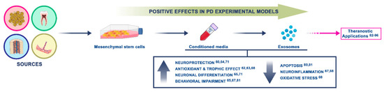
Figure 1.
1.1. Cellular and Molecular Mechanisms of PD
The underlying mechanisms of PD are still unclear, however, both environmental and genetic factors are involved in the pathogenesis [3][7]. Initially, the PD-linked mutations related to α-synuclein (A53T) [8]; subsequently, various gene mutations were identified, the most common were PINK1, DJ-1, LRRK2, and Parkin. The main cause of PD is the dopaminergic neuronal loss in the substantia nigra, which is the primary cause of motor symptoms. Dopamine metabolism, neuroinflammation, mitochondrial dysfunction, oxidative stress, and protein degradation damage are implicated in the death of dopaminergic neurons [3][9][10]. In addition, the immune system is implicated, indeed, in post-mortem brain and cerebrospinal fluid of PD patients, pro-inflammatory cytokines, including IFN-γ, TNF-α, IL-6, and IL-1β, are found to be upregulated [11].
Another key characteristic of PD are Lewy bodies, eosinophilic fibrillary intracellular deposits in neuronal bodies and appendages. The main component of Lewy bodies are proteins, polysaccharides, and fats, in particular α-synuclein, ubiquitin, parkin, neurofilaments, and synphilin. The underlying mechanism of the generation of these aggregates is still unclear [12] as is their role in neuronal death. Further, an aberrant proliferation of different glial cell types occurs, [13], thus leading to microglia activation involved in the neuroinflammation. The neuronal loss and Lewy bodies aggregation occur not only in the substantia nigra and tectum mesencephalic and basal nuclei, but also in the pedunculopontine nucleus, the locus coeruleus, parasympathetic and sympathetic postganglionic neurons, the dorsal motor nucleus of the vagal nerve, the cerebral cortices, the raphe nucleus, the olfactory bulbs, and the amygdala. Degeneration in these structures involve non-motor clinical symptoms development.
1.2. Searching for New Biomarkers and Therapeutic Approaches for PD
Human fluids are encouraging sources of molecular biomarkers, which can be categorized into molecules (e.g., elevated levels of 8-hydroxydeoxyguanosine, a byproduct of DNA oxidation, in PD patients’ urine), proteins (e.g., protein aggregates), and RNAs (e.g., noncoding microRNAs) [14]. The potential in using biomarkers isolated from body fluids (i.e., serum, cerebrospinal fluid, urine, blood, saliva, plasma) is the possibility of screening different molecules at once, while the cons are the low levels of molecules and the heterogeneity [15][16]. Exosomes have attracted a lot of interest because they are able to overcome this issue. Exosomes can be obtained from all bodily fluids, and they have a complex cargo of different RNAs (including microRNAs, ribosomal RNAs, and long noncoding RNAs), lipids, proteins, and DNA that in part depend on the tissue of origin and health conditions [17]. Catalytically active enzymes like PTEN (phosphatase and tensin homolog), and bioactive lipids such as prostaglandins, can be transferred by exosomes to target cells [5][6]. Exosomes carry different proteins, called “exosome markers”, the majority of which are associated to their biogenesis [18]. They also carry transmembrane proteins that can help in the immunoselection of exosomes with a precise cellular origin, thus increasing the sensitivity of exosomes as biomarkers. It has been reported that in neurodegenerative disorders exosomes carry misfolded proteins, such as
Human fluids are encouraging sources of molecular biomarkers, which can be categorized into molecules (e.g., elevated levels of 8-hydroxydeoxyguanosine, a byproduct of DNA oxidation, in PD patients’ urine), proteins (e.g., protein aggregates), and RNAs (e.g., noncoding microRNAs) [40]. The potential in using biomarkers isolated from body fluids (i.e., serum, cerebrospinal fluid, urine, blood, saliva, plasma) is the possibility of screening different molecules at once, while the cons are the low levels of molecules and the heterogeneity [41,42]. Exosomes have attracted a lot of interest because they are able to overcome this issue. Exosomes can be obtained from all bodily fluids, and they have a complex cargo of different RNAs (including microRNAs, ribosomal RNAs, and long noncoding RNAs), lipids, proteins, and DNA that in part depend on the tissue of origin and health conditions [7,43]. Catalytically active enzymes like PTEN (phosphatase and tensin homolog), and bioactive lipids such as prostaglandins, can be transferred by exosomes to target cells [25,26]. Exosomes carry different proteins, called “exosome markers”, the majority of which are associated to their biogenesis [44]. They also carry transmembrane proteins that can help in the immunoselection of exosomes with a precise cellular origin, thus increasing the sensitivity of exosomes as biomarkers. It has been reported that in neurodegenerative disorders exosomes carry misfolded proteins, such asα-synuclein in PD [19]. Among the different biomolecules related to exosomes, miRNAs have attracted the most attention as biomarkers.
-synuclein in PD [45]. Among the different biomolecules related to exosomes, miRNAs have attracted the most attention as biomarkers.The most relevant insight in therapeutic approaches for PD concerns the administration of the dopamine precursor
The most relevant insight in therapeutic approaches for PD concerns the administration of the dopamine precursorl
-DOPA (l-3,4-dihydroxyphenylalanine), which is able to ameliorate PD-related symptoms, increasing the level of the neurotransmitter but not replacing or counteracting dopaminergic neuron death [20]. Despite these limitations, the different side effects and the lack of improvement in nondopaminergic symptoms (such as psychiatric disorders or cognitive impairment),
-3,4-dihydroxyphenylalanine), which is able to ameliorate PD-related symptoms, increasing the level of the neurotransmitter but not replacing or counteracting dopaminergic neuron death [46]. Despite these limitations, the different side effects and the lack of improvement in nondopaminergic symptoms (such as psychiatric disorders or cognitive impairment),l-DOPA is the currently available treatment for PD patient. Indeed, to date, there is no cure [21]. Thus, among the innovative therapeutic approaches, the use of stem cells has received particular interest.
-DOPA is the currently available treatment for PD patient. Indeed, to date, there is no cure [47]. Thus, among the innovative therapeutic approaches, the use of stem cells has received particular interest.2. The Fate of MSC-Derived Secretome
2.1. Positive Effects of MSCs in PD
The first study reporting the potential of MSCs in PD demonstrated that the transplant of Wharton Jelly-derived MSCs was able to improve motor behaviors in a hemiparkinsonian rat model, indicating that the secretion of trophic factors mediated the rescue of the degenerating dopaminergic neurons [22]. Rat bone marrow-derived MSCs (BM-MSCs) intravenous administration ameliorated functional impairment and protected tyrosine hydroxylase (TH)-positive fibers in the striatum and substantia nigra in a PD rat model (6-OHDA lesioned) [23]. The authors detected chemotactic cytokine SDF-1α in the BM-MSC-derived secretome. They revealed that this cytokine inhibited the apoptosis in PC12 cells exposed to 6-OHDA, with a resultant increase of dopamine release from these cells [23]. In addition, Cova and his research group reported that human MSCs transplantation in the striatum of 6-OHDA lesioned rats protected dopaminergic neurons and induced neurogenesis, suggesting that MSCs in situ may help lesioned neurons thanks to the local release of soluble factors, such as BDNF [24].
The first study reporting the potential of MSCs in PD demonstrated that the transplant of Wharton Jelly-derived MSCs was able to improve motor behaviors in a hemiparkinsonian rat model, indicating that the secretion of trophic factors mediated the rescue of the degenerating dopaminergic neurons [57]. Rat bone marrow-derived MSCs (BM-MSCs) intravenous administration ameliorated functional impairment and protected tyrosine hydroxylase (TH)-positive fibers in the striatum and substantia nigra in a PD rat model (6-OHDA lesioned) [58]. The authors detected chemotactic cytokine SDF-1α in the BM-MSC-derived secretome. They revealed that this cytokine inhibited the apoptosis in PC12 cells exposed to 6-OHDA, with a resultant increase of dopamine release from these cells [58]. In addition, Cova and his research group reported that human MSCs transplantation in the striatum of 6-OHDA lesioned rats protected dopaminergic neurons and induced neurogenesis, suggesting that MSCs in situ may help lesioned neurons thanks to the local release of soluble factors, such as BDNF [59].These reports provide the idea that bioactive molecules released by MSCs exert neuroprotective and antiapoptotic effects. Indeed, other researchers focused on the use of MSC-derived secretome (in the form of conditioned media or an exosomal component) as a cell-free transplantation method for PD.
These reports provide the idea that bioactive molecules released by MSCs exert neuroprotective and antiapoptotic effects. Indeed, other researchers focused on the use of MSC-derived secretome (in the form of conditioned media or an exosomal component) as a cell-free transplantation method for PD.2.2. Positive Effects of MSC-Derived Conditioned Medium in PD
While the cell-free trophic consequences of MSCs transplant have been reported in different in vivo models, research on the therapeutic impacts of secretome on neurodegenerative disorders is still scant.
While the cell-free trophic consequences of MSCs transplant have been reported in different in vivo models, research on the therapeutic impacts of secretome on neurodegenerative disorders is still scant.Notably, different studies described MSC-CM positive effects in PD. In vitro studies reported the neuroprotective activity of the secretome derived from human bone marrow-MSCs and human tooth germ stem cells, in 6-OHDA-treated SH-SY5Y cells and murine differentiated neural stem cells [25]. Another research group [26] investigated the viability of dopaminergic cells from different sources upon rat bone marrow-MSC-derived secretome treatment, suggesting that prostaglandin E2 receptors represent the principal factor of neuroprotective events.
Notably, different studies described MSC-CM positive effects in PD. In vitro studies reported the neuroprotective activity of the secretome derived from human bone marrow-MSCs and human tooth germ stem cells, in 6-OHDA-treated SH-SY5Y cells and murine differentiated neural stem cells [60]. Another research group [61] investigated the viability of dopaminergic cells from different sources upon rat bone marrow-MSC-derived secretome treatment, suggesting that prostaglandin E2 receptors represent the principal factor of neuroprotective events.Other interesting research assessed the neuroprotective effect of adipose-derived mesenchymal stem cells (ASCs)-CM on neurotrophins gene expressions and TH+ cell density in 6-OHDA-lesioned rats. ASCs secrete numerous neurotrophic factors and cytokines in CM, which protect neurons by antioxidative and trophic effects. ASC-CM protected dopaminergic neurons by preserving TH+ neurons and by increasing BDNF and neurotrophin-3 expression [27]. The neurotrophic factor BDNF is crucial for neuronal survival in the substantia nigra [28]. The ability of MSC-derived secretome to reduce extracellular α-synuclein both in vitro and in vivo was also reported, mainly mediated by matrix metalloproteinase-2 [29].
Other interesting research assessed the neuroprotective effect of adipose-derived mesenchymal stem cells (ASCs)-CM on neurotrophins gene expressions and TH+ cell density in 6-OHDA-lesioned rats. ASCs secrete numerous neurotrophic factors and cytokines in CM, which protect neurons by antioxidative and trophic effects. ASC-CM protected dopaminergic neurons by preserving TH+ neurons and by increasing BDNF and neurotrophin-3 expression [62]. The neurotrophic factor BDNF is crucial for neuronal survival in the substantia nigra [63]. The ability of MSC-derived secretome to reduce extracellular α-synuclein both in vitro and in vivo was also reported, mainly mediated by matrix metalloproteinase-2 [64].2.3. Positive Effects of MSC-Derived Exosomes in PD
As we mentioned above, the secretome released by MSCs contains different bioactive molecules, including exosomes. MSCs can produce a higher amount of exosomes than can other kinds of cells [30]. Methods to isolate exosomes from the MSC-conditioned medium have been widely developed.
As we mentioned above, the secretome released by MSCs contains different bioactive molecules, including exosomes. MSCs can produce a higher amount of exosomes than can other kinds of cells [72]. Methods to isolate exosomes from the MSC-conditioned medium have been widely developed.MSC-derived exosomes are hypoimmunogenic (due to the lack of MHC-II and low expression of MHC-I) nanocarriers that comprise various immunoregulatory components. Exosomes have several advantages, they are able to cross the blood–brain barrier and blood capillaries, and are small enough to avoid being cleared by the reticuloendothelial system [31]. The mechanisms of cellular recognition and internalization are still unclear. Antigen recognition, adhesion, and free-floating are described as cellular recognition mechanism, while fusion, phagocytosis, micropinocytosis, and raft- and receptor-mediated endocytosis are indicated as exosomal internalization processes [32].
MSC-derived exosomes are hypoimmunogenic (due to the lack of MHC-II and low expression of MHC-I) nanocarriers that comprise various immunoregulatory components. Exosomes have several advantages, they are able to cross the blood–brain barrier and blood capillaries, and are small enough to avoid being cleared by the reticuloendothelial system [73]. The mechanisms of cellular recognition and internalization are still unclear. Antigen recognition, adhesion, and free-floating are described as cellular recognition mechanism, while fusion, phagocytosis, micropinocytosis, and raft- and receptor-mediated endocytosis are indicated as exosomal internalization processes [74].Exosomes have proven effective in direct MSCs transplantation, and their positive therapeutic effects have been shown in different disease models, in particular, they were beneficial for central nervous system pathologies. In a stroke animal model, MSC-derived exosomes, intravenously administered, stimulated angiogenesis and neurogenesis, neurite remodeling, and improved animal motor performances [33]. The same neuroprotective effect was shown in a traumatic brain injury model after MSC-derived exosomes administration; indeed, a reduction of neuroinflammation and better outcomes were reported [34]. Spinal cord injury rats, upon MSC-derived exosomes injection, showed reduced inflammation and increased neuronal regeneration [35][36]. In addition, in Alzheimer’s disease, the positive effects of MSC-derived exosomes with a particular impact on neuroplasticity were reported [37].
Exosomes have proven effective in direct MSCs transplantation, and their positive therapeutic effects have been shown in different disease models, in particular, they were beneficial for central nervous system pathologies. In a stroke animal model, MSC-derived exosomes, intravenously administered, stimulated angiogenesis and neurogenesis, neurite remodeling, and improved animal motor performances [75]. The same neuroprotective effect was shown in a traumatic brain injury model after MSC-derived exosomes administration; indeed, a reduction of neuroinflammation and better outcomes were reported [76]. Spinal cord injury rats, upon MSC-derived exosomes injection, showed reduced inflammation and increased neuronal regeneration [77,78]. In addition, in Alzheimer’s disease, the positive effects of MSC-derived exosomes with a particular impact on neuroplasticity were reported [79].3. MSC-Secretome: miRNA Relevance and Theranostic Applications in PD
Secretome derived from MSCs, and in particular its miRNAs component, has also been indicated as a valuable tool for targeted therapies and diagnostics. miRNAs are a highly studied class of non-coding RNAs responsible for the regulation of different genes through RNA messenger degradation or inhibition of their translation [38]. Numerous miRNAs have been indicated as α-synuclein modulators. For example, altered binding between and fibroblast growth factor 20 (FGF20) mRNA and miR-433 induced increased FGF20 levels, which consequently led to elevated α-synuclein protein levels in the cell [39]. Further, high miR-16-1 levels block the translation of the HSP70 (heat shock protein 70) mRNA, involved in α-synuclein inhibition, thus leading to an accumulation of α-synuclein [
Secretome derived from MSCs, and in particular its miRNAs component, has also been indicated as a valuable tool for targeted therapies and diagnostics. miRNAs are a highly studied class of non-coding RNAs responsible for the regulation of different genes through RNA messenger degradation or inhibition of their translation [82]. Numerous miRNAs have been indicated as α-synuclein modulators. For example, altered binding between and fibroblast growth factor 20 (FGF20) mRNA and miR-433 induced increased FGF20 levels, which consequently led to elevated α-synuclein protein levels in the cell [83]. Further, high miR-16-1 levels block the translation of the HSP70 (heat shock protein 70) mRNA, involved in α-synuclein inhibition, thus leading to an accumulation of α-synuclein [84]. Moreover, targeting miR-7, miR-153, and miR-34b/c from binding on their α-synuclein induces elevated levels of α-synuclein [40][41].
]. Moreover, targeting miR-7, miR-153, and miR-34b/c from binding on their α-synuclein induces elevated levels of α-synuclein [85,86]. In PD, a relation between miR-34b/c reduction and the resultant DJ-1 and PARKIN decline in several brain areas was found [87]. Notably, increased miR-494 and miR-4639-5p levels trigger a direct decrease of DJ-1 protein expression, making dopaminergic neurons more susceptible and predisposed to the PD phenotype [88,89]. Interestingly, it has been recently proposed that MSC-derived secretome can ameliorate different biomarkers of PD pathophysiology, thus suggesting MSC-derived secretome as a promising approach to identify and generate valuable PD biomarkers [90]. As mentioned above, MSCs secrete different biomolecules and factors, including exosomes carrying miRNA, which may represent potential biomarkers but also modulators of different pathways underlying various disorders, including PD. Theranostic applications in PD exploiting the potential of MSC-derived secretome, mainly concern the targeting of the injured brain areas and delivering miRNAs through the blood-brain barrier. Thanks to the nature of exosomes, their application in the theranostic field and in clinic have received a lot of interest. Still, different points need to be addressed, such as the “best” MSCs line and the development of valid isolation techniques and loading methods without altering the exosomal component and integrity. However, MSC-derived exosomes may represent a valuable solution. In fact, numerous experiments revealed that MSC-derived exosomes are able to transfer miRNAs to neuronal cells, for instance, exosomes enriched in miR-133b can stimulate neurite outgrowth [90,91], one of the miRNAs generally decreased in PD. Further, miR-21 and miR-143, leading players in immune response, and neuroinflammation were also observed in MSC-derived exosomes [92]. Interestingly, in MSC-derived exosomes, a miRNA cluster composed of miR-18a, miR-17, miR-20a, miR-19a/b, and miR-90a, involved in axonal growth, neurogenesis, neurite remodeling and in CNS (central nervous system) recovery, was detected [93,94].In PD, a relation between miR-34b/c reduction and the resultant DJ-1 and PARKIN decline in several brain areas was found [42]. Notably, increased miR-494 and miR-4639-5p levels trigger a direct decrease of DJ-1 protein expression, making dopaminergic neurons more susceptible and predisposed to the PD phenotype [43][44]. Interestingly, it has been recently proposed that MSC-derived secretome can ameliorate different biomarkers of PD pathophysiology, thus suggesting MSC-derived secretome as a promising approach to identify and generate valuable PD biomarkers [45]. As mentioned above, MSCs secrete different biomolecules and factors, including exosomes carrying miRNA, which may represent potential biomarkers but also modulators of different pathways underlying various disorders, including PD. Theranostic applications in PD exploiting the potential of MSC-derived secretome, mainly concern the targeting of the injured brain areas and delivering miRNAs through the blood-brain barrier. Thanks to the nature of exosomes, their application in the theranostic field and in clinic have received a lot of interest. Still, different points need to be addressed, such as the “best” MSCs line and the development of valid isolation techniques and loading methods without altering the exosomal component and integrity. However, MSC-derived exosomes may represent a valuable solution. In fact, numerous experiments revealed that MSC-derived exosomes are able to transfer miRNAs to neuronal cells, for instance, exosomes enriched in miR-133b can stimulate neurite outgrowth [46][47], one of the miRNAs generally decreased in PD. Further, miR-21 and miR-143, leading players in immune response, and neuroinflammation were also observed in MSC-derived exosomes [48]. Interestingly, in MSC-derived exosomes, a miRNA cluster composed of miR-18a, miR-17, miR-20a, miR-19a/b, and miR-90a, involved in axonal growth, neurogenesis, neurite remodeling and in CNS (central nervous system) recovery, was detected [49][50].
Another theranostic application in PD exploits the human Periapical Cyst-MSCs (hPCyMSCs) differentiated in dopaminergic neurons; thus hPCyMSC-derived exosomes may be useful therapeutic carriers for PD. hPCy-MSCs exposed to a neural-inductive medium led to functional dopaminergic neurons; the exosomes isolation from the CM of these MSCs is presently standardized [95]. The analysis of circulating exosome-derived miRNA through microarrays and gene sequencing could be related to nanotechnologies: This is a significant point to improving the capability of new smart nanomaterials to capture the small-sized biomolecules, representing a theranostic approach with elevated sensitivity and extreme specificity [96].Another theranostic application in PD exploits the human Periapical Cyst-MSCs (hPCyMSCs) differentiated in dopaminergic neurons; thus hPCyMSC-derived exosomes may be useful therapeutic carriers for PD. hPCy-MSCs exposed to a neural-inductive medium led to functional dopaminergic neurons; the exosomes isolation from the CM of these MSCs is presently standardized [51]. The analysis of circulating exosome-derived miRNA through microarrays and gene sequencing could be related to nanotechnologies: This is a significant point to improving the capability of new smart nanomaterials to capture the small-sized biomolecules, representing a theranostic approach with elevated sensitivity and extreme specificity [52].
On this basis, it is relevant to isolate and characterize the entire set of biomolecules released by MSCs and in particular hPCy-MSCs, and to dissect the cellular and molecular mechanisms regulated by miRNAs. These new understandings may allow for the development of new therapeutic approaches and offer novel evidence on functional biomarkers for early diagnosis and monitoring of neurodegenerative diseases, with particular attention to PD.
On this basis, it is relevant to isolate and characterize the entire set of biomolecules released by MSCs and in particular hPCy-MSCs, and to dissect the cellular and molecular mechanisms regulated by miRNAs. These new understandings may allow for the development of new therapeutic approaches and offer novel evidence on functional biomarkers for early diagnosis and monitoring of neurodegenerative diseases, with particular attention to PD.4. Conclusions and Future Perspectives
PD is a debilitating neurodegenerative disorder that affects millions of people worldwide; however, the molecular and cellular underlying mechanisms are still unclear. Although there are advances in the PD research field, the current therapeutic approaches improve PD patients’ quality of life, but they are not able to counteract PD progression and to stimulate dopaminergic neurons survival/differentiation. Thus, recently, MSC-derived secretome and its exosomal components have been suggested as promising therapeutic tools for numerous neurodegenerative disorders, including PD, due to their ability to promote dopaminergic neurons survival, stimulate neurogenesis, decrease neuroinflammation, promote functional recovery in in vivo models.
PD is a debilitating neurodegenerative disorder that affects millions of people worldwide; however, the molecular and cellular underlying mechanisms are still unclear. Although there are advances in the PD research field, the current therapeutic approaches improve PD patients’ quality of life, but they are not able to counteract PD progression and to stimulate dopaminergic neurons survival/differentiation. Thus, recently, MSC-derived secretome and its exosomal components have been suggested as promising therapeutic tools for numerous neurodegenerative disorders, including PD, due to their ability to promote dopaminergic neurons survival, stimulate neurogenesis, decrease neuroinflammation, promote functional recovery in in vivo models. To date, there is no cure for PD, and thus, recently, attention has been focused on cell-free approaches. MSCs have become widely used for cell-based therapy due to less scientific and ethical issues compared to the use of other kinds of cells. The ability of MSCs to release exosomes and various trophic factors makes the use of MSCs attractive for PD treatment. Thanks to their small size and/or soluble nature, these secreted molecules can cross the blood–brain barrier; moreover, exosomes are intrinsically less risky compared to live stem cell transplants. Exosomes cannot transform into harmful or malignant cells; they cannot replicate; they are less prone to activate an immunogenic response; and a virus cannot infect them. In light of the insights reported in this review, the use of MSC-derived secretome is encouraging in PD.To date, there is no cure for PD, and thus, recently, attention has been focused on cell-free approaches. MSCs have become widely used for cell-based therapy due to less scientific and ethical issues compared to the use of other kinds of cells. The ability of MSCs to release exosomes and various trophic factors makes the use of MSCs attractive for PD treatment. Thanks to their small size and/or soluble nature, these secreted molecules can cross the blood–brain barrier; moreover, exosomes are intrinsically less risky compared to live stem cell transplants. Exosomes cannot transform into harmful or malignant cells; they cannot replicate; they are less prone to activate an immunogenic response; and a virus cannot infect them. In light of the insights reported in this review, the use of MSC-derived secretome is encouraging in PD.
Further studies are needed to identify a personalized approach for the different neurodegenerative diseases and to create a new, useful, cell-free therapeutic approach towards a robust clinical outcome for PD patients. Another point that needs to be clarified is if the encouraging results are due to one or two factors or a combination of different molecules present in the secretome. To date, it is pretty clear that MSC-derived secretome exerts positive effects on neuronal cell survival, differentiation, and proliferation; however, future studies need to characterize all the bioactive molecules fully. Thus, MSC-derived secretome or their released exosomes may be used as a potential therapeutic approach or as adjuvant therapy for PD symptoms and to counteract the disease progression. Furthermore, their secretome may be used as a vehicle in cell transplantation approaches to improve the viability and survival of engrafted cells and also as a diagnostic approach. These different aspects of the knowledge about the secretome may permit the advancement of targeted secretome to fight different pathophysiological impairments in a multidisciplinary manner. In addition, since MSC-derived secretome is able to stimulate neurotrophic (i.e., BDNF, a biomarker of the majority of neurodegenerative disorders) and neuronal survival pathways and to counteract neuronal death, it could also be beneficial against other neurodegenerative conditions, including polyglutamine disorder, Alzheimer’s disease, and stroke.
Further studies are needed to identify a personalized approach for the different neurodegenerative diseases and to create a new, useful, cell-free therapeutic approach towards a robust clinical outcome for PD patients. Another point that needs to be clarified is if the encouraging results are due to one or two factors or a combination of different molecules present in the secretome. To date, it is pretty clear that MSC-derived secretome exerts positive effects on neuronal cell survival, differentiation, and proliferation; however, future studies need to characterize all the bioactive molecules fully. Thus, MSC-derived secretome or their released exosomes may be used as a potential therapeutic approach or as adjuvant therapy for PD symptoms and to counteract the disease progression. Furthermore, their secretome may be used as a vehicle in cell transplantation approaches to improve the viability and survival of engrafted cells and also as a diagnostic approach. These different aspects of the knowledge about the secretome may permit the advancement of targeted secretome to fight different pathophysiological impairments in a multidisciplinary manner. In addition, since MSC-derived secretome is able to stimulate neurotrophic (i.e., BDNF, a biomarker of the majority of neurodegenerative disorders) and neuronal survival pathways and to counteract neuronal death, it could also be beneficial against other neurodegenerative conditions, including polyglutamine disorder, Alzheimer’s disease, and stroke.