Pink Pepper (Schinus molle L.): Comparison
Please note this is a comparison between Version 1 by Sung Keun Jung and Version 3 by Catherine Yang.

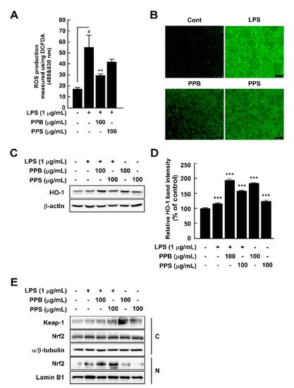
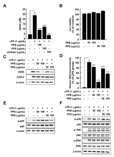
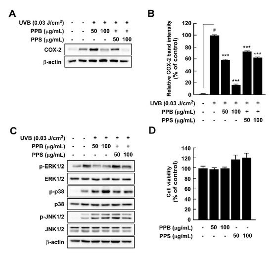
Here, we compared the chemical properties and antioxidant effects of black pepper (Piper nigrum L.) and pink pepper (Schinus molle L.). Additionally, the antioxidant and anti-inflammatory capacities of pink pepper were measured to determine nutraceutical potential. Pink peppers from Brazil (PPB), India (PPI), and Sri Lanka (PPS) had higher Hunter a* (redness) values and lower L* (lightness) and b* (yellowness) values than black pepper from Vietnam (BPV). Fructose and glucose were detected in PPB, PPI, and PPS, but not in BPV. PPB, PPI, and PPS had greater 2,2-diphenyl-1-picrylhydrazyl and 3-ethylbenzothiazoline-6-sulphonic acid radical scavenging stabilities and higher total phenolic contents than BPV. BPV had higher levels of piperine than the pink peppers. Gallic acid, protocatechuic acid, epicatechin, and p-coumaric acid were detected only in the three pink peppers. PPB significantly suppressed lipopolysaccharide-induced reactive oxygen species production with increased Nrf2 translocation from cytosol to nucleus and heme oxygenase-1 expression. PPB and PPS significantly suppressed lipopolysaccharide-induced nitrite production and nitric oxide synthase expression by suppressing phosphorylation of p38 without affecting cell viability. Additionally, PPB and PPS significantly suppressed ultraviolet B-induced cyclooxygenase-2 expression by affecting the phosphorylation of ERK1/2 without cell cytotoxicity. These results suggest that pink pepper is a potential nutraceutical against oxidative and inflammatory stress.

  • nutraceutical
  • compound compositions
  • reactive oxygen species
  • mitogen-activated protein kinase
  • nuclear factor kappa-light-chain-enhancer of activated B cells

1. Introduction

Reactive oxygen species (ROS), reactive molecules and free radicals derived from molecular oxygen, are responsible for the elimination of microbial invasion [1]. Although phagocytes, such as macrophages, are innate immune cells that are important for the elimination of pathogens, including viruses, bacteria, and parasites, the abnormal production of ROS causes oxidative stress and consequent cell death. Additionally, inflammation is closely related to ROS levels because unregulated immune response to a pathogen results in overproduction of ROS, which causes damage to the host organ [2]. Excessive production of nitric oxide (NO) also plays a critical role in inflammation. Thus, continuously high ROS and NO levels lead to chronic inflammation, which can result in the onset of various diseases, including atherosclerosis, rheumatoid arthritis, diabetes, and cancer [3][4][3,4]. Therefore, detoxification of ROS is a key step preventing inflammation and the subsequent development of chronic inflammatory diseases.
Heme oxygenase-1 (HO-1) is an Nrf2-regulated gene that has important antioxidant, anti-inflammatory, and antiapoptotic effects [5]. While Nrf2/HO-1 signaling plays a protective role in the regulation of oxidative stress and inflammation, NF-κB, a heterodimer of p65 and p50/p105, plays a central role in ROS- and pathogen-mediated inflammatory signaling cascades. In addition, mitogen-activated protein kinases (MAPKs) are involved in inflammation [6]. A previous study reported that ultrasonicated seaweed extract suppressed lipopolysaccharide (LPS)-induced nitric oxide synthase (iNOS) expression and nitrite production via regulation of MAPK phosphorylation without affecting the NF-κB signaling cascade in RAW264.7 cells [7]. Additionally, the phosphorylation of MAPKs is central in skin inflammation response to ultraviolet (UV) light exposure in vitro and in vivo [8][9][8,9]. Therefore, in addition to NF-kB, MAPKs may be a prime target for anti-inflammatory nutraceuticals.
Schinus molle L., also known as Peruvian pepper, Brazilian pepper, American pepper, Californian pepper, or molle de Peru, is a fast-growing evergreen tree [10]. Pink pepper is classified into the Californian/Peruvian type (S. molle) and the Brazilian type (S. terebinthifolius) [11]. Although it is unrelated to the black pepper (Piper nigrum), the pink fruits of S. molle are known as pink peppercorns and are used as alternatives to black pepper due to their flavor and pungency [12]. Research on pink pepper has primarily focused on the chemical composition and physicochemical characteristics of its essential oil and its antimicrobial activities [13][14][13,14]. The results of recent studies have revealed that polysaccharides from S. molle fruit have in vitro antioxidant, antigenotoxic, antidiabetic, and antihemolytic effects, as well as in vivo anti-inflammatory and antinociceptive properties [15]. However, the differences in the chemical compositions and the antioxidant and anti-inflammatory effects of S. molle fruits from different countries remain unclear.

2. Results and Discussion

2.1. Color and Sugar Content Analysis

Although S. molle is a known as American pepper, it has distinctive color and chemical compositions compared to black pepper. While the chemical and physiological properties of black pepper have been well studied [16][17][20,21], the chemical composition and physiological activity of S. molle are mainly unknown. Therefore, we compared the in vitro antioxidant effect of pink peppers from three different countries and black pepper as well as analyzed the chemical compositions of 
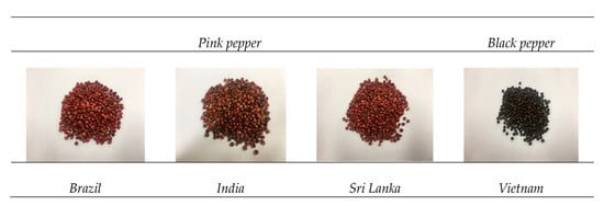
S. molle fruits. To the best of our knowledge, this is the first report of this approach and these findings. Pink and black peppers have distinctive colors and shapes. Pictures of the dried pink peppers from three countries and the BPV used in this experiment are shown in Figure 1.
Figure 1. Photographs of dried pink peppers from different countries and black pepper.
To compare the general characteristics of pink and black peppers, we analyzed the color and carbohydrate contents of the peppers. Table 1 summarizes the Hunter L, a, b values and fructose and glucose contents of PPB, PPI, and PPS. We determined fructose, glucose, sucrose, and maltose content in the pink and black peppers (S1) using HPLC assay [18][16]. While fructose and glucose were not detected in BPV, the pink peppers showed relatively high levels of both monosaccharides. PPS and PPB had the highest fructose content (11,829.82 ± 23.73 mg/100 g) and glucose content (9816.07 ± 36.51 mg/100 g). In the HPLC chromatogram of sugar analysis, we detected two peaks that were consistent with the fructose and glucose standards (Figure S1). Feriani et al. identified fucose (10.90% ± 0.024%) in S. molle fruits from Tunisia, and interestingly, glucose was not detected [15]. On the HPLC chromatogram obtained for pink pepper, we confirmed two peaks that were consistent with the standard peaks of fructose and glucose, but no peaks corresponding to fucose were detected (Figure S1). Therefore, we ruled out the hypothesis that fucose would be present in considerable amount in commercial S. molle. Further, Solis et al. reported that fresh and residue seeds of S. molle contained 16% glucose; however, they did not confirm fructose and fucose [19][22]. In our method, we used 70% EtOH for sample extraction, while Feriani et al. used hot water extraction for S. molle fruits from Tunisia. Therefore, these differences in monosaccharide contents seem to be due to the differences in the origin of the peppers, cultivation environment, and the extraction method.
Table 1. Color and sugar composition of pink and black peppers.
  Region L a b Fructose

(mg/100 g)
Glucose

(mg/100 g)
Pink Brazil 28.87 ± 0.01 b 11.94 ± 0.01 a 11.41 ± 0.004 b 11,507.21 ± 90.5 b 9816.07 ± 36.51 a
India 25.02 ± 0.09 d 6.71 ± 0.05 b 8.55 ± 0.04 d 9528.74 ± 46.67 c 6181.37 ± 315.61 b
Sri Lanka 27.36 ± 0.03 c 11.92 ± 0.06 a 10.89 ± 0.05 c 11,829.82 ± 23.73 a 9758.15 ± 330.28 a
Black Vietnam 43.65 ± 0.01 a 3.81 ± 0.02 c 13.16 ± 0.01 a N.D. N.D.
(1) Values are the average of experiments (n = 5) and represented as mean ± standard deviation; different letters (a–d) in a column indicate values that are significantly different at p < 0.05.

2.2. Total Flavonoid Contents and Total Phenolic Contents

Several studies have characterized the chemical and physiological functions of the essential oil [13][14][13,14], carbohydrates [15], and fragrance ingredients [12] of S. molle. The essential oil and fragrance ingredients are useful for food seasoning. To obtain essential oil and carbohydrates from the pink peppers, the researchers used water as an extraction solvent [12]. Because there are few studies on the application of S. molle fruits as nutraceuticals, we analyzed the phenolic and flavonoid content and antioxidant capacity of S. molle and evaluated its potential as a nutraceutical ingredient and antioxidant.
Total phenolic contents were expressed in GAE per 100 g. The highest total phenolic and flavonoid contents were found in PPB (1607.80 ± 21.11 mg GAE/100 g) and BPV (344.24 ± 3.78 mg CE/100 g), respectively (Table 2). Interestingly, PPB, PPI, and PPS had higher total phenolic and lower total flavonoid contents than BPV. In our previous study, we determined the total flavonoid and total phenolic contents of green peppers to be 1083.43 ± 8.24 mg CE/100 g and 1414.63 ± 10.56 mg CE/100 g, respectively [20][23]. Even though direct comparison of total flavonoid and total phenolic contents among green and pink peppers is limited, pink pepper may have higher total flavonoid and lower total phenolic than green and black peppers.
Table 2. Total flavonoid and total phenolic contents of pink and black peppers.
  Region Total Phenolic Content

(mg GAE/100 g)
Total Flavonoid Content

(mg CE/100 g)
Pink Brazil 1607.80 ± 21.11 a 266.67 ± 2.42 b
India 1588.29 ± 17.88 a 230.30 ± 2.10 d
Sri Lanka 1250.08 ± 10.75 b 248.89 ± 5.60 c
Black Vietnam 794.47 ± 17.19 c 344.24 ± 3.78
(1) Values are the average of experiments (n = 3) and represented as mean ± standard deviation. CE, catechin equivalents; GAE, gallic acid equivalents; different letters (a–d) in a column indicate values that are significantly different at p < 0.05.

2.3. Quantification of Major Compounds in Peppers by HPLC

Piperine, a phenolic component, is a representative component of peppers and is responsible for the pungency and flavor of black pepper [21][24]. Three repeated quantitative analyses of PPB, PPI, and PPS showed piperine concentrations of 134.6 mg/100 g, 101.1 mg/100 g, and 120.67 mg/100 g, respectively (Table 3). Meanwhile, piperine concentrations in BPV were determined to be 4097.53 mg/100 g (Table 4). Although PPB, PPI, and PPS have higher DPPH and ABTS radical scavenging capacities, their piperine concentrations were lower than that of BPV (Table 3 and Table 4). Giuffrida et al., identified a volatile compound and 10 carotenoids in the essential oil of S. molle [12]. Feuerisen et al. confirmed that anthocyanins, bioflavonoids, and gallotannins are present in S. terebinthifolia and S. molle by using UHPLC-MS/MS analysis [22][25]. In the present study, we further determined the polyphenol contents of pink peppers and compared them to those of black pepper. The results of three repeated quantitative analyses of PPB, PPI, and PPS polyphenols are listed in Table 4. The most abundant polyphenol in the pink peppers was gallic acid; its content was highest in PPI (657.59 ± 5.25 mg/100 g) and lowest in PPS (168.15 mg/100 g) (Table 4). Additionally, gallic acid, protocatechuic acid, epicatechin, and p-coumaric acid were detected in the pink peppers, but not in the black pepper. Therefore, the differences in radical scavenging capacity between pink peppers and black pepper may originate in the different chemical compositions of the peppers.
Table 3. Radical scavenging capacities of pink and black peppers.
  Region DPPH

(mg VCE/100 g)
ABTS

(mg VCE/100 g)
Pink Brazil 4015.32 ± 13.00 a
n = 3) and represented as mean ± standard deviation. VCE, vitamin C equivalents; DPPH, 2,2-diphenyl-1-picrylhydrazyl; ABTS, 3-ethylbenzothiazoline-6-sulphonic acid; different letters (a–d) in a column indicate values that are significantly different at p < 0.05.
Table 4. Individual polyphenol profiles of pink and black peppers.
  Region Piperine

(mg/100 g)
Gallic Acid

(mg/100 g)
Protocatechuic Acid

(mg/100 g)
Epicatechin

(mg/100 g)
p-Coumaric Acid

(mg/100 g)
2741.25 ± 19.69 
Pink Brazila
134.60 ± 3.20 b 526.72 ± 6.06 b 144.85 ± 0.71 b 85.91 ± 2.88 a 115.92 ± 5.00 b India 4081.92 ± 34.39 a 2845.12 ± 3.91 a
India 101.10 ± 2.84 c 657.59 ± 5.25 a 237.52 ± 0.64 a 89.24 ± 2.04 a 151.33 ± 7.07 a Sri Lanka 2812.30 ± 10.81 
Sri Lankab 120.67 ± 1.91 bc1956.96 ± 54.26 b
168.15 ± 1.43 c 29.47 ± 0.18 c 38.26 ± 1.28 b 48.24 ± 1.28  Black Vietnam 271.45 ± 12.01 861.92 ± 83.23
(1) Values are the average of experiments (
c
Black
Vietnam 4097.53 ± 46.87 a N.D N.D N.D N.D
(1) Values are the average of experiments (n = 3) and represented as mean ± standard deviation. N.D, not detected; different letters (a–d) in a column indicate values that are significantly different at p < 0.05.
Parra et al. quantified the phenolic compounds such as gallic acid (43.60 mg/100 g DW), protocatechuic acid (61.99 mg/100 g DW), and p-Coumaric acid (9.32 mg/100 g DW) in Origanum vulgare L. by using UHPLC-DAD [23][26]. Zhang et al. identified phenolic contents including gallic acid (11.30 ± 0.28 μg/g) and protocatechuic acid (39.53 ± 0.80 μg/g) from Lycium ruthenicum Murray by UPLC-Q-Orbitrap MS [24][27]. These extracts have also shown strong antioxidant activities, and the authors indicated the relevance of high phenolic compounds. However, it was not possible to directly compare whether the content of polyphenols in pink pepper contributes to the antioxidant activity as there have been no previous studies. Therefore, further research is needed for the bioactivity evaluation of pink pepper.

2.4. Radical Scavenging Effects

Phenolic compounds and flavonoids contribute to the antioxidant capacities of natural materials [25][28]. Based on the high phenolic and flavonoid contents of the peppers, we further evaluated their ability to scavenge DPPH and ABTS radicals. The radical scavenging capacities of DPPH and ABTS were measured to determine antioxidant activity and expressed as VCE. The greatest DPPH and ABTS radical scavenging capacity were found in PPI (4081.92 ± 34.39 mg VCE/100 g and 2845.12 ± 3.91 mg VCE/100 g) (Table 3). The results of a recent study showed that S. mole polysaccharide has significant DPPH and ABTS radical scavenging effects [15]. In our previous study, the range of DPPH radical scavenging capacity of the green and black peppers was found to be from 522.83 to 194.42 mg VCE/100 g, and the range of ABTS radical scavenging capacity was 1941.91 to 526.45 mg VCE/100 g [20][23]. Therefore, we suggest that the radical scavenging capacity of pink pepper is higher than that of black and green pepper.

2.5. Pink Peppers Inhibited LPS-Induced ROS Production and Increased HO-1 Expression in RAW264.7 Cells

Overproduction of ROS damages cells and host organs and subsequently causes inflammation and diseases [26][29]. Although PPB and PPI have higher DPPH and ABTS radical scavenging capacities and phenolic compound contents than those of PPS, to evaluate whether this effect is related to the antioxidant and anti-inflammatory effects in cells, we selected PPB and PPS as having the highest and lowest effects. By using DCFH-DA as a chemical probe for ROS, we determined that PPB significantly suppressed LPS-induced ROS production in RAW264.7 cells (Figure 2A,B). Increased ROS levels are affected by various factors, such as antioxidant enzymes. Among many antioxidant enzymes, HO-1 has been reported to correlate with increased intracellular ROS production [27][30]. In the present study, PPB and PPS significantly induced HO-1 expression in RAW264.7 cells independently of LPS (Figure 2C,D). Additionally, PPB and PPS increased nuclear Nrf2 translocation (Figure 2E). The results of several studies have suggested the DPPH and ABTS radical scavenging capacity of S. molle [14][15][28][14,15,31], but the effect of this pepper on cellular antioxidant and antioxidative enzyme expression is unclear. Here, we found that the antioxidant capacity of S. molle fruit is due to an increase in Nrf2 translocation from the cytosol to the nucleus and HO-1 expression in RAW264.7 cells. Additionally, we confirmed that the radical scavenging capacity at the test-tube level and the phenolic compound content highly correlated with the antioxidant activity in RAW264.7 cells.
Figure 2. Effects of pink pepper extract (PPE) on lipopolysaccharides (LPS)-induced reactive oxygen species (ROS) production and HO-1 expression in RAW264.7 cells. (A) PPE suppressed LPS-induced ROS production in RAW264.7 cells. (B) Pink peppers from Brazil (PPB) suppressed LPS-induced ROS production at 100 μg/mL. Images show representative micrographs of cells under fluorescence microscope. (C) PPB and Pink peppers from Sri Lanka (PPS) significantly elevated HO-1 expression independently of LPS presence in RAW264.7 cells. (D) Quantification of HO-1 expression. (E) PPB and PPS enhanced Nrf2 translocation from the cytosol to the nucleus. Expression levels of HO-1, Keap-1, and Nrf2 were determined by Western blot. Values represent the mean ± standard deviation of three independent experiments. # p < 0.05 between control versus LPS-exposed cells (no PPE); ** p < 0.01. *** p < 0.001 represents a significant difference compared with the control group.

2.6. Effects of PPE on LPS-Induced Nitrite Production, iNOS and COX-2 Expression, and p65 and MAPK Phosphorylation in RAW264.7 Cells

Abnormal NO production is a vital marker of inflammation in response to LPS treatment [29][32]. PPB and PPS significantly suppressed LPS-induced nitrite production without cell toxicity in comparison to pre-treatment with an NF-κB inhibitor, parthenolide (Figure 3A,B). Because NO is mainly produced by iNOS, we further explored the effect of PPB and PPS on LPS-mediated iNOS expression in RAW264.7 cells. PPB and PPS significantly inhibited iNOS expression, but not COX-2 expression (Figure 3C). Because the association between iNOS expression and upstream regulatory signaling pathways such as the NF-κB and MAPK signaling pathways is well known, we subsequently assessed the effect of PPB and PPS on the LPS-induced NF-κB and MAPK signaling pathways in RAW264.7 cells. PPE inhibited LPS-induced phosphorylation of p38, but not p65, JNK, or ERK (Figure 3E,F). Gu et al. suggested that phenolic and volatile extracts of six berries suppressed LPS-mediated abnormal NO production by 48–94% [30][33]. In the present study, the inhibitory effect of PPB and PPS on LPS-induced NO production was 86.3% and 67.7%, respectively (Figure 3A). Therefore, it seems that pink pepper exhibits inhibitory activity similar to that of berries, which are well known for their inhibition of NO production [30][33]. Interestingly, although materials that have a suppressive effect on LPS-induced NO production mainly regulated phosphorylation of p65 [20][30][31][23,33,34], a catalytic subunit of NF-κB, PPB and PPS only affected the phosphorylation of p38 in MAPKs in RAW264.7 cells. Our previous study also reported that ultrasonicated seaweed extract only affected phosphorylation of MAPKs and activator protein (AP-1), and not NF-κB [7]. Although further study is required to determine whether PPB and PPS regulate LPS-induced activator protein-1 activity, p38 is a major target of PPB and PPS in iNOS expression and NO production.
Figure 3. Effects of pink pepper extract (PPE) on lipopolysaccharides (LPS)-mediated nitrite production, cell viability, iNOS and COX-2 expression, and p65 and MAPK phosphorylation in RAW264.7 cells. (A) PPE suppressed LPS-induced nitrite production in RAW264.7 cells. (B) PPE did not affect cell viability for 24 h. (C) PPE inhibited LPS-induced iNOS expression in RAW264.7 cells, but did not alter COX-2 protein expression. (D) Quantification of iNOS expression. (E) PPE suppressed LPS-induced phosphorylation of p65 in RAW264.7 cells. (E) PPE did not affect LPS-induced phosphorylation of p65 in RAW264.7 cells (F) PPE inhibited LPS-induced phosphorylation of p38, but not JNK and ERK. Data are presented as the mean ± standard deviation of three independent experiments. # p < 0.05 between control and LPS-exposed cells (no PPE); *** p < 0.001.

2.7. Effects of PPE on UVB-Induced COX-2 Expression and MAPK Phosphorylation in HaCaT Cells

Although UV irradiation of human skin has positive effects, such as killing pathogens and aiding synthesis of vitamin D, acute and chronic exposure to UV light results in skin inflammation and skin cancer, respectively [9][32][9,35]. We investigated whether pink peppers suppressed UVB-induced COX-2 expression and phosphorylation of MAPKs, which are major regulators of cox-2 gene expression. Our results showed that PPB and PPB significantly suppressed UVB-induced COX-2 expression in HaCaT cells (Figure 4A,B). We previously suggested that botanical extracts and compounds could act as chemopreventive agents via suppression of UVB-induced COX-2 expression in vitro and in vivo [8][9][32][33][34][8,9,35,36,37]. Because PPB and PPS have significant effects on UVB-induced COX-2 expression, they could be candidates for chemopreventive materials. MAPKs are key regulators of COX-2 expression as they regulate activator protein-1 activity [35][38]. Among the MAPKs, PPB and PPS affected phosphorylation of ERK, but not p38 or JNK1/2 in HaCaT (Figure 4C). We suggest that it is due to the difference in phenolic contents by demonstrating COX-2 expression and MAPK phosphorylation activities, which are higher in PPB than in PPS, similar to antioxidant activity.
Figure 4. Effects of pink pepper extract (PPE) on UVB-induced COX-2 expression and MAPK phosphorylation in HaCaT cells. (A) PPE suppressed UVB-induced COX-2 expression in HaCaT cells. Cells were treated with the indicated concentration of PPE for 1 h and then stimulated with UVB (0.03 J/cm3) for 4 h. (B) Quantification of COX-2 expression by PPB and PPS. (C) PPE suppressed UVB-induced phosphorylation of ERK1/2, but not p38 and JNK1/2. Cells were treated with PPE for 1 h and then stimulated with UVB (0.03 J/cm3) for 30 min. (D) PPB and PPS did not affect the cell viability at the tested concentrations. The cells were treated with increasing concentration of PPB and PPS for 24 h. Phosphorylated MAPK and COX-2 expression were detected by Western blot. Data are presented as mean ± standard deviation of three independent experiments. # p < 0.05 between control and LPS-exposed cells (no PPE); *** p < 0.001.