Factors Influence Candida β-glucan Masking: Comparison
Please note this is a comparison between Version 2 by Bruce Ren and Version 1 by Ramendra Pati Pandey.

Among the several human fungal pathogens, Candida genus represents one of the most implicated in the clinical scenario. There exist several distinctive features that govern the establishment of Candida infections in addition to their capacity to adapt to multiple stress conditions inside humans which also include evasion of host immune responses. The complex fungal cell wall of the prevalent pathogen, Candida albicans, is one of the main targets of antifungal drugs and recognized by host immune cells. The wall consists of tiered arrangement of an outer thin but dense covering of mannan and inner buried layers of β-glucan and chitin. However, the pathogenic fungi adopt strategies to evade immune recognition by masking these molecules. This capacity to camouflage the immunogenic polysaccharide β-glucan from the host is a key virulence factor of C. albicans. 

  • Candida
  • MDR
  • cell wall
  • β-glucan
  • chitin
  • mannan
  • PAMP
  • PRR

Note: The following contents are extract from your paper. The entry will be online only after author check and submit it.

1. Introduction
Human fungal infections, particularly due to Candida species, are the foremost source of nosocomial life-threatening infections. Among several Candida species, Candida albicans is the primary cause of most of these infections [1]. C. albicans is a dimorphic, opportunistic fungus that causes candidiasis in immunocompromised patients, particularly among those individuals undergoing chemotherapy, organ transplants, burn injuries, etc. [2]. The current limited antifungal drug regimens include azoles, polyenes, and echinocandins [3]. However, they suffer from the drawback of emerging drug resistance, also known as multidrug resistance (MDR). Under such compelling circumstances of prevailing MDR and where synthesis of new drugs is a lengthy and costly affair, identification of novel antifungal strategies still represents the safer therapeutic option.
The ability of Candida to express virulence traits marks the success of the pathogen to cause pathogenicity. These virulence factors include biofilm formation, cell adhesins, hyphal formation, phenotypic switching, exoenzymatic activity (phospholipase, proteinase), and hemolysin production along with immune evasion [4]. To sense and adapt to the hostile niches and evade the immune responses is the basis of infectivity for any fungal pathogen affecting humans, including C. albicans. In this context, the cell wall is a necessary component of the cell that not only helps in protection from environmental insults (being the first point of contact) but also against host immune responses. The fungal cell wall is flexible to cope against local environmental inputs offered by the host viz. micronutrient depletion, acidic pH, hypoxia, and alternate carbon sources. This improves the survival of C. albicans under a range of variable conditions. This adaptability impacts host–fungus interactions by changing the antigenic determinants that are normally exposed on the cell wall and are recognized by the host immune system, which results in either immune evasion or inflammation. The primary substances reported to cause an immune response are chitin, β-glucans, and mannan, and are uniquely present in pathogenic fungi. In C. albicans, the majority of β-glucans are present in the inner cell wall layer which is masked by mannan fibrils located in the outer layer [5]. During β-glucan exposure at the surface, they are recognized by C-type lectin receptor, Dectin-1 [6]; this recognition is a crucial determinant of an antifungal response. To avoid this, the pathogen adopts strategies to evade immune recognition by masking these molecules. This masking of β-glucan polysaccharides from immune detection is a key virulence factor of C. albicans.
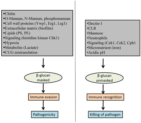
A growing appreciation in understanding the biology of host–fungi interaction is emerging. Host immune pathways involved in sensing fungal cell wall components have been characterized for C. albicans. This review is an attempt to collate various underlying factors and mechanisms (Figure 1) involved in Candida β-glucan masking from an already available pool of knowledge and provides an up-to-date understanding. The following sections will discuss various factors that are known to affect β-glucan masking, thereby presenting a resource to be utilized while designing antifungal therapeutic strategies to block fungal immune evasion.
Figure 1. Factors affecting β-glucan masking in C. albicans.

2. Factors That Influence β-Glucan Masking

2.1. Host-Pathogen Interaction

2.1.1. Dectin-1

Any invading pathogen, like Candida, faces a significant challenge from the host immune system. Pathogen recognition receptors (PRR) identify particular fungal pathogen-associated molecular patterns (PAMP), which include essential and specific fungal cell wall components such as glucan, chitin, and mannan (PRR). When fungi infect our bodies, the cell wall is crucial in triggering an immune response. The inner cell wall of fungi is high in glucan, which is responsible for triggering an inflammatory response, but immune cells are unable to recognize it. As soon as the -glucan is exposed, it is recognized by Dectin-1, a C-type lectin PRR [14,15][7][8]. When Dectin-1 recognizes β-glucan, myeloid cell signaling is activated along with the phagocytic response, and a pro-inflammatory cytokine response is introduced. Other activities for destroying fungal cells by neutrophils and macrophages are also initiated via their reactive oxygen (RO) and reactive nitrogen (RN) species [16,17][9][10]. Dectin-1 receptor shows an association with β-glucan, whereas the Dectin-2 receptor is associated with the α-mannan, which is found on the fungal cell’s outer covering [14,16][7][9]. With the aid of macrophages, dendritic cells, and neutrophils, Dectin-1 receptors boost innate immunity against Candida albicans [17,18][10][11]. Macrophages are essential in the detection of invading fungal cells. Si Min Chen et al. found that macrophages lacking Dectin-1 were unable to mount an effective response to C. albicans stimulation [19][12]. Ywp1 is one such component that helps the glucan particle remain masked and guards against immune cells’ identification [20,21][13][14]. In deprivation of O-mannan from the fungal cell wall, β-glucan gets unmasked and eventually gets recognized by Dectin-1, resulting in expanded phagosome maturation [22][15]. Nina Klippel and team showed that Chk1p is responsible for the masking of mutant β-glucan to prevent recognition by the immune cells [23][16]. Dectin-1 triggers CR 3 and S1GN-R1 to generate signaling. Dectin-2 and -3, on the other hand, have a synergistic effect on inflammatory responses [24][17]. Dectin-1 binds soluble β-glucan and particulate β-glucan, a ligand that is similar to β-glucan found in cell walls. In response to glucan-coated beads, dendritic cells (DCs) release reactive oxygen species (ROS). This response can be produced by glucan particles with a diameter of 500 nm or larger, but not by particles with a diameter of 200 nm or smaller. It indicates that glucan detection can be dependent on the physical properties of glucan at nanometric scales. However, the precise dimensions of β-glucan exposure necessary for Dectin-1 activity are still unknown. Since Dectin-1 signaling is ligand size-dependent, masked β-glucan exposures may be limited to non-stimulatory sizes, while unmasking processes may result in larger β-glucan exposures that are more effective at activating Dectin-1. This highly immunogenic molecular pattern is linked to dendritic cell, macrophage, and neutrophil activation, primarily by Dectin-1 and also by β2-integrin [17][10]. PRRs help DCs and other leukocytes identify the polysaccharides found on fungal cell walls. Dectin-1 and the DCSIGN transmembrane C-type lectins (CTLs) are the proteins bound to β-glucan and mannan, respectively. Dectin-1 detects β-glucan, triggering an immunogenic response that leads to cellular activation in innate immune cells and fungal phagocytosis. When PRRs are activated, DCs enter the picture and take care of both innate and adaptive immunity by guarding pathogen entry. As a result, controlling the amount and exposure of immunogenic cell-wall ligand is an important feature of the host-pathogen relationship that requires further investigation.

2.1.2. C-Type Lectin Receptor (CLR)

Infected patients are known to have inadequate C-type lectin receptors (CLRs) activity, and the CLRs are crucial in achieving antifungal immunity [25][18]. CARD9 is an intracellular signaling pathway for CLRs that plays an important role in preventing fungal infections [26][19]. When TLRs and CLRs are co-stimulated, the MyD88 and Syk/CARD9 pathways are activated, which improves the inflammatory response [24][17]. When these PAMPs are identified, the innate immune system is stimulated, involving phagocytic cells and the production of adaptive immunity at the same time [27][20]. A fungal invasion and an imbalance in the host defense response cause systemic candidiasis. CLRs, dendritic cell-specific intercellular adhesion molecule 3-grabbing non-integrin (DC-SIGN), Mincle and Galectin-3, mannose receptor (MR), complement receptor 3 (CR3), and Toll-like receptors (TLRs) are all PRRs that are responsible for the host immune system recognizing the fungal species. Mutations in the CLR pathway are linked to invasive fungal infections among these PRRs.

3.1.3. Mannose Receptor

This receptor can recognize mannose structure and is found on the surface of macrophages. The action of macrophages is affected by the exposure of glucan present on the fungal surface. The structure of mannan molecules plays a major role in glucan exposure. If mannan has simple molecular structure, it enhances the chances of exposure [5]. This receptor detaches from the surface of macrophages and is released into the environment to help our immune cells recognize fungi. Mannan behaves as an immune guard for macrophages that installs Dectin-1 as the primary immune recognition receptor [28][21]. Interleukin 6, 10, and INF-α, and TNF-γ are all produced by TLR 4 in conjunction with the mannose receptor. Dectin-2, -3 also acts as a dimer in RAW264.7 macrophages to identify mannans and secrete INF-α [29][22]. The primary unit found in N-mannose for immune recognition and fungus virulence is α -1, 2- mannose [30][23]. The stimulation of T-helper 1 (Th1) cytokine synthesis is thought to be mediated by mannose receptors. Macrophages ingest and kill C. albicans thanks to mannose receptors involved in TNF-α, IL-1, IL-6, and granulocyte-macrophage colony-stimulating factor (GM-CSF) responses. Human keratinocytes also express a mannose-binding receptor that causes anti-Candida activity, most likely through the development of nitrous oxide. During in vivo mouse studies of candidiasis, these receptors play a role in infection. In mannose-receptor-knockout mice, infection with Candida albicans via intraperitoneal injection leads to an increase in fungal burden [31][24].

2.1.4. Neutrophil Extracellular Trap (NET)

One aspect of the mechanisms required for altering epitope unmasking involves neutrophils causing changes in fungi’s innate pattern recognition. It sheds light on how the C. albicans cell wall reacts to the threat of a host immune response during disseminated candidiasis. This also leads to useful knowledge on host-pathogen responses during systemic infection. In a mode of neutrophil attack against C. albicans and other fungi in vivo and in vitro, neutrophils form neutrophil extracellular traps (NET) comprised of DNA and other antimicrobial components. While mouse neutrophils produce less NET than human neutrophils, neutrophil proteases are a key element of NET production in certain circumstances, and neutrophil elastase trafficking is controlled during NETosis (release and activation of neutrophil extracellular traps) toward Candida albicans. However, neutrophils from dipeptidyl peptidase (DPPI)-deficient mice—which are necessary for the activation of the three major neutrophil proteases: elastase, cathepsin G, and proteinase—showed no difference in their capacity to trigger β-glucan unmasking, chitin deposition, or streptavidin deposition. Thus, in this mechanism, these three proteases play a key part in the downstream cell wall remodelling caused by neutrophil assault. The aspartic proteases formed by C. albicans are chemotactic agents for neutrophils and are likely involved in their modulation through ROS generation. Furthermore, major components of the C. albicans cell wall can play a role in NETosis pathway activation. β-glucans, which are recognized by neutrophil Dectin-1 receptor, induce NET release, most likely through an ROS-independent mechanism [32][25]. However, the precise roles of each type of surface or secreted C. albicans components in NET formation are still unknown. Dectin-1 binding enhancement induced by neutrophils can alter the secondary immune response to Candida albicans, or the binding may not affect the response due to unnecessary recognition modalities. C. albicans use epitope masking and have developed innovative ways to escape pattern recognition receptors by covering unique epitopes. Several groups have identified how the fungal cell wall architecture restricts detection of the β-glucan sugar by immune receptors such as Dectin-1, a C-type lectin critical for fungal infection resistance [33][26]. It was discovered that patients with active symptoms had higher availability of -glucan, as well as an accumulation of neutrophils at the site. According to new research, neutrophils increase -glucan exposure in C. albicans, resulting in a stronger immune response. This means that during infections, the C. albicans cell wall will change due to environmental factors, which cause the fungal cell wall’s alterations, making it easily detectable for the Dectin-1 receptor [34][27], and the neutrophils are triggered when the fungal cell wall changes. Neutrophils begin the destruction of cell walls, assisting macrophages and cytokines in the destruction of fungal cells [33][26].
It has been demonstrated that neutrophils can detect fungi invasion early on and initiate an immune response. The phagocytosis of the fungal pathogen by innate immune cells is critical for the effective clearance of C. albicans from the host tissue. Pathogen identification triggers a multi-step mechanism called phagocytosis. This results in pathogen engulfment, phagosome maturation to remove the pathogen, and then phagolysosome resolution, which has been thoroughly reviewed by Levin et al. [35][28]. They use the MEK/ERK cascade to recognize invasive filamentous C. albicans, which causes macro type-specific neutrophil activation [36][29]. In comparison to the non-filamentous mutant fungus, these filamentous C. albicans types can manipulate phagosomal maturation [37][30]. Another study found that anoxic conditions reduced specific polymorphonuclear neutrophil (PMN) functions directed against C. albicans, such as NET formation, ROS triggering, phagocytosis, and the ability of C. albicans to form biofilms; however, as previously demonstrated for biofilms formed under normoxic conditions, anoxically formed biofilms can inhibit PMN functions. Infections in anoxic conditions confirmed the existence of ROS-independent pathways of NET induction. Fungal MAP kinase-driven responses, especially via Hog1, are triggered by NET-induced damage, limiting cell wall remodelling machinery. It has been discovered that -glucan unmasking necessitates NET moderated interference, which activates a change in the fungal cell wall that makes it easily identifiable for the Dectin-1 receptor [33][26]. NETs are more harmful to hyphae forms than they are to yeast forms. In the early stages of interaction, C. albicans stimulate NETs through autophagy and ROS [38,39][31][32]. Neutrophils have thus been established as one of the important factors for alterations in the fungal cell wall in a variety of studies.

2.2. Cell Wall Proteins

The β-glucan masking involves the role of some cell wall proteins under certain conditions. Ywp1 (yeast wall protein) is present in abundance in the cell wall of the yeast form of Candida, involved in β-glucan masking from immune recognition as it remains intact in the cell wall. Thus, there is less exposure of β-1,3-glucan by host antibodies, which favors more masking. Experimental studies showed that when antibody binding to β-1,3-glucan was more in germ tubes, very little or no Ywp1 was present, resulting in more β- glucan unmasking than in yeast forms. Ywp1 has an antiadhesive effect along with a masking effect [21][14]. Another protein, endo-1,3-β-glucanase Eng1, is also involved in β-glucan masking and studies demonstrated that in the absence of this protein, there is enhanced recognition byDectin-1 in host cells [40][33]. Lrg1is a protein encoding a GTPase-activating protein (GAP) which downregulates the GTPase Cdc42 and downstream MAPKKK and Ste11, representing a regulator of the Cek1 pathway. The study showed that hyperactivation of Cdc42in lrg1 mutant results in more exposure of β-glucan [41][34]. It has been demonstrated that deletion of this LRG1 gene leads to disruption of the Cek1 pathway and β-glucan becomes unmasked to become more prone for immune recognition by host cells. Lrg1 isa repressor of β-glucan unmasking which can in turn induce elevated levels of TNF-α secretion from murine macrophages. Rho1 is a regulatory subunit of the β-glucan synthase enzyme that directly controls biosynthesis of the cell wall through activation of catalytic subunits like Fks1 [42][35].
Kex2is a serine protease enzyme which resides in Golgi and is Ca2+-dependent and required for the proprotein dispensation step during maturation of protein prior to exiting the trans-Golgi network and does cycling through Golgi vesicles and late endosomal compartment [43][36]. It has been demonstrated that loss of the gene KEX2 leads to changes in organization of cell structure and negatively affects the N-linked mannosylation pathway. A Kex2∆ null mutant showed the amplified levels of β-glucan and thus partly exposes the surface of the cell, which accounts for the augmented IL-10 production in the interaction of cell and macrophage [44,45][37][38].

2.3. Chitin

Fungi can alter the broken structure of a cell by an alteration in chitin content to sustain cell wall integrity. In C. albicans, the immune recognition via mononuclear cells is blocked by chitin [46][39]. The unmasking of β-glucan leads to enlarged exposure of chitin content on the surface of C. albicans. Consequently, the antifungal drug echinocandin’s mechanism of resistance is by increased chitin biosynthesis [47][40]. It has been previously reported that caspofungin susceptibility is reduced with elevated chitin content in the cell surface of C. albicans in a mouse model of systemic infection. Additionally, the susceptibility of echinocandins is reduced with increased in-cell-wall chitin content of C. albicans, both in vivo and invitro [47][40]. The cell-wall-specific antifungal that targets the biosynthetic pathway disturbs the cell structure through β-glucan exposure, unmasking, or cell wall disorganization [48][41]. Caspofungin reduced the biosynthesis of β-glucan, which resulted in reduced levels of β-glucan in the cell [47][40]. This disturbance in the cell structure mechanism can cause more exposure of β-glucan. Even caspofungin-targeted cells elicited the TNF-alpha generation, which suggested that β-glucan unmasking by antifungal drug caspofungin induces the proinflammatory response. Hence, caspofungin may hit at Candida in a double way, where on one hand, it kills them at high concentrations but in lower concentrations leads to unmasking of β-glucan.

2.4. O-Mannan, N-Mannan, and Phosphomannan

In the architecture of the C. albicans cell wall, the outermost layer is composed of proteins that are covalently attached with O-linked or N-linked mannans, also known as mannoproteins. O-linked mannans are short, linear oligosaccharides which are an arrangement of seven α-1,2-mannose units. N-linked mannans are well-branched oligosaccharides incorporated with residues of mannose with several glycosidic linkages [49][42]. The importance of these N-linked and O-linked mannans’ structures lies in their involvement in the immunological distinctiveness of C. albicans and is understood as molecular patterns that interrelate with PRRs of the innate immune cells. They are immunostimulatory and involved in the identification and uptake of fungal cells by the immune cells and system. It has been reported that O-linked mannans are recognized by Toll-like receptor TLR4, whereas N-linked mannans are recognized with mannose receptors, Dectin-2, and Dectin-3, DC-SIGN [50,51][43][44]. Apart from sensing of β-glucan, it has been reported that macrophages also require the recognition of phosphomannan that negatively affects the phagocytic process. A study also reported that O-linked mannans detrimentally impact macrophage engulfment of C. albicans [44][37]. Phosphomannans are the β-1,2-mannose moiety which are bonded to branching N-glycan through a phosphodiester bond. They are recognized by immune cells and stimulate cytokine secretion [30][23]. A study [22][15] demonstrated a mutant in which there is loss of cell wall O-mannan, which results in enhanced attainment of phagosome maturation markers and weakens the hyphal growth within macrophage phagosomes. This brings macrophage change in actin dynamics and thus makes fungal cells incapable of escaping macrophages.

2.5. Extracellular Matrix

The Candida biofilms possess an extracellular matrix that is assembled from a polysaccharide complex which cooperates in the assemblage and maintenance of the matrix. In C. albicans, the polysaccharides present in the cell wall are β-glucan and mannans. It was mentioned that glucan is the major contributor for drug resistance in biofilm-mediated drug resistance [52][45]. The matrix β-1,6-glucans were linear in the biofilm matrix, in contrast with the branching structures located inside the cell wall. The role of β-1,3-glucan is vital in biofilm-mediated defense of C. albicans against host immunity and antifungal treatment [53][46].

2.6. Lipids

Lipids are an important component of membrane phospholipids in the cell membrane of Candida, so it might be interpreted that any disturbances in lipid homeostasis might affect the lipids or their related pathways, which can affect masking of β-glucan. It is known that PS (Phosphatidylserine) and PE (Phosphatidylethanolamine) make up 6% and 15% of total membrane phospholipids, respectively [54][47]. It is known that PS modulates certain signaling pathways which regulate the cell wall, such as the calcineurin signaling pathway, protein kinase C pathway, Hog1p, or Cek1p pathway [55][48]. A study demonstrated that a post-transcriptional regulator called Ccr4-Pop2 mRNA deadenylase is required for maintenance of cell wall integrity. The mutants of this regulator showed that defects in the functioning of Ccr4-Pop2 also relate to defects in phospholipid homeostasis and dysfunctional mitochondria. Additionally, Ccr4-Pop2 also plays a role in cell wall biogenesis, which correlates with the β-glucan activity and interlinks with mitochondria and phospholipid homeostasis [56][49]. A study has reported that PS is necessary for β-glucan masking against host immune system recognition. PS is encoded by PS synthase gene called Cho1. The loss of function of PS leads to decreased β-glucan masking and helps in enhancing immune recognition by host cells. There is also an increase in unmaskedβ-glucan exposure on the cell surface, evident from increased binding to Dectin-1 receptor and increased secretion of TNF-α by macrophages [57][50]. The PS synthase enzyme (Cho1) and PE are both crucial in the pathogenicity of C. albicans. Additionally, mutant CHO1 showed higher β-glucan exhibitions than non-mutant. This permits recognition to a greater extent by Dectin-1 and as a result, produces elevated pro-inflammatory responses [18][11].

2.7. Signaling Pathways

Several signaling pathways are also involved in host pathogen interaction and immune recognition by host cells. The MAPK pathways which include the cascade of Ste11-Hst7-Cek, Cek1 MAPK, have been reported to regulate β-glucan masking [58][51]. Two components of MAPK pathways such as Cek1 and Cek2 play vital roles in masking of β-glucan through disorganization of the external mannoprotein layer [59,60][52][53]. However, on the other hand, the hyperactivation of Cek1 can lead to unmasking of β-glucan. A transcription factor Cph1 encoded by CPH1 gene negatively regulates β-glucan masking and may cause unmasking upon hyperactivation [18][11]. A two-component signaling pathway called histidine kinase contributes to biosynthesis of the cell wall and virulence of C. albicans. In this pathway, several kinases are involved, such as CaSln1p, Cos1p, and Chk1p. They are also associated with the regulation of mannan biosynthesis [61,62][54][55]. However, Chk1pencoded by gene CHK1 is crucial for activation of phagocytes and represents an attractive target as there is no human homologue. A study by Klippel et al. also demonstrated that Chk1p is not only involved in maintaining the escape of C. albicans from ingestion by immune cells, but it also participates in blocking another event, such as phagosome maturation. The histidine kinase Chk1p plays a crucial role for masking of β-glucans in C. albicans from phagocyte recognition [23][16].

2.8. Micronutrient

During infection, C. albicans encounters various anatomical niches which are micronutrient depleted. One such factor is the availability of iron, as it is not freely available due to its transitional nature. It has been studied that the recognition of the fungal cell wall and stimulation of the cytokine by host immune cells is regulated by the iron, and iron-depleted conditions can induce β-glucan masking [63,64,65][56][57][58]. High iron-containing media showed higher levels of exposed β-glucan in comparison to a limited iron condition. The glucose media with a limited iron condition exhibits decreases in mitochondria activity due to the presence of various heme iron-containing proteins [66][59]. This leads to changes in cellular metabolism towards fermentation mode, which increases lactate production, while in cases of high iron-containing media, a reduction in lactate production occurs [67][60]. It is suggested that glucose-grown, high iron-containing cells have improved mitochondrial activity, which can reduce lactate-mediated β-1,3-glucan masking. Iron-rich media also reduce the level of total mannans as well as phosphomannans in the outer cell surface, thereby increasing β-1,3-glucan. Gpr1 and Crz1 genes are mediated by exogenous lactate and target β-1,3-glucan masking [68][61]. The change governed by iron that induces β-glucan exposure is affected by intracellular L-lactate. This change is GPR1-independent, but Crz1-dependent [69][62]. β-glucan masking can be affected by varying zinc concentrations, where zinc deprivation increases β-glucan exposure.

2.9. Hypoxia

Hypoxia (low oxygen) is another stress condition that infectious fungi face inside the host [70][63]. C. albicans shows a high resistance to hypoxia [16][9], and accordingly, these infectious fungi can inhabit a hypoxic environment such as the gastrointestinal region [71][64], in addition to aerobic environments including the skin and mucosa. Hypoxia conditions lead to improvements in virulence and β-glucan masking on the fungal cell surface. Additionally, Efg1 plays an important role in the regulation of the hypoxic stress in C. albicans, [72][65] and Hog1 pathway has a role in control of the hypoxic responses in Saccharomyces cerevisiae and human cells [73][66]. β-glucan masking is induced by hypoxia and is dependent and regulated by cAMP- protein kinase A signaling pathway. cAMP plays a crucial function in cell surface integrity, yeast to hyphae transformation, and stress adaption [74][67]. It is suggested that under hypoxic conditions, C. albicans enhances its survival by avoiding polymorphonuclear leukocyte attacks through β-glucan masking [75][68]
It has been reported that more polymorphonuclear leukocytes at the site of an infection promote less oxygen, which turned out to be beneficial for the fungi as phagocytic cells could not produce ROS [75][68]. Low oxygen conditions improve C. albicans’ survival due to better masking of β-glucan and avoidance of Dectin-1 recognition. As a result, less phagocytosis by macrophages occurs and there is a drop in production of IL-10 and TNF-α. This suggested that C. albicans and host interactions are remarkably affected by β-glucan masking that is promoted by hypoxia. Generally, β-glucan masking depends on cAMP-PKA, and the mitochondrial signaling pathway which regulates local immune response and supports fungal assembly [76][69].

2.10. pH

C. albicans colonizes niches like the abdomen, vagina, and gastrointestinal traits, which have varied pH C. albicans tends to show that growth within the acid atmosphere involves renovation of plasma membranes that manufacture exaggerated chitin and β-glucan on the cell wall. Vaginal mucosa has a pH ranging between 4–5, depending on age and origin [77][70]. A study on the pH alteration on C. albicans explored that exposure to acidic conditions induces the contact of β glucan and chitin, which are usually hidden below the mannan fibrils [78][71]. Both factors like β-glucan and chitin are recognized by the natural immune response [68][61] and at low pH, C. albicans activates a strong pro-inflammatory natural immune reaction and improved neutrophil recruitment [79][72]. At present, the defined cell wall remodeling because of acidic pH on cell surface unmasking is unidentified. Visualization of the configuration of C. albicans revealed that the mannan fiber was condensed due to acidic conditions, which may be the reason for less mannan production. Rim 101 is a signal transduction pathway known to be involved in cell wall remodeling by proper localization of chitin in the cell wall apart from its usual role in governing pH response. Due to the involvement of Bcr1-Rim101-dependent signal cascade in chitin localization through regulation of chitinase Cht2p, it is hypothesized that this transcription factor may also be involved in β-glucan unmasking in a pH-dependent manner [78][71].

2.11. Metabolites

The structural design of the C. albicans cell surface gets altered extensively due to carbon sources. For instance, cells grown on lactate acquire thinner, smaller amounts of flexibility in cell walls and an altered immune response compared to complex media such as glucose-grown cells [80,81][73][74]. The ratio of the β-glucan compared to chitin and mannan remains similar [80][73], as exposure of β-glucan on the cell surface might be affected by the carbon source. Further analysis exposed that C. albicans cells activate PAMP β-glucan masking at the cell wall in response to functional levels of lactate, produced either by host cells or fungal cells [82][75]. The β-glucan masking response induced by lactate is linked with an attenuation of inflammatory reaction and neutrophil recruitment. This masking, which is induced by lactate, is regulated by a signaling pathway that plays an important role in mechanisms other than well-established signaling pathways [82][75]. A recent study demonstrated that lactate-induced masking of β-glucan is governed by exoglucanase enzyme Xog1p that digests β-glucan epitopes to enhance immune evasion, thereby representing an antifungal target [14][7]. Metabolism of lactate is crucial for β-glucan masking and its stimulation depends on uptake using glyoxylate cycle enzyme isocitrate lyase [81][74] and Jen1/2 transporters [83][76]. Gpr1p receptor helps in the recognition of lactate, which is present in the cyclic AMP-protein kinase A pathway that activates lactate-induced β-glucan masking and virulence factor [84][77]. Consequently, Cag1 and Gpa2 genes may alter the function of Gpr1 signaling for masking of β-glucan and cyclic-AMP pathways through a process that remains to be defined. Lactate signaling is also influenced by involvement of the factor Crz1p, which controls cell surface integrity, drug tolerance, cell wall remodeling, and the specific response to calcium ions through calcineurin signaling [85][78]. The decreased wall β-glucan levels being less vulnerable to caspofungin signifies that environmental variation to different sources of carbon may control therapeutic options.

2.12. Mistranslation of CUG

The interaction of the host with the pathogen is also influenced by CUG mistranslation. In C. albicans, mistranslation of CUG occurs due to dual coding for serine (97%) and leucine (3%). The CUG mistranslation helps in masking the β-glucan molecule present in the cell wall, which is generally detected by the host immune system [6,86][6][79]. The studies showed that manipulation of genetic ambiguity results in increased expression of virulence factors such as morphogenesis, phenotypic switching, and cell adhesion [87][80]. The increase of leucine, a hydrophobic amino acid, results in the increased adherence of the pathogen to the host cell. The changes brought by CUG mistranslation influences the exposure of β-glucan on the surface of cells and reduces macrophage phagocytosis of C. albicans. It has been reported that the rise in adherence is interceded by two mechanisms. Firstly, CUG mistranslation increases cell surface hydrophobicity, which is crucial in the attachment of C. albicans to abiotic surfaces and host cells [88,89][81][82]. Secondly, CUG mistranslation enhances the substrate strength of adhesins like Als proteins. The presence of Als3p-Leu in yeast promotes more adherence in comparison with the expression of Als3p-Ser. CUG mistranslation enhances diversity of structures and modulates the adherence of other cell wall proteins. The functional variability of CUG mistranslation may widen the array of host substrates, of which C. albicans can bind through, manipulating the interaction of the ligand region. Previous studies reported that the extent of leucine misincorporation at CUG codons fluctuates, and greatly depends upon external conditions such as temperature and pH [87][80]. CUG mistranslation confers an advantage by increasing the capability of cells for adhering to a wide range of host substrates and minimizing connections with immune effector cells. This factor has not only provided greater diversity in proteins on the cell surface of C. albicans but also contributes to increasing the range of metabolic responses of C. albicans. Thus, CUG mistranslation expands the host micro niches in which C. albicans can thrive well to survive [90][83].

References

  1. Beardsley, J.; Halliday, C.L.; Chen, S.C.; Sorrell, T.C. Responding to the emergence of antifungal drug resistance: Perspectives from the bench and the bedside. Future Microbiol. 2018, 13, 1175–1191.
  2. Singh, S.; Fatima, Z.; Hameed, S. Predisposing factors endorsing Candida infections. Infez. Med. 2015, 23, 211–223.
  3. Perfect, J.R. The antifungal pipeline: A reality check. Nat. Rev. Drug Discov. 2017, 16, 603–616.
  4. Deorukhkar, S.C.; Saini, S.; Mathew, S. Virulence Factors Contributing to Pathogenicity of Candida tropicalis and Its Antifungal Susceptibility Profile. Int. J. Microbiol. 2014, 2014, 456878.
  5. Graus, M.S.; Wester, M.J.; Lowman, D.W.; Williams, D.L.; Kruppa, M.D.; Martinez, C.M.; Young, J.M.; Pappas, H.C.; Lidke, K.A.; Neumann, A.K. Mannan Molecular Substructures Control Nanoscale Glucan Exposure in Candida. Cell Rep. 2018, 24, 2432–2442.
  6. Goodridge, H.S.; Wolf, A.J.; Underhill, D.M. Beta-glucan recognition by the innate immune system. Immunol. Rev. 2009, 230, 38–50.
  7. Childers, D.S.; Avelar, G.M.; Bain, J.M.; Pradhan, A.; Larcombe, D.E.; Netea, M.G.; Erwig, L.P.; Gow, N.; Brown, A. Epitope Shaving Promotes Fungal Immune Evasion. mBio 2020, 11, e00984-20.
  8. Wester, M.J.; Lin, J.; Neumann, A.K. A computational model for regulation of nanoscale glucan exposure in Candida albicans. PLoS ONE 2017, 12, e0188599.
  9. Pradhan, A.; Avelar, G.M.; Bain, J.M.; Childers, D.; Pelletier, C.; Larcombe, D.E.; Shekhova, E.; Netea, M.G.; Brown, G.D.; Erwig, L.; et al. Non-canonical signalling mediates changes in fungal cell wall PAMPs that drive immune evasion. Nat. Commun. 2019, 10, 5315.
  10. Lin, J.; Wester, M.J.; Graus, M.S.; Lidke, K.A.; Neumann, A.K. Nanoscopic cell-wall architecture of an immunogenic ligand in Candida albicans during antifungal drug treatment. Mol. Biol. Cell 2016, 27, 1002–1014.
  11. Chen, T.; Jackson, J.W.; Tams, R.N.; Davis, S.E.; Sparer, T.E.; Reynolds, T.B. Exposure of Candida albicans β (1,3)-glucan is promoted by activation of the Cek1 pathway. PLoS Genet. 2019, 15, e1007892.
  12. Chen, S.M.; Zou, Z.; Qiu, X.R.; Hou, W.T.; Zhang, Y.; Fang, W.; Chen, Y.L.; Wang, Y.D.; Jiang, Y.Y.; Shen, H.; et al. The critical role of Dectin-1 in host controlling systemic Candida krusei infection. Am. J. Transl. Res. 2019, 11, 721–732.
  13. Granger, B.L. Propeptide genesis by Kex2-dependent cleavage of yeast wall protein 1 (Ywp1) of Candida albicans. PLoS ONE 2018, 13, e0207955.
  14. Granger, B.L. Accessibility and contribution to glucan masking of natural and genetically tagged versions of yeast wall protein 1 of Candida albicans. PLoS ONE 2018, 13, e0191194.
  15. Bain, J.M.; Louw, J.; Lewis, L.E.; Okai, B.; Walls, C.A.; Ballou, E.R.; Walker, L.A.; Reid, D.; Munro, C.A.; Brown, A.J.; et al. Candida albicans hypha formation and mannan masking of β-glucan inhibit macrophage phagosome maturation. mBio 2014, 5, e01874.
  16. Klippel, N.; Cui, S.; Groebe, L.; Bilitewski, U. Deletion of the Candida albicans histidine kinase gene CHK1 improves recognition by phagocytes through an increased exposure of cell wall beta-1,3-glucans. Microbiology 2010, 156, 3432–3444.
  17. Childers, D.S.; Avelar, G.M.; Bain, J.M.; Larcombe, D.E.; Pradhan, A.; Budge, S.; Heaney, H.; Brown, A. Impact of the Environment upon the Candida albicans Cell Wall and Resultant Effects upon Immune Surveillance. Curr. Top. Microbiol. Immunol. 2020, 425, 297–330.
  18. Hopke, A.; Brown, A.J.; Hall, R.A.; Wheeler, R.T. Dynamic fungal cell wall architecture in stress adaptation and immune evasion. Trends Microbiol. 2018, 26, 284–295.
  19. Marakalala, M.J.; Vautier, S.; Potrykus, J.; Walker, L.A.; Shepardson, K.M.; Hopke, A.; Mora-Montes, H.M.; Kerrigan, A.; Netea, M.G.; Murray, G.I.; et al. Differential adaptation of Candida albicans in vivo modulates immune recognition by dectin-1. PLoS Pathog. 2013, 9, e1003315.
  20. Brown, A.J.; Gow, N.A.; Warris, A.; Brown, G.D. Memory in fungal pathogens promotes immune evasion, colonisation, and infection. Trends Microbiol. 2019, 27, 219–230.
  21. Yadav, B.; Mora-Montes, H.M.; Wagener, J.; Cunningham, I.; West, L.; Haynes, K.; Brown, A.J.P.; Gow, N.A.R. Differences in fungal immune recognition by monocytes and macrophages: N-mannan can be a shield or activator of immune recognition. Cell Surf. 2020, 6, 100042.
  22. Cottier, F.; Hall, R.A. Face/off: The interchangeable side of Candida albicans. Front. Cell. Infect. Microbiol. 2020, 9, 471.
  23. Hall, R.A.; Bates, S.; Lenardon, M.D.; MacCallum, D.M.; Wagener, J.; Lowman, D.W.; Kruppa, M.D.; Williams, D.L.; Odds, F.C.; Brown, A.J.; et al. The Mnn2 mannosyltransferase family modulates mannoprotein fibril length, immune recognition and virulence of Candida albicans. PLoS Pathog. 2013, 9, e1003276.
  24. Lee, S.J.; Zheng, N.Y.; Clavijo, M.; Nussenzweig, M.C. Normal host defense during systemic candidiasis in mannose receptor-deficient mice. Infect. Immun. 2003, 71, 437–445.
  25. Zawrotniak, M.; Bochenska, O.; Karkowska-Kuleta, J.; Seweryn-Ozog, K.; Aoki, W.; Ueda, M.; Kozik, A.; Rapala-Kozik, M. Aspartic Proteases and Major Cell Wall Components in Candida albicans Trigger the Release of Neutrophil Extracellular Traps. Front. Cell Infect. Microbiol. 2017, 7, 414.
  26. Hopke, A.; Nicke, N.; Hidu, E.E.; Degani, G.; Popolo, L.; Wheeler, R.T. Neutrophil Attack Triggers Extracellular Trap-Dependent Candida Cell Wall Remodeling and Altered Immune Recognition. PLoS Pathog. 2016, 5, e1005644.
  27. Pericolini, E.; Perito, S.; Castagnoli, A.; Gabrielli, E.; Mencacci, A.; Blasi, E.; Vecchiarelli, A.; Wheeler, R.T. Epitope unmasking in vulvovaginal candidiasis is associated with hyphal growth and neutrophilic infiltration. PLoS ONE 2018, 13, e0201436.
  28. Levin, R.; Grinstein, S.; Canton, J. The life cycle of phagosomes: Formation, maturation, and resolution. Immunol. Rev. 2016, 273, 156–179.
  29. Wozniok, I.; Hornbach, A.; Schmitt, C.; Frosch, M.; Einsele, H.; Hube, B.; Löffler, J.; Kurzai, O. Induction of ERK-kinase signalling triggers morphotype-specific killing of Candida albicans filaments by human neutrophils. Cell. Microbiol. 2008, 10, 807–820.
  30. Seider, K.; Heyken, A.; Lüttich, A.; Miramón, P.; Hube, B. Interaction of pathogenic yeasts with phagocytes: Survival, persistence and escape. Curr. Opin. Microbiol. 2010, 13, 392–400.
  31. Small, A.G.; King, J.R.; Rathjen, D.A.; Ferrante, A. The role of phagocytes in immunity to Candida albicans. In Candida Albicans; IntechOpen: London, UK, 2018.
  32. Last, A.; Maurer, M.; Mosig, A.S.; Gresnigt, M.S.; Hube, B. In vitro infection models to study fungal-host interactions. FEMS Microbiol. Rev. 2021, 1, fuab005.
  33. Yang, M.; Solis, N.; Marshall, M.; Garleb, R.; Tingting, Z.; Wang, D.; Swidergall, M.; Pearlman, E.; Filler, S.; Liu, H. Control of β-glucan exposure by the endo-1,3-glucanase Eng1 in Candida albicans modulates virulence. bioRxiv 2020.
  34. Chen, T.; Wagner, A.S.; Tams, R.N.; Eyer, J.E.; Kauffman, S.J.; Gann, E.R.; Fernandez, E.J.; Reynolds, T.B. Lrg1 Regulates β (1,3)-Glucan Masking in Candida albicans through the Cek1 MAP Kinase Pathway. mBio 2019, 10, e01767-19.
  35. Qadota, H.; Python, C.P.; Inoue, S.B.; Arisawa, M.; Anraku, Y.; Zheng, Y.; Watanabe, T.; Levin, D.E.; Ohya, Y. Identification of yeast Rho1p GTPase as a regulatory subunit of 1,3-beta-glucan synthase. Science 1996, 272, 279–281.
  36. Fuller, R.S.; Brake, A.J.; Thorner, J. Intracellular targeting and structural conservation of a prohormone-processing endoprotease. Science 1989, 246, 482–486.
  37. McKenzie, C.G.; Koser, U.; Lewis, L.E.; Bain, J.M.; Mora-Montes, H.M.; Barker, R.N.; Gow, N.A.; Erwig, L.P. Contribution of Candida albicans cell wall components to recognition by and escape from murine macrophages. Infect. Immun. 2010, 78, 1650–1658.
  38. Gómez-Gaviria, M.; Lozoya-Pérez, N.E.; Staniszewska, M.; Franco, B.; Niño-Vega, G.A.; Mora-Montes, H.M. Loss of Kex2 Affects the Candida albicans Cell Wall and Interaction with Innate Immune Cells. J. Fungi 2020, 6, 57.
  39. Mora-Montes, H.M.; Netea, M.G.; Ferwerda, G.; Lenardon, M.D.; Brown, G.D.; Mistry, A.R.; Kullberg, B.J.; O’Callaghan, C.A.; Sheth, C.C.; Odds, F.C.; et al. Recognition and Blocking of Innate Immunity Cells by Candida albicans Chitin. Infect. Immun. 2011, 79, 1961–1970.
  40. Lee, K.K.; Maccallum, D.M.; Jacobsen, M.D.; Walker, L.A.; Odds, F.C.; Gow, N.A.; Munro, C.A. Elevated cell wall chitin in Candida albicans confers echinocandin resistance in vivo. Antimicrob. Agents Chemother. 2012, 56, 208–217.
  41. Gow, N.A.; Netea, M.G.; Munro, C.A.; Ferwerda, G.; Bates, S.; Mora-Montes, H.M.; Walker, L. Immune recognition of Candida albicans beta-glucan by dectin-1. J. Infect. Dis. 2007, 196, 1565–1571.
  42. Martínez-Duncker, I.; Díaz-Jímenez, D.F.; Mora-Montes, H.M. Comparative Analysis of Protein Glycosylation Pathways in Humans and the Fungal Pathogen Candida albicans. Int. J. Microbiol. 2014, 2014, 1–16.
  43. Netea, M.G.; Gow, N.A.; Munro, C.A.; Bates, S.; Collins, C.; Ferwerda, G.; Hobson, R.P.; Bertram, G.; Hughes, H.B.; Jansen, T.; et al. Immune sensing of Candida albicans requires cooperative recognition of mannans and glucans by lectin and Toll-like receptors. J. Clin. Investig. 2006, 116, 1642–1650.
  44. Cambi, A.; Netea, M.G.; Mora-Montes, H.M.; Gow, N.A.; Hato, S.V.; Lowman, D.W.; Kullberg, B.J.; Torensma, R.; Williams, D.L.; Figdor, C.G. Dendritic cell interaction with Candida albicans critically depends on N-linked mannan. J. Biol. Chem. 2008, 283, 20590–20599.
  45. Zarnowski, R.; Westler, W.M.; Lacmbouh, G.A.; Marita, J.M.; Bothe, J.R.; Bernhardt, J.; Lounes-Hadj Sahraoui, A.; Fontaine, J.; Sanchez, H.; Hatfield, R.D.; et al. Novel entries in a fungal biofilm matrix encyclopedia. mBio 2014, 5, e01333-14.
  46. Sheppard, D.C.; Howell, P.L. Biofilm Exopolysaccharides of Pathogenic Fungi: Lessons from Bacteria. J. Biol. Chem. 2016, 291, 12529–12537.
  47. Chen, Y.L.; Montedonico, A.E.; Kauffman, S.; Dunlap, J.R.; Menn, F.M.; Reynolds, T.B. Phosphatidylserine synthase and phosphatidylserine decarboxylase are essential for cell wall integrity and virulence in Candida albicans. Mol. Microbiol. 2010, 75, 1112–1132.
  48. Monge, R.A.; Román, E.; Nombela, C.; Pla, J. The MAP kinase signal transduction network in Candida albicans. Microbiology 2006, 152, 905–912.
  49. Dagley, M.J.; Gentle, I.E.; Beilharz, T.H.; Pettolino, F.A.; Djordjevic, J.T.; Lo, T.L.; Uwamahoro, N.; Rupasinghe, T.; Tull, D.L.; McConville, M.; et al. Cell wall integrity is linked to mitochondria and phospholipid homeostasis in Candida albicans through the activity of the post-transcriptional regulator Ccr4-Pop2. Mol. Microbiol. 2011, 79, 968–989.
  50. Davis, S.E.; Hopke, A.; Minkin, S.C., Jr.; Montedonico, A.E.; Wheeler, R.T.; Reynolds, T.B. Masking of β(1-3)-glucan in the cell wall of Candida albicans from detection by innate immune cells depends on phosphatidylserine. Infect. Immun. 2014, 82, 4405–4413.
  51. Galán-Díez, M.; Arana, D.M.; Serrano-Gómez, D.; Kremer, L.; Casasnovas, J.M.; Ortega, M.; Cuesta-Domínguez, A.; Corbí, A.L.; Pla, J.; Fernández-Ruiz, E. Candida albicans beta-glucan exposure is controlled by the fungal CEK1-mediated mitogen-activated protein kinase pathway that modulates immune responses triggered through dectin-1. Infect. Immun. 2010, 78, 1426–1436.
  52. Roman, E.; Correia, I.; Salazin, A.; Fradin, C.; Jouault, T.; Poulain, D.; Liu, F.T.; Pla, J. The Cek1-mediated MAP kinase pathway regulates exposure of α-(1,2) and β-(1,2)-mannosides in the cell wall of Candida albicans modulating immune recognition. Virulence 2016, 7, 558–577.
  53. Correia, I.; Prieto, D.; Román, E.; Wilson, D.; Hube, B.; Alonso-Monge, R.; Pla, J. Cooperative Role of MAPK Pathways in the Interaction of Candida albicans with the Host Epithelium. Microorganisms 2019, 8, 48.
  54. Kruppa, M.; Goins, T.; Cutler, J.E.; Lowman, D.; Williams, D.; Chauhan, N.; Menon, V.; Singh, P.; Li, D.; Calderone, R. The role of the Candida albicans histidine kinase [CHK1) gene in the regulation of cell wall mannan and glucan biosynthesis. FEMS Yeast Res. 2003, 3, 289–299.
  55. Kruppa, M.; Jabra-Rizk, M.A.; Meiller, T.F.; Calderone, R. The histidine kinases of Candida albicans: Regulation of cell wall mannan biosynthesis. FEMS Yeast Res. 2004, 4, 409–416.
  56. Ramanan, N.; Wang, Y. A high-affinity iron permease essential for Candida albicans virulence. Science 2000, 288, 1062–1064.
  57. Almeida, R.S.; Wilson, D.; Hube, B. Candida albicans iron acquisition within the host. FEMS Yeast Res. 2009, 9, 1000–1012.
  58. Potrykus, J.; Stead, D.; Maccallum, D.M.; Urgast, D.S.; Raab, A.; van Rooijen, N.; Feldmann, J.; Brown, A.J. Fungal iron availability during deep seated candidiasis is defined by a complex interplay involving systemic and local events. PLoS Pathog. 2013, 9, e1003676.
  59. Grahl, N.; Demers, E.G.; Lindsay, A.K.; Harty, C.E.; Willger, S.D.; Piispanen, A.E.; Hogan, D.A. Mitochondrial activity and Cyr1 are key regulators of Ras1 activation of C. albicans virulence pathways. PLoS Pathog. 2015, 11, e1005133.
  60. Finch, C.A.; Gollnick, P.D.; Hlastala, M.P.; Miller, L.R.; Dillmann, E.; Mackler, B. Lactic acidosis as a result of iron deficiency. J. Clin. Investig. 1979, 64, 129–137.
  61. Amarsaikhan, N.; Sands, E.M.; Shah, A.; Abdolrasouli, A.; Reed, A.; Slaven, J.E.; Armstrong-James, D.; Templeton, S.P. Caspofungin Increases Fungal Chitin and Eosinophil and γδ T Cell-Dependent Pathology in Invasive Aspergillosis. J. Immunol. 2017, 199, 624–632.
  62. Lamaris, G.A.; Lewis, R.E.; Chamilos, G.; May, G.S.; Safdar, A.; Walsh, T.J.; Raad, I.I.; Kontoyiannis, D.P. Caspofungin-mediated beta-glucan unmasking and enhancement of human polymorphonuclear neutrophil activity against Aspergillus and non-Aspergillus hyphae. J. Infect. Dis. 2008, 198, 186–192.
  63. Grahl, N.; Shepardson, K.M.; Chung, D.; Cramer, R.A. Hypoxia and fungal pathogenesis: To air or not to air? Eukaryot. Cell 2012, 11, 560–570.
  64. Synnott, J.M.; Guida, A.; Mulhern-Haughey, S.; Higgins, D.G.; Butler, G. Regulation of the hypoxic response in Candida albicans. Eukaryot. Cell 2010, 9, 1734–1746.
  65. Sellam, A.; van Het Hoog, M.; Tebbji, F.; Beaurepaire, C.; Whiteway, M.; Nantel, A. Modeling the transcriptional regulatory network that controls the early hypoxic response in Candida albicans. Eukaryot. Cell 2014, 13, 675–690.
  66. Stichternoth, C.; Ernst, J.F. Hypoxic adaptation by Efg1 regulates biofilm formation by Candida albicans. Appl. Environ. Microbiol. 2009, 75, 3663–3672.
  67. Hickman, M.J.; Spatt, D.; Winston, F. The Hog1 mitogen-activated protein kinase mediates a hypoxic response in Saccharomyces cerevisiae. Genetics 2011, 188, 325–338.
  68. Lopes, J.P.; Stylianou, M.; Backman, E.; Holmberg, S.; Jass, J.; Claesson, R.; Urban, C.F. Evasion of Immune Surveillance in Low Oxygen Environments Enhances Candida albicans Virulence. mBio 2018, 9, e02120-e18.
  69. Pradhan, A.; Avelar, G.M.; Bain, J.M.; Childers, D.S.; Larcombe, D.E.; Netea, M.G.; Shekhova, E.; Munro, C.A.; Brown, G.D.; Erwig, L.P.; et al. Hypoxia Promotes Immune Evasion by Triggering β-Glucan Masking on the Candida albicans Cell Surface via Mitochondrial and cAMP-Protein Kinase a Signaling. mBio 2018, 9, e01318-e18.
  70. Linhares, I.M.; Summers, P.R.; Larsen, B.; Giraldo, P.C.; Witkin, S.S. Contemporary perspectives on vaginal pH and lactobacilli. Am. J. Obstet. Gynecol. 2011, 204, 120.e1–120.e5.
  71. Sherrington, S.L.; Sorsby, E.; Mahtey, N.; Kumwenda, P.; Lenardon, M.D.; Brown, I.; Ballou, E.R.; MacCallum, D.M.; Hall, R.A. Adaptation of Candida albicans to environmental pH induces cell wall remodelling and enhances innate immune recognition. PLoS Pathog. 2017, 13, e1006403.
  72. Wheeler, R.T.; Kombe, D.; Agarwala, S.D.; Fink, G.R. Dynamic, morphotype-specific Candida albicans beta-glucan exposure during infection and drug treatment. PLoS Pathog. 2008, 4, e1000227.
  73. Ene, I.V.; Adya, A.K.; Wehmeier, S.; Brand, A.C.; MacCallum, D.M.; Gow, N.A.; Brown, A.J. Host carbon sources modulate cell wall architecture, drug resistance and virulence in a fungal pathogen. Cell Microbiol. 2012, 14, 1319–1335.
  74. Ene, I.V.; Walker, L.A.; Schiavone, M.; Lee, K.K.; Martin-Yken, H.; Dague, E.; Gow, N.A.; Munro, C.A.; Brown, A.J. Cell wall remodeling enzymes modulate fungal cell wall elasticity and osmotic stress resistance. mBio 2015, 6, e00986.
  75. Ballou, E.R.; Avelar, G.M.; Childers, D.S.; Mackie, J.; Bain, J.M.; Wagener, J.; Kastora, S.L.; Panea, M.D.; Hardison, S.E.; Walker, L.A. Lactate signalling regulates fungal β-glucan masking and immune evasion. Nat. Microbiol. 2016, 2, 16238.
  76. Vieira, N.; Casal, M.; Johansson, B.; MacCallum, D.M.; Brown, A.J.; Paiva, S. Functional specialization and differential regulation of short-chain carboxylic acid transporters in the pathogen Candida albicans. Mol. Microbiol. 2010, 75, 1337–1354.
  77. Maidan, M.M.; De Rop, L.; Serneels, J.; Exler, S.; Rupp, S.; Tournu, H.; Thevelein, J.M.; Van Dijck, P. The G protein-coupled receptor Gpr1 and the Galpha protein Gpa2 act through the cAMP-protein kinase A pathway to induce morphogenesis in Candida albicans. Mol. Biol. Cell 2005, 16, 1971–1986.
  78. Karababa, M.; Valentino, E.; Pardini, G.; Coste, A.T.; Bille, J.; Sanglard, D. CRZ1, a target of the calcineurin pathway in Candida albicans. Mol. Microbiol. 2006, 59, 1429–1451.
  79. Miranda, I.; Silva-Dias, A.; Rocha, R.; Teixeira-Santos, R.; Coelho, C.; Gonçalves, T.; Santos, M.A.; Pina-Vaz, C.; Solis, N.V.; Filler, S.G.; et al. Candida albicans CUG mistranslation is a mechanism to create cell surface variation. mBio 2013, 4, e00285-e13.
  80. Miranda, I.; Rocha, R.; Santos, M.C.; Mateus, D.D.; Moura, G.R.; Carreto, L.; Santos, M.A. A genetic code alteration is a phenotype diversity generator in the human pathogen Candida albicans. PLoS ONE 2007, 2, e996.
  81. Gomes, A.C.; Miranda, I.; Silva, R.M.; Moura, G.R.; Thomas, B.; Akoulitchev, A.; Santos, M.A. A genetic code alteration generates a proteome of high diversity in the human pathogen Candida albicans. Genome Biol. 2007, 8, R206.
  82. Almeida, R.S.; Brunke, S.; Albrecht, A.; Thewes, S.; Laue, M.; Edwards, J.E.; Filler, S.G.; Hube, B. The hyphal-associated adhesin and invasin Als3 of Candida albicans mediates iron acquisition from host ferritin. PLoS Pathog. 2008, 4, e1000217.
  83. Rauceo, J.M.; Gaur, N.K.; Lee, K.G.; Edwards, J.E.; Klotz, S.A.; Lipke, P.N. Global cell surface conformational shift mediated by a Candida albicans adhesin. Infect. Immun. 2004, 72, 4948–4955.
More