Polydatin/Resveratrol interference in ACE2:Spike recognition: Comparison
Please note this is a comparison between Version 1 by Domenica Musumeci and Version 2 by Amina Yu.

In the search for new therapeutic strategies to contrast SARS-CoV-2, we here studied the interaction of polydatin (PD) and resveratrol (RESV)—two natural stilbene polyphenols with manifold, well known biological activities—with Spike, the viral protein essential for virus entry into host cells, and ACE2, the angiotensin-converting enzyme present on the surface of multiple cell types (including respiratory epithelial cells) which is the main host receptor for Spike binding. Molecular Docking simulations evidenced that both compounds can bind Spike, ACE2 and the ACE2:Spike complex with good affinity, although the interaction of PD appears stronger than that of RESV on all the investigated targets. Preliminary biochemical assays revealed a significant inhibitory activity of the ACE2:Spike recognition with a dose-response effect only in the case of PD. 

  • SARS-CoV-2
  • polydatin
  • resveratrol
  • molecular docking
  • protein-binding
  • ACE2:Spike binding-inhibition

1. Introduction
Coronaviruses (CoV) are a large family of viruses that may cause disease in animals or humans [1][2][3][1,2,3]. They can provoke respiratory infections ranging from the common cold to more severe illnesses [3]. The novel coronavirus, called SARS-CoV-2, which emerged in December 2019 causing coronavirus disease 2019 (COVID-19), can lead to serious, even fatal, disease [4][5][6][4,5,6], and was declared a global pandemic by the World Health Organization on 11 March 2020.
All coronaviruses possess an enveloped, positive-sense, single-stranded RNA genome encoding for 4 structural membrane proteins, i.e., Spike (S), envelope (E), membrane (M) and nucleocapsid (N) proteins [7]. The Spike proteins S are essential for viral entry into host cells, which occurs essentially through binding to the angiotensin-converting enzyme ACE2 [8][9][10][11][8,9,10,11]. ACE2 is present on the surface of multiple cell types, including respiratory and intestinal epithelial cells, endothelial cells, kidney cells (renal tubules), cerebral neurons, and immune cells, such as alveolar monocytes/macrophages [12][13][12,13].
Therefore, bioactive compounds able to inhibit the interaction between the COVID-19 S protein and the ACE2 receptor may be precious drugs for effective antiviral therapeutic strategies [14]. Indeed, human neutralizing antibodies targeting S protein and blocking SARS-CoV-2 cellular entry are promising therapeutic tools [15][16][17][18][19][15,16,17,18,19].
After attachment of the virus, a proteolytic enzyme of the host cell, mainly type II transmembrane serine protease TMPRSS2, cleaves and activates the receptor-attached Spike macromolecule [20]. This protease, anchored in the cell membrane near ACE2 receptors, and expressed in the epithelial cell lining of the nose, trachea and distal airways, cleaves SARS-CoV-2 S protein into two subunits, S1 and S2, respectively. The N-terminus of S1 subunit represents the receptor-binding domain (RBD) which binds to ACE2, whereas S2 subunit serves to promote fusion activity via its C-terminus [20].
Drugs able to bind key regions of the selected targets with high affinity and specificity could in principle sterically block the binding sites of the viral/host proteins or induce conformational switches in the biomolecules avoiding their correct recognition. Various works have already investigated, experimentally or in silico, the effects of natural compounds or synthetic drugs on COVID-19-related targets [21][22][23][24][25][21,22,23,24,25]. Several natural products endowed with significant biological activities, especially extracted from plants, have been thus identified as potentially able to contrast the dissemination of Coronavirus and, at the same time, enhance immunity, stimulating further screenings to discover new candidate drugs.
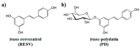
Natural polyphenols are an abundant and widely distributed family of bioactive molecules, whose structure is generally constituted by one or more aromatic rings carrying one or more hydroxyl groups [26]. Two natural stilbene polyphenols that have attracted much attention, especially for their manifold biological properties, are trans-resveratrol (here named RESV, 3,5,4′-trihydroxystilbene) [27] and trans-polydatin (here named PD, 3,5,4′-trihydroxystilbene-3-β-D-glucoside, Figure 1) [28]. These polyphenols were originally isolated from the root and rhizome of Polygonum cuspidatum, a plant used in traditional Chinese medicine for its analgesic, antipyretic and diuretic properties. Resveratrol is a phytoalexin produced by more than 70 plants in response to various stresses and is found in a variety of foods, including red grapes, peanuts, pistachios, red wine, blueberries, cranberries, and even cocoa and dark chocolate [29]. Polydatin is a glycosylated form of RESV and the most abundant derivative of resveratrol in nature [30].
Figure 1. Chemical structures of (a) trans-resveratrol (RESV) and (b) trans-polydatin (PD).
Many studies have been carried out on the beneficial effects of these polyphenols on the human body (e.g., anti-oxidant, anti-inflammatory, antitumor, antiviral, neuroprotective, hepatoprotective and ischemia preventing activities), and on their mechanisms of action [27][28][31][32][33][27,28,31,32,33].
Analogously to other polyphenols, RESV has limited bioavailability and poor water solubility [34]. On the other hand, PD displays higher water solubility and metabolic stability, as well as better oral absorption than RESV and is used in clinics with no side effects [35][36][35,36].
These compounds were recently proposed as potential drugs against COVID-19-related targets as indicated by preliminary in silico studies and cellular assays [37][38][39][37,38,39]. Furthermore, polydatin and resveratrol treatments could be beneficial for COVID-19 infection also due to their anti-inflammatory activities particularly in the respiratory tract [40][41][42][43][44][45][46][47][48][49][40,41,42,43,44,45,46,47,48,49].
On these bases, we here investigated—by means of detailed in silico studies and preliminary biochemical assays—the potential of RESV and PD to bind ACE2 and/or Spike proteins interfering with their interaction, essential for virus host-cell entry. To the best of our knowledge, this is the first report exploring, with preliminary experimental assays, the interference of PD/RESV on the binding of a COVID-19 key protein to a host target.
In particular, we here studied the interactions of PD and RESV with both Spike and ACE2 as separated proteins as well as with their complex through a molecular docking-based computational approach, using the available molecular structures as deposited in the PDB database. Furthermore, preliminary biochemical assays, i.e., ELISA-like assays employing the target recombinant proteins (Spike S1 subunit and ACE2) and the tested small-molecules, were performed to evaluate the ability of PD/RESV to inhibit/block the ACE2 recognition by Spike.

2. Molecular docking simulation.

Spike-protein pre-fusion conformation [50,76] is a trimer constituted of two subunits, S1 and S2, which are cleaved following receptor binding [77]. S1 Receptor Binding Domains (RBDs) host the binding motifs (RBMs) able to recognize ACE2. The high RBD flexibility allows the Spike to sample open or closed conformations, in which RBMs are respectively exposed or hidden inside the protomers interface [77–81].

Therefore, the binding to SARS-CoV-2 Spike structure with one RBD in an open conformation (PDB ID: 6VSB [50]) has been investigated by molecular docking simulations. The pockets on the RBD surface appear able to accommodate both PD and RESV ligands (Figure 2). A slightly lower affinity for the RBD domain was found for RESV (−6.5 kcal/mol for the best docking pose, Table 1) with respect to PD (top-ranked pose −6.9 kcal/mol).

Table 1. Docking scores (i.e., the approximate binding energy estimated by the docking scoring function, kcal/mol units) for PD and RESV top-ranked docked poses.


PD

RESV

Spike RBD

−6.9

−6.5

ACE2

−8.4

−6.9

S:ACE2 region I

−8.1

−7.6

S:ACE2 region II

−6.9

−6.5

ACE2 homodimer (PDB ID: 6M18, [51]) is constituted by an N-terminal protease domain, involved in the interaction with SARS-CoV-2 Spike RBD [51], and a C-terminal collectrin-like domain, comprising the ACE2 transmembrane helix. Molecular docking simulations were performed near ACE2 protease domain α1 and α2 helices, thus far from the catalytic site related to ACE2 physiological function. Despite its size, PD can be easily accommodated in a deep groove behind the two helices (Figure 3a,b) producing a high docking score (−8.4 kcal/mol, Table 1). RESV can also bind to the pockets near α1 and α2 helices, but with a ~1 kcal/mol lower score with respect to PD (−6.9 kcal/mol, Table 1) (Figure 3c,d).
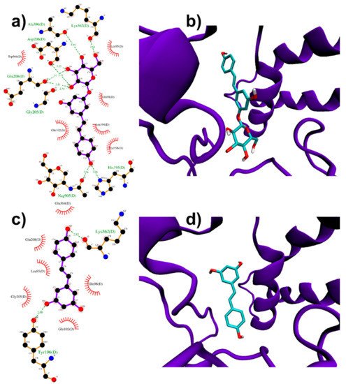
Figure 3. PD (a,b) and RESV (c,d) top-ranked poses docked to ACE2.

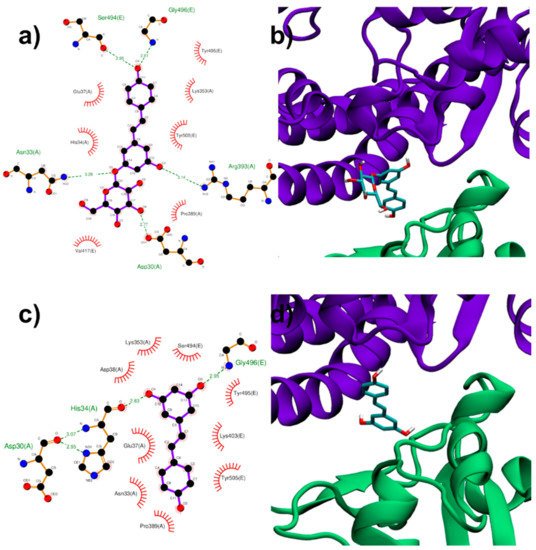
Both RESV and PD showed a good estimated binding affinity in the groove between the Spike RBD and ACE2. Due to the high spatial extent of the Spike:ACE2 interface, docking simulations were performed in two distinct regions (named regions I and II). In the first interfacial region investigated (region I), the most stable RESV binding mode involves recognition of one of the interfacial pockets through polar and hydrophobic contacts (Figure 4c,d). PD binds in the same pocket as RESV (Figure 4a,b) but interacting with more surrounding residues, thus showing a higher docking score (−8.1 kcal/mol, Table 1), compared to that of RESV (−7.6 kcal/mol).

Table 1. Docking scores (i.e., the approximate binding energy estimated by the docking scoring function, kcal/mol units) for PD and RESV top-ranked docked poses.
 PDRESV
Spike RBD−6.9−6.5
Spike A/B interface−7.3>−6.5
Spike A/C interface−7.3>−6.5
ACE2−8.4−6.9
S:ACE2 region I−8.1−7.6
S:ACE2 region II−6.9−6.5
Figure 4. PD (a,b) and RESV (c,d) top-ranked poses docked to Spike:ACE2 interface region I.

In the second region (region II) investigated at the interface between viral Spike RBD and human ACE2, the top-ranked RESV conformer fits into a large ACE2 cavity far from the interaction sites between the two protein domains. On the contrary, in the best PD binding mode, the ligand binds onto a pocket located at one end of the ACE2:Spike complex interface with a docking score 0.4 kcal/mol lower than RESV (−6.9 kcal/mol, Table 1). It is interesting to note the fundamental role played by the glucosidic moiety which interacts with the residues of the main chains that are involved in the formation of the ACE2:Spike dimer. Weakening of these interactions could, in principle, contribute to the dissociation of the dimeric complex.
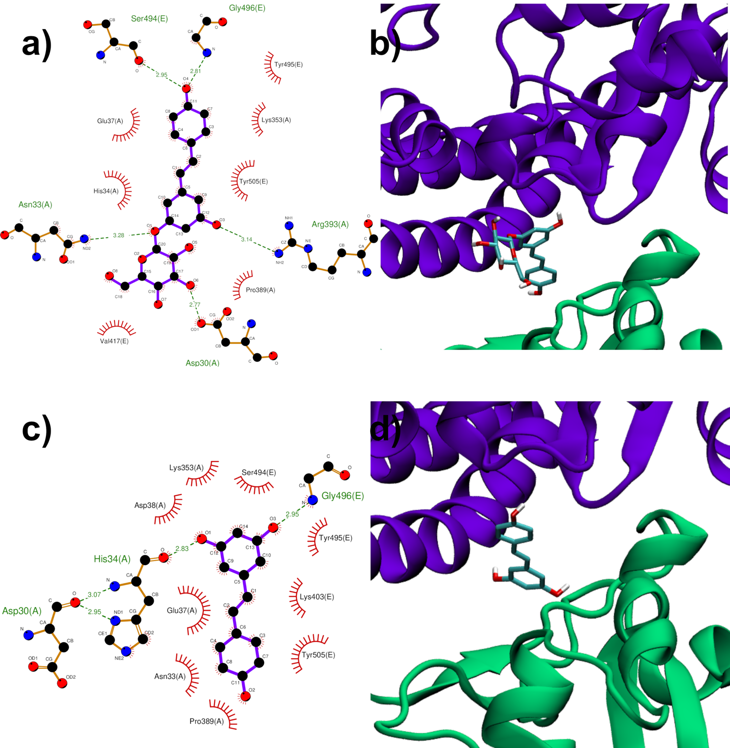
Figure 4. PD (a,b) and RESV (c,d) top-ranked poses docked to Spike:ACE2 interface region I. Two-dimensional interaction maps (ac): C, N and O atoms are reported in black, blue and red, respectivelts from docky. Hydrogen bonds are depicted as green dashed lines, while hydrophobic interactions as red cogwheels. Hydrogen atoms are not depicted for ease of illustration. The names of proteins’ residues involved in interactions with the ligand are reported. Three-dimensional representations of PD and RESV docked poses (bd): proteins’ backbone is represented as a cartoon, while the lig siand as sticks.

Results from docking simulations on the already assembled Spike:ACE2 complex reveal the potential capability of PD, but also RESV, to insert themselves into the extended adduct interface. This leads to the hypothesis of a ligand-induced dissociation or weakening effect, through allosteric (such as for region I) or direct (region II) interference.

3. To establish if RESV and PD can experimentally interfere with the binding of the Spike protein with ACE2 receptor, as suggested by the molecular docking simulations, binding inhibition assays were performed.
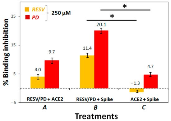
The assay we carried out was based on the following steps: (1) immobilization of the purified ACE2 protein, labelled with a His-tag (ACE2-His), on a Ni-coated 96-well plate; (2) attachment of biotinylated SARS-CoV-2 Spike S1 protein (from here on named just Spike) on the ACE2-functionalized plate, exploiting the high affinity of Spike for ACE2; (3) binding of streptavidin-horseradish peroxidase (HRP) to the bound Spike, thanks to the high recognition affinity between biotin and streptavidin; (4) treatment of the so-prepared plate with an HRP-substrate to produce chemiluminescence, measured at the end of the assay using a luminescence reader. Chemiluminescence intensity is correlated with the amount of Spike attached to the plate. If an inhibitor of the ACE2:Spike interaction is added to the plate, a reduction in chemiluminescence can be observed, proportional to the efficacy of the inhibition. Chemiluminescence reading in wells treated in the same way as the samples but without Spike and inhibitors (RESV/PD) were called “Blanks”, whereas those without inhibitors were named “Positive controls”. The chemiluminescence reduction observed in the wells treated with PD and RESV was converted in % inhibition of the ACE2:Spike binding by the stilbenoids; this was obtained by subtracting the chemiluminescence value of the blank from that of all the other treatments and calculating the complementary percentage relative to the positive control.
We optimized the assay protocol using a high concentration of the two polyphenols (250 μM). We explored three different treatments: (1) Treatment A, i.e., the protocol suggested in the datasheet of the used assay kit, with a pre-incubation of RESV/PD with ACE2-coated plate; (2) Treatment B, involving a pre-incubation of RESV/PD with Spike S1 in solution; (3) Treatment C, involving a pre-incubation of Spike S1 with ACE2-coated plate.
In all cases, we noted that RESV always produced a smaller effect than PD. This observation is consistent with the Molecular Docking results, always showing a higher docking score of PD for all the investigated targets (Table 1). Moreover, we evidenced that the more convenient treatment, among the three, explored ones, was B, consisting of the pre-incubation of RESV/PD with Spike in solution before addition of ACE2 (Figure 5 and Figure S9 for the chemiluminescence data). In particular, the percentage of ACE2:Spike binding-inhibition produced by PD, in this case, was about 20%.
Figure 5. ACE2:Spike inhibition binding assay. In Treatment A, the polyphenols were pre-incubated with ACE2 on the plate, and then Spike was added; in Treatment B, the polyphenols were pre-incubated with Spike in solution, and this mixture was then added to ACE2 on the plate; in Treatment C, ACE2 and Spike were pre-incubated on the plate and then the polyphenols were added. Percentages of binding inhibition were calculated correlating the chemiluminescence intensity readings (Figure S9) to that of the positive control. p-Values have been calculated using the Student’s t-test (* p ≤ 0.05).
Treatments A and B were also repeated at 200 μM concentration of the two natural compounds confirming the observed trend (Figure S10).
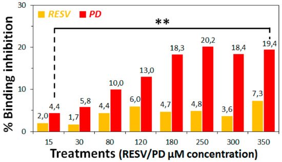
Subsequently, a range of suitable concentrations (0–350 μM) of RESV and PD were explored for the ACE2:Spike-binding inhibition assay under the optimal conditions found (Treatment B). This experiment afforded the chemiluminescence data reported in Figure S11a, evidencing a dose-response effect in the case of PD treatments in the range 0–250 μM, while higher concentrations did not produce additional effects. RESV, in our hands, did not seem to afford significant effects. Conversion of the chemiluminescence data in percentages of ACE2:Spike binding-inhibition by RESV and PD was reported in Figure 6 and Figure S11b.
Figure 6. ACE2:Spike inhibition binding assay. In all treatments, the polyphenols were pre-incubated with Spike in solution. Chemiluminescence intensities were measured on the 96-well plate with a luminescence reader and converted in percentages of ACE2:Spike-binding inhibition with respect to the positive control. p-values have been calculated using the Student’s t-test (** p ≤ 0.01).
Analysis of these data evidenced that the highest effect was obtained at 250 μM PD concentration, with a binding-inhibitory activity of ca. 20%.
Thus, in the conditions of this specific assay, we could not calculate the IC50 value for PD since we did not reach the 100% binding inhibition. This behaviour could be probably due to solubility and aggregation issues of the two polyphenols, especially RESV [50][51][82,83], in the assay buffer conditions.
These preliminary experimental assays directly revealed a PD inhibitory action of the ACE2:Spike interaction, in agreement with the Molecular Docking simulations on the surface regions of ACE2, Spike and their complex (corresponding to the experimental conditions here named Treatments A, B and C), demonstrating some binding capabilities by PD. RESV in turn did not produce a significant binding inhibition under the assay conditions. This could be mainly due to solubility and aggregation issues of RESV, which are more critical than for PD. In addition, even if the binding of RESV occurs, this could not impede the interaction between ACE2 and Spike proteins. Indeed docking simulations predicted a lower binding score by RESV for both isolated Spike and ACE2 and their complex.

4. Conclusions

In this work, the binding abilities of the natural compounds polydatin (PD) and resveratrol (RESV) towards two key targets involved in SARS-CoV-2 viral infection—Spike viral protein and ACE2 host receptor—were investigated by molecular docking simulations.
In particular, we here studied the interactions of PD/RESV with both Spike and ACE2 as separated proteins, as well as with their complex, through a molecular docking-based computational approach, using the PDB available molecular structures.
Molecular docking targeted at Spike and ACE2 surface pockets near their interaction sites and the interface of the already assembled ACE2:Spike complex revealed potential binding and insertion capabilities by both PD and RESV ligands. In all cases, the predicted binding with PD appeared stronger than with RESV. These Molecular Docking data thus encourage further computational investigations aimed at verifying PD and RESV interference or weakening effects in the ACE2:Spike recognition.
Furthermore, aiming at supporting the data obtained from molecular docking simulations, preliminary biochemical assays were performed to experimentally evaluate the ability of PD/RESV to interfere with the binding of the Spike protein with the ACE2 receptor. Our assays evidenced a dose-response effect in the case of PD reaching a maximum of >20% ACE2:Spike binding inhibition at 250 μM PD concentration.
Even if high concentrations were required to obtain a significant effect in this kind of experiment, we were encouraged from the obtained results due to the known absence of side effects and toxicity of PD even at high dosage, as demonstrated by its use as a nutraceutical product (as a human food supplement, the recommended dose of polydatin is 160 mg/day for assumption cycles of at least three months [52][84]) and in clinical applications [53][54][85,86].
In addition, we have here showed a biochemical assay not considering (i) several biological aspects of ACE2-Spike binding only identifiable by cellular assays, e.g., the role of biological multimerization [55][51], (ii) solubility issues and aggregation state of the studied polyphenols, especially RESV [50][51][82,83], in the assay buffer conditions (not considered by the modelling studies), (iii) synergistic effects deriving from the interaction of these polyphenols with other key viral proteins or other host targets, which could reinforce the overall result.
From the current picture, PD emerges as a potential candidate drug/protective agent, which can act as a sort of “biological mask”. It can inhibit the binding of Spike to ACE2 and therefore reduce viral entry into host cells, also being well-known its favourable properties like high water solubility and metabolic stability, good oral absorption and absence of side effects, as well as beneficial and protective effects during inflammation particularly of the respiratory tract [56][87].
Video Production Service