Seizures and Newborn Infants: Comparison
Please note this is a comparison between Version 1 by Alistair Gunn and Version 2 by Lily Guo.

Seizures are common in newborn infants with hypoxic-ischemic encephalopathy and are highly associated with adverse neurodevelopmental outcomes. The impact of seizure activity on the developing brain and the most effective way to manage these seizures remain surprisingly poorly understood, particularly in the era of therapeutic hypothermia. Critically, the extent to which seizures exacerbate brain injury or merely reflect the underlying evolution of injury is unclear. Current anticonvulsants, such as phenobarbital and phenytoin have poor efficacy and preclinical studies suggest that most anticonvulsants are associated with adverse effects on the developing brain. Levetiracetam seems to have less potential neurotoxic effects than other anticonvulsants but may not be more effective. Given that therapeutic hypothermia itself has significant anticonvulsant effects, randomized controlled trials of anticonvulsants combined with therapeutic hypothermia, are required to properly determine the safety and efficacy of these drugs. Small clinical studies suggest that prophylactic phenobarbital administration may improve neurodevelopmental outcomes compared to delayed administration; however, larger high-quality studies are required to confirm this. In conclusion, there is a distinct lack of high-quality evidence for whether and to what extent neonatal seizures exacerbate brain damage after hypoxia-ischemia and how best to manage them in the era of therapeutic hypothermia. 

  • hypoxic-ischemic encephalopathy
  • asphyxia
  • seizures
  • antiepileptic drugs
  • anticonvulsants
  • therapeutic hypothermia
  • phenobarbital
  • levetiracetam

1. Neonatal Seizures after Hypoxic-Ischemic Encephalopathy

Loss of oxygen (hypoxia) and blood supply (ischemia) to the brain can occur before, during or shortly after birth. Moderate to severe hypoxic-ischemic encephalopathy (HIE) occurs in ~1 to 3/1000 live births in developed nations and can lead to death, or in survivors, brain damage with lifelong disability, including cerebral palsy and epilepsy [1]. Induced mild “therapeutic” hypothermia (cooling) is now established to significantly improve survival without disability, but many infants still survive with disability despite treatment [2]. There are many unanswered questions about how to manage infants during therapeutic hypothermia, particularly whether add-on anticonvulsant therapy is necessary or beneficial [3].
Seizures are common in newborn infants with HIE. For example, in a study of electroencephalographic (EEG) monitoring in 47 neonates with HIE, 62% had seizures [4]. Seizure burden is consistently associated with both brain injury and adverse neurodevelopmental outcome [5][6][5,6]. However, the direction of causality is unclear, and the consequences of seizure activity for the developing brain and the most effective way to manage these seizures remain surprisingly poorly understood. Although seizures in the neonate are largely regarded as a neurologic emergency requiring prompt management, in practice there is little evidence supporting a beneficial effect of anticonvulsants and there is concerning preclinical evidence that many anticonvulsants may have neurotoxic effects on the developing brain [7]. Furthermore, it is not clear how the relative benefits and risks of anticonvulsants have changed in the era of therapeutic hypothermia [7]. Thus, optimal management of seizures remains controversial.

2. The Biology of Neonatal Seizures

The mechanisms of seizures in the neonate appear to be different in many ways to those in the adult or even during childhood. The majority of neonatal seizures occur as a transient response to acute cerebral pathology and are most commonly associated with HIE (38%), ischemic stroke (18%) and intracranial hemorrhage (11%) [8]. In infants with HIE, seizures usually start hours after birth, their intensity reflects the severity of underlying cerebral injury and they tend to resolve over hours to days irrespective of seizure management [4].
Biologically, there are differences in neurotransmitter function during development that may increase the susceptibility of the immature brain to seizures compared to adults and have the potential to affect the efficacy of anticonvulsants. Early in development, gamma-aminobutyric acid (GABA) receptors may respond to stimulation with excitation, rather than the inhibition observed in the adult [9]. This difference is due to a high intracellular Cl- concentration resulting from high Na-K-2Cl (NKCC1) expression, which mediates Cl- entry and low K-Cl cotransporter KCC2 expression, which mediates Cl- exit from cells [10][11][10,11]. Following the upregulation of the potassium-chloride co-transporter KCC2 during the early postnatal period, Cl- can be extruded from cells and therefore GABA and glycine become inhibitory. This relative increase in excitability is important for the development of neural circuitry, but likely increases susceptibility to seizures [12]. Further, there is evidence in juvenile rats that hypoxia-ischemia late in gestation reduced neuronal KCC2 expression, impairing hippocampal CA3 inhibitory tone [13]. Impaired GABAergic signaling after hypoxia-ischemia may lower the seizure threshold, contributing to reduced efficacy of GABAergic agonists such as phenobarbital in neonates.
Moreover, the immature brain has a greater density of calcium permeable, GluR2-subunit-deficient AMPA receptors, which contribute to a lower threshold for seizures, and expression of NMDA glutamate receptor subunits (GluN) such as GluN2B, which promote prolonged excitatory post-synaptic potentials and GluN3A [14]. Further, hypoxia-ischemia can change the expression of NMDA receptor subunits, therefore promoting seizures in the developing brain [15].
These biological differences in the neonatal brain compared to the adult are still being explored, but they may have consequences for the initiation and propagation of seizure activity as well as the efficacy and long-term neurodevelopmental effects of anticonvulsants. Thus, it is vital to better understand the pathophysiology of neonatal seizures and develop and test anticonvulsant protocols specifically in neonates, and not rely on evidence from studies in adults.

3. Do Seizures Exacerbate Brain Damage after HIE?

Understanding whether seizures independently contribute to brain damage after HIE, or merely reflect the underlying evolution of injury resulting from a period of hypoxia-ischemia is crucial for determining how aggressively seizures should be managed. The effects of seizure activity on the severity of brain damage after HIE are complex and poorly understood, with much conflicting evidence. Much of this uncertainty stems from the inherent difficulty in trying to detangle the direct effects of seizures on brain injury from indirect effects mediated by the severity of the underlying hypoxic-ischemic injury that triggered the seizures in the first place.
There is some, limited clinical evidence that seizures may exacerbate injury in infants with HIE. A clinical magnetic resonance spectroscopy study suggested that seizures were associated with a mismatch in oxygen supply and demand, such that increased seizure burden was associated with elevated lactate and reduced NAA/choline ratio, a marker of neuronal injury [16]. This seems to indicate that the severity of seizures in human newborns with HIE is independently associated with brain injury [16]. Consistent with this hypothesis, a small prospective study found that infants with clinical seizures had worse motor and cognitive outcomes after controlling for injury severity seen on magnetic resonance imaging (MRI) 5 days after birth [17], and further, that greater seizure severity was associated with worse motor and cognitive outcomes. More recently, two small, randomized trials of management of clinical vs. EEG-proven seizures have suggested that higher seizure burden is associated with greater brain injury and worse neurodevelopmental outcomes [18][19][18,19]. Although these data seem to suggest that higher seizure burden may be harmful, the effect of greater management during EEG monitoring in these studies on seizures was not statistically significant. Recently, a study of predictive models for death and neurodevelopmental impairment for infants with HIE, found that seizures were not independently predictive of outcome, due to collinearity with injury severity [20]. More generally, the clinical impact of a mild to moderate seizure burden is still unclear and the threshold that should prompt management in routine clinical care is unknown.
A limitation of these studies is that imaging reflects a single time point, while seizures reflect a dynamic, evolving process. Critically, these studies are not able to determine the direction of causality. That is to say, it is possible that more severe underlying injury could have reduced NAA expression [16], and increased both seizure burden and risk of adverse neurodevelopmental outcome. Supporting this hypothesis, a secondary analysis of the National Institute of Child Health and Human Development (NICHD) whole body hypothermia trial found that after adjusting for treatment group and the severity of HIE, there was no significant association between clinical seizures and death, or moderate or severe disability or lower Bayley Mental Development Index score at 18 months of life [21]. Acknowledging that this was a post hoc analysis, these data suggest the hypothesis that among infants with HIE, the mortality and morbidity often attributed to seizures could be better explained by the underlying severity of encephalopathy.
There has also been speculation that neonatal seizures prime the brain for later development of epilepsy. In rodents, neonatal seizures are associated with long-lasting changes including reduced neurogenesis, sprouting of mossy fibers, and altered cell firing, as previously reviewed [22]. Clinically, in a study of 92 term infants with HIE, there was a significant association between neonatal seizures and subsequent development of epilepsy [23]. However, this association was no longer significant after adjusting for the severity of HIE, such that only the presence of severe HIE was independently associated with the later development of epilepsy [23]. Supporting this hypothesis, there is evidence from a cohort study of reduced rates of epilepsy in infancy and childhood after treatment of HIE with therapeutic hypothermia [24].

4. Should We Try to Prevent Seizures?

There is a lack of evidence that phenobarbital or any other anticonvulsant improves outcomes after neonatal seizures have already started. However, there is some clinical and preclinical evidence that prophylactic phenobarbital administration, that is to say administered before the appearance of seizure activity, may be beneficial [25][68]. Any benefit could be mediated by improved seizure control as phenobarbital suppressed seizures when administered before, but not after hypoxia-ischemia in P11 rats (Table 1) [26][38]. Although brain injury was not assessed in this study [26][38], there is intriguing evidence that phenobarbital may have neuroprotective effects independent of its anticonvulsant properties. For example, a small prospective study, conducted in the pre-hypothermia era, found that prophylactic high-dose phenobarbital administration (40 mg/kg) was associated with improved neurological outcome at three years of age, despite only showing a 27% reduction in seizures compared to neonates who received phenobarbital after seizures had started [27][59]. Further supporting the efficacy of prophylactic phenobarbital, a recent Cochrane review of prophylactic barbiturates (8/9 studies used phenobarbital) for infants with perinatal HIE [25][68], found that prophylactic barbiturate therapy reduced the risk of seizures after hypoxia-ischemia, with no reduction in mortality. However, there was little data on long-term outcomes. They concluded that “the results of the current review support the use of prophylactic barbiturate therapy as a promising area of research” although they could not recommend routine clinical use at this stage due to the low quality of evidence.
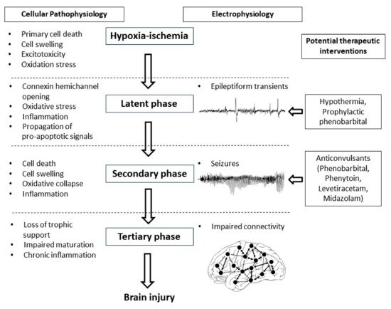
The potential for early but not later administration of phenobarbital to be neuroprotective, likely relates to the well-characterized progressive evolution of hypoxic-ischemic brain injury (Figure 1) [3]. During the period of hypoxia-ischemia itself, there is often only limited cell death. Many brain cells partially or even completely recover after reperfusion, in a latent phase that lasts approximately 6 h after hypoxia-ischemia, only to undergo secondary deterioration as shown by delayed onset of seizures, cell swelling and bulk cell death from 6–72 h after hypoxia-ischemia [3]. It is now well established that therapeutic hypothermia needs to be started during this latent phase, before the onset of seizures (and secondary cell death), in order to be effective [28][69]. This suggests that when phenobarbital is administered after the onset of seizures (i.e., during the secondary phase), the window of opportunity for neuroprotection has already closed and so phenobarbital can only act as an anticonvulsant. It is plausible but unproven that administration of prophylactic phenobarbital during the latent phase, corresponding with the established window of opportunity for therapeutic hypothermia [28][69], may offer direct neuroprotection, and so reduce secondary cell death, leading to fewer seizures.
Figure 1. Flow diagram showing the phases of injury including hypoxia-ischemia, the latent phase, the secondary phase and the tertiary phase leading to the development of brain injury. Intervention during the latent phase with therapeutic hypothermia or prophylactic phenobarbital has the potential to reduce the development of brain injury as well as the occurrence of seizures. Intervention during the secondary phase with anticonvulsants such as phenobarbital, phenytoin, levetiracetam and midazolam may reduce seizure activity but their effects on long-term outcome are not clear.
There is some evidence to suggest that early phenobarbital can augment the beneficial effects of therapeutic hypothermia. In P7 rats exposed to hypoxia-ischemia induced by single carotid artery ligation and inhalation hypoxia, phenobarbital augmented hypothermic neuroprotection [29][39]. Similarly, in P10 rat pups, Krishna et al. also found augmentation of hypothermic neuroprotection with prophylactic phenobarbital subjected to unilateral hypoxia-ischemia [30][40]. Although these studies are limited by the use of sub-optimal durations of hypothermia (3 to 4 h instead of the 72 h in established clinical protocols), they support the concept that prophylactic phenobarbital administration may have additive neuroprotective effects with therapeutic hypothermia. Strikingly, there no large animal, translational studies to date have tested whether there is benefit from combined treatment with prophylactic phenobarbital and hypothermia.

5. Conclusions

Current anticonvulsant protocols are clearly far from optimal for use in neonates—they show limited efficacy, have high potential for adverse effects and there is a striking lack of evidence that they improve long-term outcomes. Preclinical neurophysiological studies are essential to help to better understand the pathophysiology of seizures occurring in the neonatal brain compared to the mature brain and identify specific anticonvulsant treatment strategies for the neonatal brain. High quality translational studies will help to answer questions around whether prophylactic anticonvulsant treatment is neuroprotective and more effective than delayed administration and whether anticonvulsants are associated with neurotoxic effects when given in combination with therapeutic hypothermia. Well-designed randomized controlled trials, taking into account the use of therapeutic hypothermia, are essential to develop evidence-based treatment strategies for the neonate with seizures.