The V-ATPase a3 Subunit: Comparison
Please note this is a comparison between Version 1 by Anh Chu and Version 3 by Rita Xu.

This rentryview focuses on one of the 16 proteins composing the V-ATPase complex responsible for resorbing bone: the a3 subunit. The rationale for focusing on this biomolecule is that mutations in this one protein account for over 50% of osteopetrosis cases, highlighting its critical role in bone physiology. Despite its essential role in bone remodeling and its involvement in bone diseases, little is known about the way in which this subunit is targeted and regulated within osteoclasts. To this end, this review is broadened to include the three other mammalian paralogues (a1, a2 and a4) and the two yeast orthologs (Vph1p and Stv1p). By examining the literature on all of the paralogues/orthologs of the V-ATPase a subunit, we hope to provide insight into the molecular mechanisms and future research directions specific to a3. This review starts with an overview on bone, highlighting the role of V-ATPases in osteoclastic bone resorption. We then cover V-ATPases in other location/functions, highlighting the roles which the four mammalian a subunit paralogues might play in differential targeting and/or regulation. AuthorWe review the ways in which the energy of ATP hydrolysis is converted into proton translocation, and go in depth into the diverse role of the a subunit, not only in proton translocation but also in lipid binding, cell signaling and human diseases. Finally, the therapeutic implication of targeting a3 specifically for bone diseases and cancer is discussed, with concluding remarks on future directions.

  • V-ATPase
  • osteoclasts
  • bone
  • osteoporosis
  • osteopetrosis
  • anti-resorptive therapeutics
  • signalosome
  • TCIRG1
  • V-type proton ATPase 116 kDa subunit a3
  • OC-116 kDa
  • ATP6V0A3
  • ATP6V1C

1. Bone

Bone is a remarkable dynamic tissue which is involved in a variety of roles besides providing structural support. Bone exhibits endocrine, immune, mineral storage, growth factor, organ protection and repair functions [1][2][3][1,2,3]. Most of these functions can be attributed to the presence of three distinct major cell types, the osteoblast (OB), the osteoclast (OC) and the osteocytes. Osteoblasts are derived from mesenchymal stem cells during embryogenesis, and are responsible for the secretion of a proteinaceous matrix, including growth factors, which becomes mineralized [1][4][1,4]. OBs are found lining the bone surface and also become encased in the mineralized matrix, where they differentiate into osteocytes [5][6][5,6]. Osteocytes communicate with each other and other cell types via canaliculi found in bone. Osteocytes are capable of detecting stresses on the skeleton, and are able to activate OBs lining the bone surface, as well as OCs to start the repair process [7]. Osteoclasts have been thought to arise from hematopoietic cells exclusively; however, recent lineage tracing studies using mice have shown that there is also an extraembryonic component to this [8][9][8,9]. Cells derived from erythromyeloid-progenitors (EMP) in the embryonic yolk sac are the first wave of OC to differentiate, followed later by a distinct second wave derived from hematopoietic stem cells (HSCs). These two stem cell populations occupy two different niches in the adult, with the EMP homing to the spleen while the HSCs seed the bone marrow [8][9][8,9]. OCs are capable of resorbing bone via their ability to secrete acid to dissolve the mineral component and proteinases in order to digest the now exposed proteinaceous matrix [10][11][10,11]. This is a highly organized process that involves pre-OC cells fusing with each other, the formation of a sealed bone compartment underneath the now multinucleated OC sequestered by the sealing zone, and the formation of a ruffled membrane contained within the sealing zone [12][13][14][12,13,14]. The ruffled border acts as the gateway for the secretion of the acid and proteinases, and allows for the uptake of the dissolved mineral and digested proteins, which are mostly transcytosed by vesicles to the apical cell membrane for eventual disposal via the circulation [15]. Experiments performed in RAW 264 cells showed that the formation of an actin ring redirects intracellular vesicles, mostly secretory lysosomes, to transport large quantities of proteinases (e.g., Cathepsin K, alkaline phosphatase) and the acid generating machinery (made up from chloride channel 7 and the V-ATPase) to the OC plasma membrane adjacent to the bone surface [16][17][16,17]. Bone remodeling is a highly coordinated process that involves constant communication between OBs and OCs, and any interference with this can lead to disease [10][18][19][10,18,19]. V-ATPases are involved in pre-pro-protein processing (including glycosylation) [20], secretion [21], the internalization and degradation of molecules [22], vesicle transport and fusion [23][24][23,24], modulate signaling complexes, participate in distinct signalosomes [25], and promote cell migration in cancer [26]. To this end, mutations that interfere with V-ATPase function underlie diseases affecting a number of organ systems.

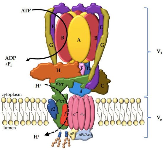
2. V-ATPase Functions

V-ATPases are ATP-driven proton pumps found in the endomembrane of the intracellular compartments in all of the eukaryotic cells and the plasma membrane of several specialized cells [27]. V-ATPases are responsible for acidifying and maintaining the pH of intracellular organelles, including the Golgi apparatus, endosome, lysosome and secretory vesicles [28][29][28,29]. V-ATPases pump protons into the Golgi apparatus, which become more acidic from the cis-Golgi to the trans-Golgi [20]. As newly synthesized proteins traverse the Golgi apparatus, they undergo post-translational modification including glycosylation, sulfation and phosphorylation. The maintenance of the pH gradient in the Golgi apparatus by V-ATPases is crucial for the function and localization of the glycosyltransferases required for the modification processes [30]. V-ATPase activity in the intracellular membrane is important for membrane trafficking processes such as receptor-mediated endocytosis [21][22][21,22]. The V-ATPase-dependent acidification of the endocytic compartments is required for the dissociation of ligand–receptor complexes, allowing the receptors to recycle to the cell surface. The released ligands are subsequently targeted to the lysosomes, where the low pH maintained by V-ATPases facilitates their degradation [31][32][31,32]. This process is important for the continued uptake of ligands such as low-density lipoprotein (LDL), a main carrier of plasma membrane cholesterol [33]. Many pathogens employ the V-ATPase-mediated acidification of the endocytic compartments to gain entry into cells, including diphtheria and anthrax toxins, as well as viruses such as influenza and Ebola [34][35][34,35]. After entering the host cells, viruses also require a low pH to trigger fusion and to deliver their viral genome into the host. V-ATPases are also involved in the intracellular trafficking of lysosomal enzymes by establishing a luminal pH gradient between compartments [21][32][21,32]. Lysosomes are more acidic than late endosomes, which in turn are more acidic than the trans-Golgi network (TGN). This gradient allows the binding of lysosomal proteases to the mannose-6-phosphate receptor at the TGN, facilitating the enzyme delivery to the lysosomes, and the dissociation of enzyme-receptor complexes in late endosomes, allowing the receptors to recycle to the TGN [36]. V-ATPases play a key role in cellular nutrient homeostasis by providing the acidic environment within lysosomes which is necessary for proteolysis, which is a major way in which cells generate free amino acids [37][38][39][37,38,39]. In addition to maintaining the lysosomal pH, V-ATPases also associate with the nutrient-sensing machinery in the lysosomal membrane, and are involved in the recruitment of the metabolic regulators mTORC1 and AMPK [38][39][40][38,39,40]. Within secretory vesicles, V-ATPases generate a proton gradient driving the uptake of small molecules such as the neurotransmitter glutamate [41][42][43][41,42,43], and they facilitate the processing of prohormones like proinsulin [33][44][45][33,44,45].
V-ATPases are targeted to the plasma membrane of specialized cells such as kidney intercalated cells [46][47][46,47], epididymis clear cells [48][49][48,49] and osteoclasts [50][51][50,51], where they function to transport protons from the cytoplasm to the extracellular space [30][52][53][54][30,52,53,54]. In the kidneys, V-ATPases are localized to the apical membrane of the alpha-intercalated cells to facilitate the secretion of protons into the urine in order to maintain pH homeostasis [54]. Osteoclasts rely on V-ATPases at the ruffle border for the demineralization of bone and the activation of the proteolytic enzymes required for bone resorption [55][56][55,56]. V-ATPases targeting the plasma membrane of epididymis clear cells are involved in the establishment of the acidic luminal pH necessary for sperm maturation and storage [49]. Recently, plasma membrane V-ATPases have been shown to be overexpressed in breast cancer cells, and to facilitate invasion by promoting the activity of acid-dependent proteases that degrade the extracellular matrix [57]. The inhibition of V-ATPases by concanamycin in prostate cancer cells results in a decreased level of mRNA for prostate-specific antigens [58]. Increasing evidence implicates the important role of V-ATPases in cancer cells’ growth and metastasis, and suggests a potential therapeutic treatment of metastatic cancer by the inhibition of V-ATPases activity.
In addition to the conventional functions of V-ATPases in intracellular signalling and membrane trafficking by generating pH gradients, recent findings suggest novel emerging roles of V-ATPases in the modulation of the function of receptors and their regulatory complexes through direct protein–protein interactions. For example, it was recently uncovered that Wnt/β-catenin signal transmission requires the interaction of co-receptor LRP6 with V-ATPase lysosomal accessory protein-2 (ATP6AP2) in late endosomes [59]. In Drosophila, V-ATPases have been suggested to be involved in the membrane fusion of synaptic vesicles via direct interaction with calmodulin [60]. Emerging studies propose the importance of V-ATPases in modulating various signalling pathways, including Notch, mTOR and AMPK via unconventional mechanisms [39][61][39,61].
In summary, V-ATPase-dependent acidification is essential for cellular metabolism, membrane trafficking and intracellular signalling. Moreover, the importance and novel emerging roles of V-ATPases in many signalling pathways and diseases, including cancers, makes them promising targets for drug development.

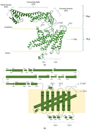
3. V-ATPase Structure

V-ATPases share their structure with mitochondrial and chloroplast F-type ATPases [27]. Both enzymes are composed of a peripheral catalytic sector (V1 or F1) and a membrane-bound proton channel sector (VO or FO). They are evolutionarily related, and are functionally conserved as rotary proton pumps [44]. The eukaryotic V-ATPase is a 900 kDa complex consisting of sixteen subunits: A3B3CDE3FG3H comprising the V1 sector, and ac9cdefAP1AP2 forming the membrane-bound VO (the subscript numbers represent the subunits’ stoichiometry in the complex) (Figure 1) [62][63][62,63]. Subunits A and B are arranged in a hexameric configuration and contain the nucleotide binding sites responsible for ATP hydrolysis [64]. ATP hydrolysis creates a driving force to induce the rotation of the central stalk composed of subunits D, F and d, and the membrane-bound proteolipid c-ring c9c[65].
Figure 1. Mammalian V-ATPase complex. Cytosolic sector V1, comprised of A3B3CDE3FG3H, is responsible for the ATP hydrolysis, which generates the force required to drive the rotation of the proteolipid c-ring (c9c) of the membrane-bound VO consisting of ac9cdefAP1AP2. The aCT forms two half-channels that create a pathway for protons to cross the lipid bilayer as the c-ring rotates. Both a2 and a3 orthologs are glycosylated twice on the first luminal loop within the C-terminus (depicted here), whereas a1 and a4 are only glycosylated once [66][82].
Each proteolipid subunit c, c has a conserved glutamate residue which is essential for proton translocation (E139 in c, and E98 in c) [64][67][68][64,66,67]. The glutamate residues are protonated when the subunit rotates past the membrane-embedded C-terminal domain of the a subunit (aCT). The aCT forms two half-channels that create a pathway for protons to cross from cytoplasm to the organellar lumen or the extracellular space [64]. Protons access the glutamate residue of subunit c upon entering the cytosolic half-channel, and the protonated glutamate residue carries the proton through the lipid bilayer as the c-ring rotates. The proton is released through the luminal half-channel following the deprotonation of the glutamate residue and stabilization by the critical arginine residue R740 in the a subunit (R735 in Vph1p, an S. cerevisiae ortholog of the a subunit) [69][68]. The AB hexamer is held stationary relative to the a subunit by three peripheral stalk EG heterodimers which connect the V1 sector to subunits C and H, and the N-terminal domain of the a subunit (aNT) [70][69].
V-ATPase activity is tightly controlled both spatially and temporally. One example of the temporal modes of V-ATPase regulation is the reversible assembly/disassembly upon environmental cues, which was first described in yeast [71][70]. The dissociation of V1-VO sectors is regulated by nutrient availability, as the dissociated complex is inactive in both ATP hydrolysis and proton translocation, reflecting the cells’ attempt to conserve cellular ATP. In yeast, the dissociation occurs in response to glucose starvation, involves an intact microtubular network, and is reversible without new protein synthesis [72][71]. Upon V1–VO disassembly, the C subunit dissociates from the V-ATPase complex, and the H subunit undergoes a conformational change resulting in the loss of the interaction with aNT [73][72]. The reassembly of the complex requires the RAVE complex (Rav1, Rav2 and Skp1). The RAVE complex binds to subunits E and G, the dissociated C subunit of V1, and to the VO subunit a, thereby positioning them to promote assembly [71][70]. The glucose-induced reassembly of V-ATPases requires the interaction of the protein complexes with regulatory proteins, such as the RAVE complex. Moreover, studies in yeast and RAW 264 osteoclast-like cells suggest a direct interaction between the glycolytic enzyme aldolase and V-ATPase subunits in a glucose-dependent manner [74][73]. The deletion of the aldolase gene in yeast resulted in V-ATPase disassembly and a reduction in V-ATPase activity [75][74]. In the presence of glucose, aldolase and V-ATPase interactions increase, inducing the reassembly of V1 and VO; hence, aldolase can act as a glucose sensor mediating V-ATPase assembly [76][75]. Several other determinants of V-ATPase assembly have been identified, including the membrane environment [77][76] and the interaction with regulatory factors such as HRG-1 [78][77] and viral infection [34]. The spatial regulation of V-ATPases is observed in the luminal pH gradients between compartments [79][80][78,79]. This mechanism of controlling V-ATPase activity is through the regulation of the trafficking of the complex, which is facilitated by different isoforms of the a subunit [81][82][80,81].

4. The V-ATPase a Subunit

Each V-ATPase complex contains one copy of the ~100 kDa a subunit, which exists as two isoforms in yeast (Vph1p and Stv1p) and four isoforms (a1, a2, a3 and a4) in mammals [27][81][83][27,80,83]. The a subunit has a bipartite structure, with a cytoplasmic N-terminal half (aNT) and a membrane-integrated C-terminal half (aCT) which consists of eight transmembrane helices (Figure 2) [62][84][62,84]. As described above, two of the eight helices in aCT are tilted and interact with the proteolipid c-ring to form the two hemichannels for proton translocation [62][85][62,85]. Even though ATP hydrolysis-coupled proton translocation can tolerate numerous a subunit mutations, the arginine residue in aCT (R735 in Vph1p, and R740 in TCIRG1 encoding the mammalian a3 isoform) is absolutely essential [69][68]. The dominant R740S missense mutation of this critical arginine in mice uncouples the proton pumping activity from ATP hydrolysis, resulting in mice with a high bone mineral density [86]. The aNT, oriented parallel to the membrane, is essential for V-ATPase function as it couples V1 ATP hydrolysis to VO proton translocation [44].
Figure 2. Mammalian V-ATPase a3 subunit. (a) Homology model of the a3 isoform generated using the Phyre2.0 server with constraint coordinates from the mammalian brain a1 isoform (PDB: 6vqc_3); (b) topology of the a3 isoform. The a subunit contains a cytoplasmic N-terminal half (aNT), which can be divided into three sub-domains—a distal domain (DD), connecting stalk (CS) and a proximal domain (PD)—and a membrane-bound C-terminal half (aCT) consisting of eight transmembrane helices (TM1-8), two of which are tilted and form the two hemichannels with the proteolipid c-ring. Cytosolic loops (CL1-3) connect TM2 and 3, TM4 and 5, and TM6 and 7, respectively; luminal loops 1 and 2 (LL1 and LL2) connect TM3 and 4, and TM5 and 6, respectively. Within luminal loop 1, a2 and a3 orthologs are glycosylated twice, whereas a1 and a4 are glycosylated once [66][82].
Studies with chimeric forms of Vph1p and Stv1p suggest that organelle targeting information is located in aNT [87]. In yeast, V-ATPases are targeted to the vacuole and Golgi by Vph1p and Stv1p, respectively; when chimeric a subunits were made, the targeted organelles were determined by the aNT. Furthermore, mutagenesis studies revealed that the signal sequence W83KY within the aNT of Stv1p is necessary for V-ATPase Golgi localization [88].
Similarly, in mammalian cells, different isoforms of the a subunit are enriched in specific organelles or cell types. However, the specific targeting signal of mammalian a isoforms has not been determined. V-ATPases containing the a1 isoform are found in the synaptic vesicles of neurons, and are relocated to the presynaptic plasma membrane at the nerve terminals [60][89][60,89]. The a2 isoform targets Golgi [90], and the a3 isoform is expressed in late endosomes and lysosome [16][91][16,91]. The a3 and a4 isoforms are also found on the plasma membrane of specialized cells, with a3 targeting the ruffle border of osteoclasts [50][92][50,92]; a4 is found in the apical membrane of kidney alpha intercalated cells and epididymal cells [49][93][49,93]. The upregulation of both a3 and a4 have been linked to the invasiveness of metastatic breast cancer cells [26]. Recently, the a4 isoform was shown to localize to the membrane of the invapodia of mouse breast cancer cells, where it plays a crucial role in the invasion and migration of the cancer cells [94]. While it is ubiquitously expressed in different organelles and cell types, the expression of a3 is approximately 100-fold greater in osteoclasts than in other cell types [95]. V-ATPases containing a3 are enriched in the membrane of the ruffled border, where they actively pump acid to dissolve bone and provide an acidic environment to activate the secreted proteases required for bone resorption. Furthermore, mutations in the a3 isoform in mammals—for example, the R740S in mice, mentioned above [86]—are associated with V-ATPase-related autosomal recessive osteopetrosis [53][96][97][53,96,97]. To this end, it is clear that the a3 isoform plays a crucial role in bone resorption by osteoclasts; therefore, the a3 isoform is a potential drug target for osteoporosis treatment, in which the excessive bone loss associated with this disease could be controlled by inhibiting a3-containing V-ATPases [50][98][99][50,98,99].