1q21.1 CNV in Neuropsychiatric Disorders: Comparison
Please note this is a comparison between Version 1 by Joy Yoon and Version 2 by Peter Tang.

The 1q21.1 CNVs, rare and large chromosomal microduplications and microdeletions, are detected in many patients with NDs. Phenotypes of duplication and deletion appear at the two ends of the spectrum. Microdeletions are predominant in individuals with schizophrenia (SCZ) and microcephaly, whereas microduplications are predominant in individuals with autism spectrum disorder (ASD) and macrocephaly.

  • copy number variation
  • microdeletion
  • microduplication
  • schizophrenia
  • autism spectrum disorder
  • microcephaly
  • macrocephaly
  • neurodegeneration
  • synaptic plasticity

1. Introduction

Rare CNVs, such as chromosomal deletions and duplications, have raised much scientific interest in etiological studies of NDs. It has been suggested that genetics play a major role in NDs, with ~52.4% and ~80% of inheritability in ASD and SCZ, respectively. A genetic study has shown that rare and large CNVs are likely to be causative, as they can lead to numerous gene imbalances [1]. Case–control studies have demonstrated that rare CNVs occur at higher frequency in cases than in controls, suggesting that patients bear a high CNV burden [2][3][2,3]. Moreover, 17.1% of those who presented abnormal clinical presentations carried pathogenic CNVs [4]. Approximately 40% of carriers had de novo mutations, and the majority of the de novo mutations (91%) were pathogenic [4]. These patterns show up in most ND studies, including ASD, SCZ, intellectual disability (ID) and attention deficit hyperactivity disorder (ADHD) [5][6][7][5,6,7]. These findings shed light on the contribution of CNVs to the risks of different NDs.
In general, CNVs are pleiotropic and have variable expressivity, in that different patients carrying CNVs at the same chromosomal regions can show the symptoms of different psychiatric disorders; for example, many ASD-associated CNVs are also found in SCZ patients [3][4][8][9][3,4,8,9]. Despite having the same CNV carriers, phenotypes and severity range diversely, and show incomplete penetrance [10]. This suggests that there must be other factors involved, such as other genetic components (the two-hit model) [11] or environmental factors [12]. Hence the complexity of CNVs has been underscored in the etiology of ND.
A recent GWAS has identified risk loci prevalent in NDs, which are rare CNVs seen in cases but not in controls [2]. At least eight distinct CNVs,1q21.1, 2p16.3, 3q29, 7q11.23, 15q13.2, 16p11.2, 22q11.2 and NRXN1, have been consistently reported as risk factors for many NDs [6][7][8][13][14][15][6,7,8,13,14,15]. Deletions are less frequent but more pathogenic than duplications. Therefore, an increased odds ratio (OR) was found for deletions (i.e., ORs of 1q21.1 = 11.82 (del) and = 6 (dup)) [15]. The abnormal clinical presentations are postulated to be a result of carrying those pathogenic CNVs. Many genetic studies have attempted to identify the relationships between genetic rearrangements in the regions and clinical phenotypes. As little is known about their effect size, penetrance and genetic predisposition towards a certain phenotype, it is too early to use those rare CNVs for diagnoses of any NDs.
Among the aforementioned associated CNVs, this paper aimed to focus on the 1q21.1 CNV that is found with high incidence in ASD, SCZ, ADHD, ID and epilepsy [16]. Due to its structural complexity and inconsistent clinical phenotypes, this genetic locus has been understudied. A significant and popular finding in 1q21.1 is its mirror effect on neurodevelopment: microdeletions are widely found in the cases of SCZ, and microduplications are widely found in the cases of ASD [17].

2. Chromosomal Mapping and Genetic Pathway of 1q21.1

2.1. Chromosomal Structure

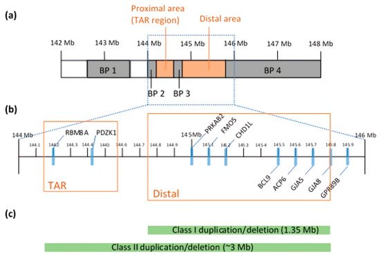
The 1q21.1 CNV is found within a 144 to 148 Mb region [18] (Figure 1a). In contrast to small CNVs, which are less detrimental, larger CNVs (>500 kb in size) can alter the expression levels of multiple genes [19]. It is a complex locus to study in that it not only spans 20–40 putative genes, but the region is also susceptible to genomic rearrangements due to the numbers of low copy repeats (LCRs). The more LCRs in the region, the more prone it is to frequent non-allelic homologous recombination (NAHR) during meiosis [20]. Clustered with LCRs, breakpoints (BPs) divide the locus into four possible segmental blocks and complicate the mapping and prediction of phenotypic expressivity [18]. Many of the LCRs and BPs are located adjacent to the crossing over points, making it difficult to estimate the phenotypes or genomic sequences in any given persons [21]. Through this mechanism, the CNVs, emerging in chromosomal duplications or deletions, can alter some of the dosage-sensitive genes and create a broad range of phenotypic variability [22]. Array comparative genomic hybridization and fluorescent in-situ hybridization analyses mapped out the overall structure of the 1q21.1 in great detail. The 1q21.1 region is associated with mental retardation, autism [23], schizophrenia [24] and microcephaly [21]. Duplication of 1q21.1 is strongly associated with autism [21].
Figure 1. (a) Chromosomal structure of 1q21.1, mapped with four BPs (gray) and two distinct regions (red). (b) An enlargement of the region between 144 Mb and 146 Mb. Known genes commonly found in microduplication and microdeletion carriers are marked with blue bars. The reference locations on the chromosome are based on the March 2006 human reference sequence (NCBI build 36.1). The two distinct regions—TAR and Distal—are indicated by red blocks. (c) The two classes of duplications and deletions are shown with green bars. The size of the bars represents the minimally affected region in each class.
Duplications and deletions are classified into two classes: Class I and Class II. Class I duplication/deletion involves only the distal 1q21.1 region between BP3 and BP4 (1.35 Mb in size), whereas Class II duplication/deletion extends from the distal 1q21.1 to the proximal 1q21.1 commonly detected between BP2 and BP4 (~3 Mb) [21] (Figure 1c). Combined data show enrichment in Class I deletions and duplications with a parental origin, but the components of genes and BPs can be varied after generations [25]. Both analyses discovered two distinct regions: proximal and distal 1q21.1, where a genomic gain or loss occurs (Figure 1b) [21][26][21,26]. Microdeletions at proximal 1q21.1 are mainly associated with thrombocytopenia-absent radius (TAR) syndrome and this region is often referred to as the TAR region. In particular, a core exon junction complex gene, 
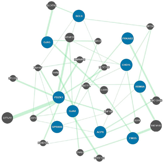
RBM8A, is located in the TAR region and compound mutations in the RBM8A gene cause the TAR syndrome [27] that is comorbid with ID [28]. Other brain dysfunctions, including psychosis, agenesis of the corpus callosum and hypoplasia of the cerebellar vermis, are present in TAR patients [28][29][30][28,29,30]. Consistent with human patient studies, knockdown and knockout of Rbm8a in a mouse model revealed the critical role of RBM8A in neural progenitor cell (NPC) proliferation, neuronal migration and interneuron development, and loss of function in RBM8A in NPCs causes microcephaly [31][32][33][31,32,33]. Moreover, RBM8A plays a key role in adult neurogenesis and in regulating anxiety-related behavior [34], further supporting the important role of RBM8A in psychiatric diseases.

2.2. Genetic Architecture

The recent advanced genomic assay has deciphered the genes encoded in the region and the position on the locus. The core genes commonly affected in the 1q21.1 CNV carriers are PRKAB2, FMO5, CHD1L, BCL9, ACP6, GJA5, GJA8, GPR89B and PDZK1 [25][35][36][25,35,36] (Figure 2; Table 1). However, the genetic study of the risk genes is far from clear as to the phenotypic consequences. Reported clinical phenotypes of the 1q21.1 duplication and deletion are not consistent, and no single gene has been confirmed to cause a pathologic effect in human studies [36].
This complex expressivity can be explained by a cis-epistasis genetic model. In contrast to a single gene CNV model, the gene expression is regulated by one or more CNV drivers and multiple modifiers [37]. Gain or loss of a single gene contributes only a small effect to trigger explicit clinical phenotypes [38]. This was confirmed in a number of genotype–phenotype association studies. A correlation analysis between gene expression and the copy number of 1q21.1 indicated that the candidate genes drew a positively correlated trend, in which a duplication CNV model was likely to have increased gene expression and vice versa [25], but the clinic severity may not have been correlated with the level of gene expression [39]. Harvard et al. conducted a family-based study of 1q21.1 microdeletion and microduplication and showed that individuals with the same CNV exhibited different levels of severity despite the identical gene components and almost identical BPs. Entangled chromatids are increased in lymphoblast cells derived from patients carrying both duplication and deletion of 1q21. To narrow down the causal gene, they identified two candidate genes, CHD1L and PRKAB2. Knockdown of CHD1L led to increased micronuclei in response to a topoisomerase II inhibitor, ICRF-193. However, both deletion and duplication carriers show the same cellular phenotype, suggesting that the gene dosage difference may not correlate with severity of symptoms. These findings once again emphasize the characteristic of the variable expressivity and the cis-epistasis model of the 1q21.1 CNVs [40][41][40,41]. Nevertheless, understanding of a linkage between genetic imbalance and apparent phenotypes is still incomplete.
Figure 2. A genetic map of the associated genes observed in 1q21.1 microduplications and microdeletions. Blue circles are the 10 affected genes; black circles are the 20 related genes. None of the 10 core risk genes interacts directly with another. A total of 49 genetic linkages are drawn with different widths of green lines and were generated by the GeneMANIA program [42]. Gene expression of the candidate genes is positively correlated with the copy number of 1q21.1 but not with phenotypic severity. Even within the same genetic components, clinical presentations are shown to a different extent in cases, which denies the one gene–one phenotype module. The blue circles are the major genes discussed in the paper. Eight top-ranked genes in the correlation study are not directly linked to each other but are indirectly connected via subtype genes.
Table 1. Genetic function and known phenotypes of dosage-sensitive genes associated with 1q21.1.

Function 1

Molecular/Cellular Phenotypes

References

CHD1

Chromatin remodeling and DNA damage response

Impaired decatenation checkpoint activation

[25]

PRKAB2

AMPK regulatory subunit; maintaining energy homeostasis

Neurodegeneration; learning and memory impairment

[42]

GJA8

Gap junction protein; Connexin50

Cataracts; cardiac myopathy; increased risk of SCZ

[43][44]

[43,44]

GJA5

Gap junction protein; Connexin40

Cataracts; cardiac abnormalities

[18][45][46]

[18,45,46]

PDZK1

Ion transporter protein; regulates second messenger cascades

Increased risk of ASD and psychosis

[36]

GPR89B

Voltage dependent anion channel

Unknown


BCL9

Wnt signaling pathway

Increased risk of SCZ

[47]

FMO5

Modulator of metabolic aging


[48][49]

[48,49]

ACP6

Histidine acid phosphatase protein

Unknown


1 The Genecards Human Gene Database.

2.3. Pathogenesis of Proximal 1q21.1

These clinical manifestations are associated with the genomic segmental regions on 1q21.1. The frequency of the chromosomal abnormalities was highly skewed to distal regions compared with proximal regions. Minimal deletions in BP2-BP3, known as the TAR syndrome region, however, raised a question of whether this region is benign or pathogenic. The overall chromosomal abnormalities in the proximal region were less frequent than in the distal region. However, the relative enrichment of proximal 1q21.1 in microduplication, especially with a low ratio of de novo inheritance compared with the microdeletions, suggests that the proximal BP2–BP3 region is responsible for clinical microduplication aberrations and is mild enough to maintain fecundity [50][51][50,51]. Bearing in mind that developmental delay (DD) is a common history in microdeletions and microduplications, the genes within the proximal BP2–BP3 region account for cerebral development in addition to TAR syndrome [51]. On the other hand, even though the head size was a notable phenotype by dosage, head sizes between the proximal microdeletions and microduplications were not found to be discrete, suggesting that the genes in the proximal region are not sufficient or not responsible for microcephaly/macrocephaly [51]. These findings confirmed the pathogenicity of the proximal 1q21.1 region; this should be re-evaluated on a large scale to be supportive.

3. Molecular and Cellular Mechanisms Associated with 1q21.1 CNVs

3.1. Effect Range of 1q21.1 CNVs

Studies of CNV pathogenesis have shown that deletions have deleterious effects, while duplications exhibit mild phenotypes [4]. Consistent with its pathogenicity, individuals with deletions have low fecundity and therefore undergo negative selection pressure [52][53][74,75]. These features of pathogenic CNVs appear in populations with low frequency and high mutation rates [8][52][8,74]. In light of this fact, it has become mainstream in genetic studies to distinguish distinct effect sizes in each ND. In line with the comparable burden of each structural variant, duplications exhibit a smaller burden than deletions in the synaptic pathway; functional clusters of duplications are enriched in NMDA receptor signaling, while functional clusters of deletions are enriched in the nervous system or behavioral phenotypes [15].
Examination of the cellular phenotype is a crucial step in the study of pathogenesis. Because many implicated risk genes in 1q21.1 CNVs are responsible for different cellular processes, including cell signaling, sensing and repair, impairment of these gene functions is expected to disrupt the cellular functions specifically involved with brain development and, in turn, to cause diseases [25]. However, a systematic pathological analysis of postmortem brains carrying 1q21.1 CNVs is still lacking. Due to the clinical manifestation reported among patients, animal models that mimic the genetic deficiency of 1q21.1 CNV could be good tools to provide some mechanistic insights and cellular and molecular targets for further therapeutic development [8][54][55][8,72,76].

3.2. Synaptic Signaling Pathway

Genes for cell signaling are enriched in 1q21.1 [56][67]. Cell signaling in the brain is impeded by abnormal synaptic plasticity. The dopamine hypothesis has been proposed in many ND studies, including ASD [57][77] and SCZ [58][59][78,79]. A 1q21.1 deletion mouse model recapitulated the function of 1q21.1 CNV in cellular phenotypes [55][76]. The 1q21.1 CNV accounts for the increased sensitivity to psychostimulants (e.g., amphetamine) and increased dopamine cell firing, and hypersensitivity is not mediated by a different number of D1/D2 receptors [55][76]. Thus, the findings are consistent with previous studies showing that 1q21.1 deletion shows a higher prevalence in SCZ patients than in ASD [58][78].
Alteration of the potassium channel function can impair in the whole neural network. Disruption of potassium ion homeostasis often becomes an initiator of the cells’ pathological cascade. In light of the crucial function of the potassium channel in neurodevelopment, GWAS has revealed a genetic overlap between rare risk CNVs (e.g., 1q21.1) and genes (e.g., KCNN3) encoding the potassium pump, transporter and channel [60][61][62][80,81,82]. The longer CAG repeats within the KCNN3 gene seem to be associated with SCZ patients [61][62][81,82]. However, other studies did not confirm this association [63][83]. Interestingly, a mutant KCNN3 channel found in a SCZ patient was localized in the nucleus and inhibited the current mediated by another potassium channel, KCNN2 [64][84]. Therefore, the SCZ KCNN3 variant can function as a dominant-negative mutant to suppress endogenous small-conductance K currents and interfere with neuronal firing. Consistent with this notion, dysfunction in astrocyte differentiation derived from SCZ patient-derived induced pluripotent cells (iPSCs) was a result of excessive downregulation of potassium transporters in SCZ glia [60][80].

3.3. Mitochondrial Functions

Mitochondrial diseases are often associated with ASD children [65][85]; as a result, creatine kinase, ammonia and aspartate aminotransferase have been used biomarkers for mitochondrial dysfunction in ASD [66][86]; however, the scale of these studies is still small. In animal studies, AMP-activated protein kinase (AMPK) function is modulated by one of the highest correlated genes, PRKAB2 [25] in a Drosophila model of 1q21.1 [42]. A study confirmed that decreased AMPK activity impaired synaptic plasticity, which is critical for working memory and learning, and leads to sleep dysregulation and shortened lifespan [42]. Loss of AMPK activity also has been associated with the neurodegeneration phenotypes in a fly model of mitochondrial dysfunction [67][87]. Intriguingly, transcriptomic analyses of the three CNV mouse models—hemizygous deletions in corresponding regions of 1q21, 15q13 and 22q11—have identified that neuronal mitochondrial genes are consistently downregulated across three mutant genotypes and are shared with the transcriptomic changes observed in both SCZ and ASD postmortem brains [68][88]. This study suggests a previously understudied mitochondrial hypothesis underlying neuropsychiatric diseases associated with CNVs [69][70][89,90].

3.4. The WNT Signaling Pathway and BCL9

Epidemiological studies have revealed that the prenatal period is vulnerable to ASD [71][72][73][74][75][76][91,92,93,94,95,96] and SCZ [77][78][79][80][81][82][83][84][85][97,98,99,100,101,102,103,104,105]. Among the key signaling pathways regulating fetal brain development, Wnt proteins play indispensable roles in angiogenesis [86][87][88][89][90][106,107,108,109,110], neurogenesis [91][92][93][94][95][96][97][98][111,112,113,114,115,116,117,118], cell survival [99][100][101][102][119,120,121,122], synaptogenesis [103][104][105][123,124,125] and neurite outgrowth [106][107][126,127]. The canonical pathway is well known to play a major role in neural development [108][128]. WNT signaling is regulated by several key components of the canonical Wnt pathway, including β-catenin, whose level determines the activity of canonical Wnt signaling. Recently, mutations in β-catenin have been identified as a frequent cause of ID (OMIM #615075), known as CTNNB1 syndrome [109][110][111][112][113][114][129,130,131,132,133,134], with some individuals also being diagnosed with ASD [115][116][117][118][119][135,136,137,138,139]. CTNNB1 syndrome patients are characterized by low IQ, microcephaly and facial dysmorphism that cannot be attributed to a known clinical syndrome [109][110][111][112][113][114][129,130,131,132,133,134]. A β-catenin conditional KO mouse specifically in PV interneurons showed that β-cat cKO mice have increased anxiety, impaired social interactions and elevated repetitive behaviors, which mimic some core symptoms of patients with ASD [120][140]. In addition, several mouse models with KO of Wnt regulators have shown consistent ASD-like behavioral deficits, including APC [121][141], DVL1 [122][142] and PTEN [123][124][125][126][143,144,145,146]. These data provide compelling evidence that an abnormal Wnt pathway is involved in the development of mental illness.
The BCL9 gene is located within the 1q21 region and encodes a nuclear retention factor for β-catenin, a critical part of the WNT signaling pathway [127][128][129][147,148,149]. BCL9 is essential for activation of the Wnt signaling in adult myogenic progenitors and regulates muscle regeneration [130][150]. To determine whether common variants in 1q21 can function as a candidate risk of SCZ, a large-scale GWAS comprising 5772 control and 4187 SCZ patients and 1135 patients with bipolar disorder was conducted in the Chinese Han population [47]. Interestingly, multiple SNPs within the BCL9 gene are significantly associated with SCZ. Consistently, other GWAS and integrative analyses suggest that BCL9 is associated with negative symptoms in SCZ [131][132][151,152] and is one of top risk genes in CNV [133][153]. As disruption of the BCL9–β-catenin interaction inhibits Wnt activation [134][154], which has been proposed as a therapeutic target for cancer [135][136][155,156], it remains to be tested if increasing BCL9 levels or fine-tuning WNT signaling could reverse the deficits caused by 1q21 CNV. In addition, several components of the Wnt signaling show an association with SCZ [137][138][139][140][141][157,158,159,160,161] and other psychiatric disorders [115][142][143][135,162,163]. Among the genetic factors associated with schizophrenia, the DISC1 [144][164] gene is a genetic risk factor for major mental illness [145][146][147][148][149][165,166,167,168,169]. DISC1 is a key regulator of NPC proliferation and mouse behavior through modulating the canonical Wnt signaling pathway [150][170]. DISC1 regulates cortical NPC proliferation and neuronal differentiation via inhibition of GSK3β. Treatment with pharmacological inhibitors of GSK3β can completely ameliorate the DISC1 loss-of-function-induced progenitor proliferation defects and behavioral abnormalities, which illustrates the exciting opportunity to develop small-molecule modulators of the Wnt pathway as prototypical drug treatments for psychiatric diseases.