
Melanoma is the most aggressive type of skin cancer, the incidence of which has been increasing annually worldwide.
- vanadium
- vanadium complexes
- vanadium nanoparticles
1. Introduction
The past decades have marked important advances in the traditional view of many roles that metals and their compounds play in biological systems. In fact, metals, inorganic compounds and/or metals-organic frameworks show a diversity of properties that allowed them to present several and distinct biological, environmental and health applications [1][2][3][4][5][6][7][8][9][10][11][12][13][14][15][16][17][18][19]. Recent insights into metals applications includes for instance, zinc nanoparticles as feed additives showing more efficiency than zinc salts, increasing growth not only in fish [1] but also in plants [2], besides preventing metal contaminants accumulation [2] and normalizing antioxidant biomarkers [1][2]. Very recently, zinc salts have been referred with a potential use in medicine such for the prevention and treatment of SARS-CoV-2 infection [3][4]. Polyoxotungstates (POTs), such as decatungstate (W
10
324−), have shown, by photocatalytic activity, to decompose antibiotics namely sulfasalazine and sulfapyridine with different specificities and rates [5]. Besides green biotechnology applications, it was suggested that some POTs hamper melanoma cancer cells growth through inhibition of aquaporin-3 activity [6], whereas others POTs as well as gold compounds showed specific inhibitory activities for P-type ATPases [7][8].
+
+
+
+
+
+-ATPase activity observed in chorea-acanthocytosis patients [13]. Essential elements such as cobalt could be a good choice in hip prosthesis [14]. However, it was described that cobalt is accumulated and affects differently astrocytes and neurons, inducing cytotoxicity in brain cells [14], whereas functionalized cobalt nanoflakes were described with anticancer activities [15]. Finally, a large number of different vanadium salts and complexes have been investigated and reported to have insulin enhancing, as well as anticancer properties [16][17]. Regarding vanadium and cancer, the number of articles found are higher for lung (n = 80), breast (n = 73) and liver (n = 70) cancer, medium for colon (n = 32), leukemia (n = 26) and bone (n = 21), whereas lower numbers of studies were found for brain (n = 10) and skin (n = 8), after a research in the Web of Science.
Although vanadium studies in skin cancer have being scarce, melanoma is the most aggressive type of skin cancer, and its incidence has been increasing annually worldwide at a faster rate compared to any other type of malignant tumor [20][21]. Usually, this pathology is diagnosed early and treated by surgery. On the other hand, its ability to metastasize makes this pathology dangerous [21], which along with patients’ relapse driven by the acquisition of therapy resistance [22], makes the search for novel therapeutic targets and options for melanoma treatment a priority [23].
This disease develops from melanocytes, cells found predominantly in the basal layer of the epidermis [20][24][25]. Melanocytes derive embryologically from pluripotent neural crest stem cells, which have high migratory potential [25][26]. This migratory embryonic origin of the melanocytes explains why melanoma is a type of cancer with a high capacity for metastasis [25]. The homeostasis of these melanocytes is controlled by epidermal keratinocytes. These last cells produce a hormone called MSH (melanocyte stimulating hormone), which allows the binding between MC1R (melanocortin 1 receptor) and melanocytes, controlling melanocytes proliferation and preventing the appearance of changes in DNA, through the production of melanin [20]. When skin cells are exposed to excessive ultraviolet (UV) radiation, the formation of malignant melanocytes can be induced through two different mechanisms: direct transformation of normal melanocytes into cancerous melanocytes and the transformation of melanocytes into benign nevi, which subsequently become malignant (
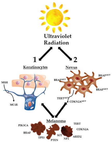
Figure 1.
2. Methodology
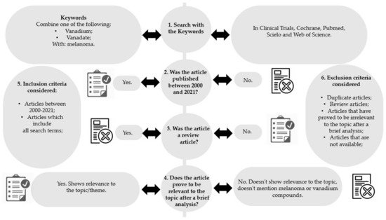
Figure 2.
-
Removal of articles not published between 2000 and 2021 (Figure 2, step 2);
-
Removal of review articles (Figure 2, step 3);
-
Evaluation of titles/abstracts and obtaining all articles potentially relevant;
-
Confirmation of the relevance or irrelevance of the articles obtained, checking for factors that would imply inclusion/exclusion, through the reading of the full articles (Figure 2, step 4);
-
Organization of selected studies in a reference management program.
-
Evaluation of titles/abstracts and obtaining all articles potentially relevant;
-
Confirmation of the relevance or irrelevance of the articles obtained, checking for factors that would imply inclusion/exclusion, through the reading of the full articles (Figure 2, step 4);
-
Organization of selected studies in a reference management program.
3. Types of Vanadium Based-Compounds Used in Studies against Melanoma
3
7
2
1
5
6
8
9, 10
11
4). The structure of these vanadium compounds and/or vanadium materials [27][28][29][30][31][32][33][34][35] are represented in
6
4
5 described in two articles [30][31], while compounds
5
8
9
Table 1. Representation of the structures of vanadium compounds and/or vanadium materials included in the entry.
| Vanadium Compound/Material | Structure | Year |
|---|---|---|
| VOSO4 | (VO2+) |
2021 |
| Vanadyl sulfate (Abreviated VO) | ||
| 1 | ||
| V2CTz-ox24/ V2CTz-ox48 (V2CTz) |  |
2020 |
| Vanadium carbides (MXenes) | ||
| 2 | ||
| V2O5 | 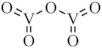 |
2020 |
| Vanadium pentoxide (VP) | ||
| 3 | ||
| H2VO4— |  |
2019, 2017 |
| monomeric vanadate (VN) | ||
| 4 | ||
| VIVO(dhp)2 (VS2) | 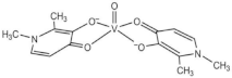 |
2019, 2017 |
| dhp: 1,2-dimethyl-3-hydroxy-4(1H)-pyridinonate | ||
| 5 | ||
| Xyloglucan oxovanadium (IV/V) (XGCVO) | 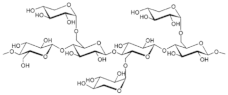 +  |
2018 |
| 6 | ||
| YIIIVVO4: EuIII (YVEu NPs) | 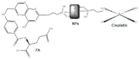 |
2018 |
| Europium(III)-doped yttrium vanadate nanoparticles | ||
| 7 | ||
| [VIVO (mpp)2] (VS3) | 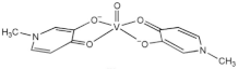 |
2017 |
| mpp: 1-methyl-3-hydroxy-4(1H) pyridinonate | ||
| 8 | ||
| [VIVO(ppp)2] (VS4) | 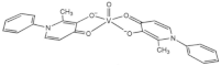 |
2017 |
| ppp: 1-phenyl-2-methyl-3-hydroxy-4(1H)-pyridinonate | ||
| 9 | ||
| [Ru-(pbt)2(tpphz)VO(sal-L-tryp)]Cl2 | 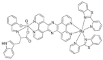 |
2013 |
| (RuVO) | ||
| pbt = 2-(2′-pyridyl)benzothiazole | ||
| tpphz = tetrapyrido [3,2-a:2′,3′-c:3′′,2′′-h:2′′′,3′′′-j]phenazine | ||
| sal-L-tryp = N -salicylidene-L-tryptophanate | ||
| 10 | ||
| N,N’-ethylenebis (pyridoxylideneiminat) | 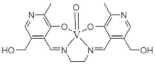 |
2013 |
4. Vanadium Species, Complexes and/or Materials Effects in Melanoma Cells
4.1. Cell Viability Effects
4
1
4
4
4
4
2
z
24
2
z
48
2
2
z
48
2
z
24
2
z
24
2
z
48
2
5
3
2
5
4
5
8
9 (VN, VS2, VS3, and VS4, respectively) was evaluated by MTT assay in A375 and CN-mel (human noncutaneous metastatic melanoma [37]) cells. Both cell lines were treated for 72 h at three different concentrations (1, 10 and 100 µM) of each vanadium compound [30][31]. At the lowest concentration, cells did not show accentuated signs of decreased cell viability. On the other hand, at 10 and 100 µM, A375 cells underwent a marked decrease in cell viability, presenting viability values never greater than 2% for all compounds, regardless of the concentration tested. CN-mel showed a less accentuated decrease, having viability values between 52% and 59% at 10 µM but even so, at the highest concentration (100 µM), there was a more pronounced decrease viability, with viability values between 1.5% and 13%. In these conditions, IC50 values between 2.4 and 4.7 µM were found for A375 cells and between 6.5 and 14 µM for CN-mel cancer cells. The relative order of potency for human noncutaneous metastatic melanoma was found to be compound
4
8
5
9
8
5
9
4
6
11
2
50
2
4.2. Cell Morphology and Apoptosis Effects
2
2
z
24
2
z
48
2
3
2
5) and with SU-5416 (semaxanib) for 18 h [29]. SU-5416 was used as a positive control, since this substance is an inhibitor of angiogenesis and leads to suppression of tumors through the induction of apoptosis [35][38]. Flow cytometry assay showed that cells treated with V
2
5
2
5
4
5 (VN and VS2, respectively) in two different articles [30][31], having counted the live, apoptotic and necrotic cells. In Pisano, et al. [30] untreated and treated cells were also treated with NAC (N-acetylcysteine), a ROS inhibitory compound [39], as control. The Annexin V apoptosis assays suggested that treatments with the longer duration (48 h) and higher doses cause a larger apoptotic effect in A375 cells. VN increased apoptotic population up to 48%, while VS2 increased apoptotic population up to 52%, at the highest concentration used (20 µg/mL) in 48 h assays. In Rozzo, el al. [31], the Annexin V apoptosis assays suggested that treatments with the longer duration (72 h) and higher doses cause larger apoptotic effects in A375 cells, as well. VN increased apoptotic population up to 70%, while VS2 increased apoptotic population up to 67%, at the highest concentration used (20 µg/mL) in 72 h assays.
6
10
10
2
10
Table 2.
| Compound | Cell Viability | Cell Morphology and Apoptosis Effects | Cell Cycle Effects | ROS Production | Mitochondrial Effects | Protein Expressions Studies | In Vivo Anticancer Activity |
|---|---|---|---|---|---|---|---|
| VO 1 | B16F10 cells | Mice Tumor regression upon VOSO4 (40 mg/kg), 96 h |
|||||
| 200 µM produces 90% inhibition, after 48 h | |||||||
| V2CTz 2 | A375 cells | A375 cells The population of apoptotic cells was negligible, for 24 h |
A375 cells Cellular cycle arrest in the G0/G1 phase, triggering apoptosis |
A375 cells V2CTz-ox24 increased ROS to 225%, (100 µg/mL) V2CTz-ox48 increased ROS to 140% (100 µg/mL) |
A375 cells s-V2CTz-ox48, slight increase mitochondrial membrane potential (ΔΨm) |
||
| 50% inhibition for 1–5 µg/mL V2CTz-ox48 and for 25–50 µg/mL V2CTz-ox24 | |||||||
| VP 3 | B16F10 cells | B16F10 cells Increase of apoptosis (10 µg/mL), for 18 h DNA damage (10 µg/mL), for 18 h |
B16F10 cells Anion superoxide formation (10 µg/mL) |
B16F10 cells Upregulation of p53 downregulation of anti-apoptotic survivin (10, 20 µg/mL) |
Mice The survival rate increased up to 47 days, (10 mg/kg) No changes: body weight; in feed intake. No toxicity in vital organs |
||
| 50% inhibition for 10 µg/mL |
|||||||
| VN 4 | A375 cells IC50 = 4.7 µM, 72 h |
A375 cells Pisano, et al. Increased apoptotic population to 48%, (20 µg/mL), 48 h. Rozzo, et al. Increased apoptotic population to 70%, (20 µg/mL), 72 h |
A375 cells Cells did not go through the G2/M phase, dying through apoptosis |
A375 cells ROS increased to 80% (20 µg/mL), 48 h. |
A375 cells ph-ERK, ph-Rb and ph-Cdc25c levels decreased to 20% (20 μM), 24 h, p21Cip1 rised up to 10-14 times (20 μM) |
||
| CN-mel IC50 = 6.5 µM, 72 h |
|||||||
| VS2 5 | A375 cells IC50 = 2.6 µM, 72 h |
A375 cells Pisano, et al. Increased apoptotic population up to 52%, at the highest concentration used (20 µg/mL) in 48 h assays. Rozzo, et al. Increased apoptotic population up to 67%, at the highest concentration used (20 µg/mL) in 72 h assays |
A375 cells Cell cycle arrested in G0/G1 phase, showing not to be able to enter the S phase. |
A375 cells ROS levels increasing up to 80% as well, at the highest duration (48 h) and dose (20µg/mL). |
Pisano, et al. A375 cells ph-ERK, ph-Rb and ph-Cdc25c levels decreased to 20% (20 μM), p21Cip1 levels incresed up to 18 times (20 μM) Rozzo, et al. Increasing of the cleaved PARP band (85 kD) |
||
| CN-mel IC50 = 12.4 µM, 72 h |
|||||||
| XGCVO 6 | B16F10 cells | B16F10 cells 50% increase of apoptosis (200 µg/mL) |
B16F10 cells Cell cycle has not changed |
B16F10 cells Mitocondrial respiration decreased to 43% (5 μg/mL) 34% pyruvate decrease No effects lactate production |
|||
| 50% inhibition at 300 μg/mL | |||||||
| YVEu NPs 7 | Mice Tumor regression upon YIIIVVO4:EuIII: CPTES:FA: CDDP (15 mg/kg), 5 days; CDDP toxicity reduced |
||||||
| VS3 8 | A375 cells IC50 = 2.4 µM, 72 h |
||||||
| CN-mel IC50 = 10.4 µM, 72 h |
|||||||
| VS4 9 | A375 cells IC50 = 4.2 µM, 72 h |
||||||
| CN-mel IC50 = 14.0 µM, 72 h |
|||||||
| RuVO 10 | Amelanotic melanoma In the absence of light, changes in cell morphology (20 µM). In the presence of light, apoptosis observed, 20 µM. |
Mice The survival rate was 100%. Tumor weight reduction. Proliferative activity reduction |
|||||
| Pyr2enVO 11 | A375 cells | A375 cells Increase of G0/G1 cells to 60% (100 µM), 72 h |
A375 cells ROS increased to 23% (100 µM), 24 h |
A375 cells Percentage of cells with loss of ΔΨm increased up to 35% (100 μM), 48 h, and 73%, after 72 h |
|||
| IC50 = 61.5 (24 h) IC50 = 13.0 (48 h) IC50 = 6.0 (72 h) | |||||||
4.3. Cell Cycle Effects
2
2
2
4
5
4
4
5
11
2
4.4. Effects on ROS Production
2
2
2
2
2
3
2
5
2
5
4
5,
11
2
2
2
4.5. Effects on Mitochondria Function
2
2
2
2
6
11
2
4.6. Protein Expressions Effects
2
5
4
5
4
5
4.7. Anticancer Effects In Vivo
1
4
4
4
4
7
7
2
2
5
3
2
5
2
5
2
5
2
5
2
5
7
III
V
4
III
3
III
V
4
III
III
V
4
III
III
V
4
III
III
V
4
III
III
V
4
III
III
V
4
III
III
V
4
III
III
V
4
III
III
V
4
III
III
V
4
III
III
V
4
III
III
V
4
III
III
V
4
III