Pomegranate Extract and Skin: Comparison
Please note this is a comparison between Version 1 by Lucía Yepes Molina and Version 2 by Vivi Li.

Pomegranate extract (PG-E) has been reported to exert a protective effect on the skin due to its antioxidant activity. Ingredients rich in phenolic compounds are unstable in extract solutions, and, therefore, the use of a suitable nanosystem to encapsulate this type of extract could be necessary in different biotechnological applications. Thus, we investigated the capacity of Brassica oleracea L. (cauliflower) inflorescence vesicles (CI-vesicles) to encapsulate PG-E and determined the stability and the antioxidant capacity of the system over time. In addition, the protective effect against UV radiation and heavy metals in HaCaT cells was also tested. The CI-vesicles had an entrapment efficiency of around 50%, and accelerated stability tests did not show significant changes in the parameters tested. The results for the HaCaT cells showed the non-cytotoxicity of the CI-vesicles containing PG-E and their protection against heavy metals (lead acetate and mercuric chloride) and UV-B radiation through a reduction of oxidative stress. The reduction of the percentage of deleted mtDNA (mtDNA4977, “common deletion”) in UV-treated HaCaT cells due to the presence of CI-vesicles containing PG-E indicated the mechanism of protection. Therefore, the effects of CI-vesicles loaded with PG-E against oxidative stress support their utilization as natural cosmeceuticals to protect skin health against external damage from environmental pollution and UV radiation.

  • pomegranate
  • antioxidant capacity
  • membrane vesicles
  • cauliflower
  • keratinocytes
  • oxidative stress
  • cytotoxicity

1. Introduction

Pomegranate (Punica granatum L.), a fruit of the Punicaceae family, is considered a fruit with high pharmaceutical value since its bioactive compounds have been shown to have biological activities in the treatment of several human diseases [1]. The main benefit is due to the antioxidant potential derived from the high concentrations of phenolic compounds, such as galloylglucose, punicalagin, punicalin, ellagic acid, and gallic acid [2][3][2,3]. Besides, anthocyanins and other nutraceutical components, such as sterols, γ-tocopherol, punicic acid, and hydroxybenzoic acids, have been found in the different parts of pomegranate [4][5][4,5]. Thus, functional products enriched with pomegranate extract (PG-E) have been reported to be useful for the treatment of certain diseases—such as diabetes mellitus, obesity, and cardiovascular and gastrointestinal diseases [1][6][7][1,6,7]—since their antioxidant potential gives protection from inflammation because it reduces the activity of cytokines, such as tumor necrosis factor-α (TNF-α) or interleukin-6 (IL-6) [8][9][10][8,9,10], as well as the levels of total cholesterol, low density lipoprotein (LDL), and lipid peroxidation [11]. Further, beneficial and protective effects of PG-E in the skin are also due to antioxidant activity, as reported in different studies [12][13][12,13]. In this regard, it is important to focus on the keratinocytes, as they comprise much of the outermost layer of skin (epidermis) [14]. Therefore, keratinocytes suffer damage due to extrinsic stimuli (UV exposure or pollutants, such as heavy metals) [15][16][15,16]. These stimuli trigger an excessive production of reactive oxygen species (ROS), which entails a loss of cellular functions and even cell death [17][18][17,18]. It is well known that ROS is a threat to cellular integrity, as it causes damage to essential macromolecules, including DNA, lipids, and proteins [19]. Regarding DNA damage, it has been observed to be more persistent in mitochondrial DNA (mtDNA) than in nuclear DNA due, among other causes, to limited repair mechanisms [20]. An indicator of DNA damage is a large deletion of 4977 bp from mtDNA called “common deletion”, which is considered an early marker for mutations induced by high levels of ROS [21][22][21,22]. It has been reported that PG-E can reduce the H2O2 overproduction as well as the cytotoxicity and the inflammatory stress induced by UV exposure [13][23][13,23].
Based on the above, this type of extract is of great interest as a natural cosmeceutical for skin health. But, one problem is that phenolic compounds are unstable in extract solutions, and, therefore, it is necessary to remove the solvents of the extracts to stabilize them. The shelf life of the phenolics could be enhanced in the dry extracts, but the stability of the formulated liquid extracts is very limited. Thus, procedures to prolong the stability of the final product, such as the addition of pectins for jelly formation, have been investigated [24]. Similarly, microencapsulation has been reported as a suitable option to stabilize the phenolics of the PG-E [25]. In this procedure, the phenolics are surrounded by a maltodextrin matrix in order to produce small capsules, with significant improvement of the antioxidant and α-glucosidase inhibitory activities.
Recently, new technologies of encapsulation, such as the use of membrane vesicles derived from natural sources, have been studied for different applications, such as cosmetics or therapy, such as treatment of colitis or melanoma [26][27][28][29][30][26,27,28,29,30]. The most profitable sources may well be those of plant origin since, in many crops, by-products are produced, which can be used to obtain membrane vesicles. The latest research in our group has focused on the study of stable natural membrane vesicles from brassicas. Plasma membrane vesicles from broccoli (Brassica oleracea L. var. italica) are characterized by their potential to stabilize the bioactive glucosinolate glucoraphanin [31]; the stability of this type of vesicle was studied in other work [32] and was found to be related to aquaporins. Recent work confirmed the potential of these vesicles as carriers in cosmetic or therapeutic applications [28]. Besides, in this study, an interaction between plant and human cell membranes was shown, revealing their potential in numerous applications in nanotechnology. In addition to broccoli-derived vesicles, membrane vesicles from cauliflower inflorescence have been well characterized [33]. The vesicles described in this study had sizes between 300 and 400 nm, appropriate for use in various biotechnological applications [34]. Besides, the osmotic permeability (Pf) values are related to vesicle functionality and membrane integrity, and high values of Pf have been determined in vesicles from Brassica oleracea L. var. botrytis inflorescences [33]. These types of vesicles are defined by their versatility since, in addition to their use in cosmetics, applications in agriculture are being studied [35][36][35,36]. All these findings lead us to propose these membrane vesicles as nanocarriers, whose advantages are based on their specific lipid/protein composition, their biodegradability, and their ability to carry the encapsulated substance to the target cells.

2. Physicochemical and Morphological Characterization

Table 12 
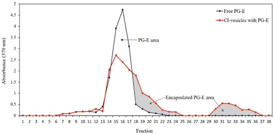
shows DLS analysis performed in a similar way to that previously reported [37][38][39][38,39,40]. The CI-vesicles had an average hydrodynamic diameter around 620.7 nm, which increased when PG-E was encapsulated in CI-vesicles (797.5 nm). TEM picture of the shape of CI-vesicles with PG-E is shown in Supplemental material (Figure S1). Zeta potential values of −21 mV were obtained in both CI-vesicles and CI-vesicles with PG-E, indicating adequate stability of the formulations [40][52] and a negative electric charge on the surface of the vesicles. Regarding free PG-E, it was not possible to measure the size by DLS, and the zeta potential value was −15 mV.
Table 12. Characteristics of CI-vesicles, CI-vesicles with PG-E, and PG-E: Particle Size, Polydispersity Index, and Zeta Potential.
  CI-Vesicles CI-Vesicles with PG-E PG-E
Z-average (nm) 620.72 ± 25.17 a 797.50 ± 38.93 b -
Polydispersity index (0–1) 0.70 ± 0.03 a 0.76 ± 0.12 a -
Z-potential (mV) −21.56 ± 0.38 a −21.65 ± 0.24 a −15.04 ± 0.40 b
PG-E: pomegranate extract, CI-vesicles: cauliflower inflorescence vesicles. Data are means ± SE (n = 3). Different letters indicate significant differences between groups for each variable.

3. Pomegranate Extract Entrapment Efficiency (EE)

The entrapment efficiency (EE) was determined by absorbance measurements at 370 nm, the wavelength in the visible spectrum at which the absorbance by samples containing PG-E is maximum. Both free PG-E and PG-E encapsulated in CI-vesicles were passed through a Sephadex column, and different fractions were collected to measure the absorbance. The CI-vesicles were disrupted to allow the release of the encapsulated extract. Figure 1 shows the absorbance at 370 nm of different fractions collected after passing free PG-E and CI-vesicles containing PG-E through a Sephadex column. The free PG-E appeared in fractions 13 to 19. For the samples with vesicles, absorbance also appeared from fraction 13 but remained until fraction 24, with a second peak between fractions 30 and 36. The colored areas correspond to fractions where proteins appeared, that is, those fractions containing the CI-vesicles with encapsulated PG-E. The first colored area corresponds to small vesicles that appeared together with the last molecules of the free PG-E, and the second area corresponds to vesicles retained in the column and disrupted by chloroform.
Figure 1. Absorbance (370 nm) of each fraction obtained after passing through a Sephadex column the samples of free PG-E (blue line) and CI-vesicles with encapsulated PG-E (red line). The grey area indicates the proportion of PG-E encapsulated in CI-vesicles. PG-E, pomegranate extract; CI-vesicles, cauliflower inflorescence vesicles.
The data regarding the areas under the curves and the protein concentration are shown in Table 23. Taking into account the total area under the free PG-E curve and corresponding to fractions with proteins, an EE of 46.50 ± 1.62% was estimated. No significant differences appeared between the total areas under the curve of the two samples, and, therefore, no extract residues were retained in CI-vesicles without being determined. Besides, the sum of the protein contents of all the fractions collected was the same as the protein content in the sample previous to elution through the Sephadex column. Thus, both the entire extract and all the vesicles passed through the column.
Table 23. Entrapment efficiency calculated from absorbance data and the protein content (mg) in samples before and after passage through a Sephadex column and fractions collection.
  Free PG-E CI-Vesicles with PG-E
The total area under the curve (a.u.) 3160 ± 33.20 3357 ± 161.40
Encapsulated area (a.u.) - 1561 ± 234.15
Entrapment efficiency (%) - 46.50 ± 1.62
Protein before column (mg) 0 0.22 ± 0.02
Total protein collected (mg) 0 0.21 ± 0.01
PG-E: pomegranate extract, CI-vesicles: cauliflower inflorescence vesicles, a.u.: arbitrary unit. Data are means ± SE (n = 3).

4. Discussion

Due to the variety of beneficial effects of pomegranate extract (PG-E) enriched in punicalagin [1][41][1,55], a vehicle is needed to improve its functionality in therapeutic and cosmetic applications, with the focus on its use as a protective agent against damaging environmental factors, such as UV radiation or pollution [42][43][56,57]. In the last few years, the use of vehicles or carriers from natural sources has been an object of study [44][45][58,59]; specifically, the use of membrane vesicles from plant materials as nanocarriers has shown promising results in different fields, such as cosmetics [28], medicine [26], or agriculture [35][36][35,36]. This work highlights the potential use of cauliflower inflorescence membrane vesicles (CI-vesicles) to encapsulate PG-E for cosmetic applications. The preparation of these vesicles consists of the purification of the plant membrane fraction, as described in Rios et al. [35], which allows a reproducible preparation of vesicles in terms of yield and size. The entrapment efficiency (EE) of the nanocarrier will depend on factors, such as the integrity and chemical composition of the vesicles, but also on the chemical and physical properties of the encapsulated compound [34][46][34,60]. In this work, an EE of 46.5% was obtained for PG-E encapsulated in CI-vesicles, similar to that reported previously with broccoli-derived plasma membrane vesicles, for which EE values around 50% were obtained when encapsulating two dyes [28]. Regarding other works where PG-E was encapsulated in nanocarriers, values of EE similar to that obtained in our study were reported. Marin et al. [47][61] showed an EE of 63% for a PG-E encapsulated in liposomes and established that punicalagin (a polar phenolic compound of PG-E) is located in the aqueous core of the liposomes, while ellagic acid intercalates into the aliphatic-chain zone of the membrane since it is poorly soluble in polar solvents. Besides, it is known that phenolic compounds interact with the polar head and also intercalate into the bilayer membrane [48][62]. This knowledge, together with the EE percentages, gives us an idea of how our system, based on membrane vesicles and a PG-E, is composed and arranged. After obtaining acceptable EE data, the stability over time of the system (CI-vesicles with PG-E) was the next point to study. The stability was addressed on several fronts: protein content, color, and antioxidant activity. None of these parameters was affected by the passage of time (3 months) at any of the storage temperatures (20 °C, 4 °C, and 40 °C), which evidences the suitability of the system for its final purpose. The CI-vesicles with PG-E are maintained in a solution based on a polyalcohol protector, which promotes high stability during storage as it decreases the surface tension of water [49][63]. In this way, the polyalcohol also protects the non-encapsulated extract. This type of assay is an accelerated storage-stability test and is commonly used to determine the long-term behavior of the system [50][64]. Other studies also demonstrated that the antioxidant activity of materials derived from pomegranate persists over time without changes. Mali et al. [51][65] showed that the antioxidant activity of pomegranate peel powder had not changed after 90 days at room temperature, but degradation had occurred in aqueous solutions. The color stability is related to oxidation, with the concomitant implications for cell functionality. Therefore, our stability assays showed that this is a system suitable for use in further cosmeceutical applications. The protective effect of PG-E encapsulated in CI-vesicles was assayed in a keratinocyte cell line (HaCaT). Keratinocytes form the majority of the epidermis, the outermost layer of the skin; therefore, these cells are part of the first defense barrier against harmful external stimuli, such as UV radiation or pollution [52][66]. The cytotoxicity of CI-vesicles containing PG-E was assayed at different concentrations to find the working concentration. Application of the highest concentration induced a significant decrease in viability with respect to the control, but in such trials, cell viabilities around 80% are not considered to represent cytotoxicity [53]. Hence, this concentration was chosen to ensure clear effects in subsequent assays. This cytotoxicity was due to the CI-vesicles because free PG-E did not show cytotoxicity and even increased cell viability. There are no previous reports of cytotoxicity caused by this type of membrane vesicle, and several works have reported that such vesicles have zero cytotoxicity at suitable concentrations in cell cultures [54][55][67,68]. In other studies, null cytotoxicity of PG-E has been reported for HaCaT cells; for example, in Liu et al. [13], for concentrations of PG-E from 6.25 to 100 µg/mL. In our work, the highest concentration of PG-E was 13 µg/mL; thus, our results are in line with what has been reported previously. These good results obtained in the cytotoxicity tests, together with previous results from our group, confirm the suitability of the system. Previous work [28] showed an interaction between plant and human cell membranes, with plasma membrane vesicles from broccoli exhibiting a high fusion ability with human keratinocytes. The protective effect of our system (PG-E encapsulated in CI-vesicles) against heavy metals and UV-B radiation was determined in this work. Heavy metals like lead and mercury are common air pollutants and have been shown to trigger health risks [56][69]. To analyze the effectiveness of CI-vesicles containing PG-E with regard to decreasing the damage caused by heavy metals in keratinocytes, different concentrations of lead acetate and mercuric chloride and a mixture of both were applied to HaCaT cells. Individually, both metals caused a reduction in cell viability (data not shown), as reported previously in other studies [57][58][70,71], but in this work, assays were also carried out with a mixture of lead acetate and mercuric chloride. An important reduction in cell viability was shown after 24 h of incubation of HaCaT cells with different concentrations of the metals and in the same way as previously reported, the reduction being highly dose-dependent [58][71]. Oxidative stress has been revealed as one of the actors in heavy metal-induced cytotoxicity [18][59][60][18,72,73]; thus, due to their antioxidant capacity, the protective effects of CI-vesicles with PG-E against heavy metals were tested. As described in the Results section, an improvement in cell viability appeared when PG-E was applied in encapsulated form. This could be due to a better entry of the extract into the cells when it is encapsulated due to the fusion of the vesicles with the cell membranes [28]. Studies were carried out to determine the effectiveness of CI-vesicles with PG-E against UV-B radiation (290–320 nm), which is the main UV component that causes a wide variety of skin disorders, including skin cancers [61][74]. The cell viability after irradiation with UV-B was higher when CI-vesicles with PG-E were applied 24 h before irradiation. In addition, the TBARS release into the medium after irradiation with UV-B was lower for cells previously treated with PG-E encapsulated in CI-vesicles than for untreated cells, and this response is probably related to the antioxidant capacity of the PG-E. A cellular environment with increased lipid peroxidation can produce immune and inflammatory responses [62][75]. The reduction of these inflammatory responses, thereby maintaining or restoring cell homeostasis, could be achieved with our PG-E treatment. The results obtained in our experiments showed that the encapsulated PG-E provided protective effects against the UV-B-induced oxidative stress, suggesting prevention of membrane damage. In this way, our results with encapsulated PG-E are similar to those obtained in other work with free pomegranate extracts but using higher concentrations [63][76]. These previous studies also showed promising results regarding protection against UV-B radiation: the protection by CI-vesicles with PG-E against the oxidative stress triggered by UV-B radiation in HaCaT cells coincided with higher cell viability, an enhanced intracellular glutathione (GSH) content, and a decline in membrane damage (analyzed as lipid peroxidation). Besides, in other work, inhibition of UV-B-mediated activation and phosphorylation of the mitogen-activated protein kinase (MAPK) and factor nuclear kappa B (NF-κB) pathways by a pomegranate fruit extract was revealed [64][77]. Our results showed a protective effect of CI-vesicles with PG-E against the mutations in mtDNA induced by UV-B radiation, measured as common deletion mtDNA4997. These types of mutations are considered as markers for ROS-mediated genotoxicity [65][78], and several previous studies have shown an increase in these mutations (deletions) in the mtDNA of cells exposed to UV-B radiation [66][67][48,50]. This is in accordance with our results because an increase in mtDNA4997 in HaCaT cells that were not treated with the protective agent was found after UV irradiation. The antioxidant capacity of PG-Es could play an important role in preventing the deletion since, as we stated above, this mutation is related to an increase in ROS generation. In this sense, melatonin, an endogenous antioxidant, has been reported to prevent mtDNA4997 under both basal conditions and induced oxidative stress in cybrids [68][79]. Our results, along with others published previously [64][69][77,80], support the capacity of antioxidant compounds to prevent damage related to aging and UV-B exposure [70][81]. The fact that the protective effect was higher for encapsulated PG-E could be due to its greater penetrability in cells, as reported previously [28].