Extracellular vesicles (EVs) are membranous structures, which are secreted by almost every cell type analyzed so far. In addition to their importance for cell-cell communication under physiological conditions, EVs are also released during pathogenesis and mechanistically contribute to this process. Here we summarize their functional relevance in asthma, one of the most common chronic non-communicable diseases. Asthma is a complex persistent inflammatory disorder of the airways characterized by reversible airflow obstruction and, from a long-term perspective, airway remodeling. Overall, mechanistic studies summarized here indicate the importance of different subtypes of EVs and their variable cargoes in the functioning of the pathways underlying asthma, and show some interesting potential for the development of future therapeutic interventions. Association studies in turn demonstrate a good diagnostic potential of EVs in asthma.
1. Introduction
Chronic non-communicable diseases (NCDs) are inflammatory conditions, which are not caused by infectious agents (e.g., bacteria, viruses, parasites). To name a few, these diseases include respiratory disorders such as asthma or chronic obstructive pulmonary disease (COPD), chronic inflammatory bowel diseases, cardiovascular disorders such as coronary artery disease/ischemic heart disease, peripheral vascular disease or stroke, all based on atherosclerosis, inflammatory disease conditions in the skin (e.g., atopic dermatitis, psoriasis), metabolic diseases such as obesity, metabolic syndrome, and diabetes, different forms of cancer, adverse mental outcomes, etc. The burden of NCDs is high in western countries and still rising, in particular in less developed areas. To effectively face this challenge, novel diagnostic and therapeutic approaches should be established based on the growing knowledge on pathobiological mechanisms underlying the development and the clinical course of NCDs.
This specifically applies also to asthma as one of the most prominent NCDs, for which, despite substantial progress, current diagnostic and therapeutic approaches remain suboptimal. One of the major reasons behind this is the heterogeneity of asthma, with a complex etiology and multiple clinical representations, requiring the development of stratified diagnosis and treatment strategies. These can only be achieved on the basis of novel cellular and molecular insights based on innovative methods.
2. Extracellular Vesicles and Asthma: Cellular Level
2.1. Airway Epithelial Cells and Fibroblasts
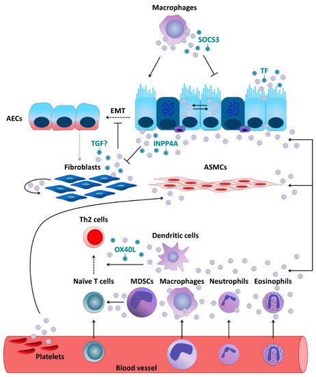
EVs are involved in asthma-related interactions between different cell types. Additionally, for airway epithelial cells (AECs), the exchange of EV cargo seems to be an important way of communicating with each other, as well as with other cell types. For example, in primary human tracheobronchial cells and cultured Calu-3 cells, a respiratory epithelial cell line, the reciprocal transfer of EV-associated proteins and microRNAs (miRNAs) was shown to be sufficient to qualitatively and quantitatively alter the profiles of airway secretions including miRNA cargo of EVs of the target cells and cause mucin hypersecretion. This mechanism may play an important role in epithelial remodeling and other pathologic processes in the airways involved in chronic inflammatory disorders of the respiratory tract, such as asthma, cystic fibrosis, and bronchogenic carcinoma. In a mouse study, it was shown that the composition of the pool of extracellular miRNAs in the lung was very similar to that of the airway epithelium, with 80% of the EVs detected in bronchoalveolar lavage fluid (BALF) being of epithelial origin. However, the number of miRNAs selectively expressed by immune cells, including miR-223 and miR-142a, and hematopoietic cell-derived EVs increased significantly following the induction of allergic airway inflammation (AAI), showing the importance of alterations in the EV miRNA pool for the development of allergic inflammation. Another group reported that EV secretion and production of EV-associated proteins were both higher in the lungs of mice in which AAI was induced compared to the control animals. These EVs, which were released during asthma/AAI by AECs under the influence of type-2 cytokines such as IL-13, triggered the proliferation and chemotaxis of undifferentiated macrophages. Not surprisingly, the use of GW4869, an inhibitor of exosome production, resulted in a reduction in the population of proliferating monocytes in the AAI mouse model and the alleviation of various asthmatic features.
Additionally, primary human fibroblasts were demonstrated to secrete exosomes, which undergo subsequent internalization by normal human bronchial epithelial cells (NHBECs). Moreover, compared to healthy controls, exosomes derived from fibroblasts which were obtained from severe asthmatics showed lower levels of transforming growth factor beta 2 (TGF-β2) and significantly increased the proliferation of NHBECs. These results are intriguing, given that TGF-β is considered to be a major driver of abnormal epithelial-mesenchymal transition (EMT). During EMT, epithelial cells demonstrate enhanced motility and invasive capacity through the downregulation of epithelial markers and higher expression of mesenchymal proteins, being this way a source of migrating myofibroblasts and fibroblasts. In turn, these cells promote extracellular matrix deposition and subepithelial fibrosis, which strongly contributes to the establishment of a persistent asthma phenotype. Moreover, fibroblasts themselves can also be recipients of EVs. In vitro experiments using cell lines demonstrated that AECs were able to secrete enzymatically active inositol polyphosphate 4-phosphatase type I A (INPP4A) in EVs and as a soluble free form. INPP4A was then transferred to lung fibroblasts, and inhibition of such transfer resulted in increased fibroblast proliferation. Moreover, in mice with or without AAI neutralization of extracellular INPP4A-induced AHR, with prominent airway remodeling, subepithelial fibroblast proliferation, and collagen deposition.
Figure 1. Extracellular vesicle- (EV-) mediated communication between cells crucial for asthma pathobiology. If not otherwise stated, EVs are thought to carry their usual content such as microRNAs, proteins, lipids, etc.
2.2. Antigen-Presenting Cells (APCs)
APCs, such as dendritic cells (DCs), macrophages, monocytes, and others can communicate through EVs with other types of cells involved in asthma development. A study performed in primary human macrophages and DCs demonstrated that they can secrete exosomes which contain enzymes for leukotriene biosynthesis and thus contribute to chronic inflammation, for example through granulocyte recruitment. Primary human DCs activated with TSLP, an epithelial cell-derived cytokine, release exosomes expressing OX40 ligand (OX40L), which was able to promote proliferation and differentiation of CD4+ T cells towards a Th2 phenotype. Resident alveolar macrophages were, in turn, demonstrated to dampen inflammatory signaling in AECs and thus AAI in a mouse model through transcellular delivery of suppressor of cytokine signaling 3 (SOCS3) within EVs. Air pollutants such as particulate matter are well-known contributors to the pathogenesis of chronic inflammatory airway diseases including asthma. In vitro exposure to particulate matter stimulated human macrophages to release more EVs in a dose-dependent manner. Moreover, those EVs were able to induce secretion of pro-inflammatory cytokines, such as IL-6 and tumor necrosis factor-α (TNF-α), by pulmonary epithelial cells.
2.3. Granulocytes and Mast Cells
Likewise, human eosinophils were found to be able to secrete exosomes, the production of which was higher by cells deriving from asthmatics. In addition, exosomes secreted by the eosinophils of patients with asthma could, in an autocrine manner, modify several specific eosinophil functions related to asthma pathogenesis including an increase in reactive oxygen species and nitric oxide synthesis and an augmentation of eosinophil migration and adhesion, suggesting that they could fundamentally contribute to the development and maintenance of asthma. Further, asthmatic eosinophil-derived exosomes could enhance the apoptosis of primary AECs and delay the repair of established epithelial damage, as well as increase the proliferation of primary bronchial smooth muscle cells and perpetuate airway inflammation status . Upon in vitro stimulation with LPS, horse neutrophil-derived exosomes, carrying proteins associated with immune response and positive regulation of cell communication, were rapidly internalized by equine airway smooth muscle (ASM) cells and enhanced their proliferative capabilities. The effects of neutrophil-derived exosomes on ASM proliferation [might play an important role in the neutrophil-mediated progression of asthma and promotion of airway remodeling in severe and corticosteroid-insensitive patients with asthma. Based on in vitro data obtained using human cells, exosomes were also suggested to partially contribute to mast cell-mediated pro-inflammatory modulation of ASM cells, although it was undisputed that soluble, extra-exosomal factors such as IL-8 were critical for the effect.
2.4. Platelets
Moreover, platelets, which are known to contribute to the pathophysiology of asthma as well, can exert their effects through EVs. Plasma EVs, a substantial portion of which is of platelet origin, isolated from asthmatics were found to be able to reduce the endothelium-dependent relaxation in response to bradykinin and increase the acetylcholine-induced contraction of the mouse trachea, which is suggestive of their potential role in ASM dysfunction typical for asthma. Moreover, the levels of circulating platelet microparticles (PMPs) were reported to be increased in asthmatics.
3. Conclusions and Perspectives
CThe sturrently available studiesdies presented in this review, whatever their nature, i.e., in vitro, in vivo on animals, in human studies, etc., clearly demonstrate the existence of EV communication between cells known as the crucial players in asthma pathology and, moreover, they also strongly suggest an importance of EV-mediated communication mechanisms for the pathobiology of the disease. This includes the mediation of etiopathogenic effects of environmental factors, e.g., microbes or pollutants, and the role of EVs from external origins, e.g., those present in cow’s milk or secreted by certain bacteria. Some of the studies already characterized components of the cargo of EVs and identified molecules responsible for asthma-related effects, mainly small RNAs, proteins, and lipid mediators. Based on this continuously expanding knowledge, the high diagnostic potential of EVs has been highlighted in a variety of studies. It has been shown that EV-based asthma diagnostics effectively targeting miRNA, proteins, or lipids could be performed in different types of biomaterials such as bronchoalveolar lavage fluid (BALF), sputum, nasal lavage fluid (NLF), or serum. Considering the access to biomaterials and the methodological rationale, the analytical approach based on the analysis of serum miRNAs seems particularly promising, irrespective of whether and how the miRNAome patterns in blood and lung correspond to each other. These approaches will certainly be expanded in the future to more precisely identify asthma phenotypes particularly by means of non- or minimally-invasive diagnostic sampling techniques. Finally, several in vitro and animal studies reviewed in this article already show that EVs, not only, but especially those secreted by MSCs, can exert beneficial immunomodulatory effects with anti-asthmatic capacity, suggesting a promising potential for EV-based therapeutic approaches. Although procedures regarding targeting specific cell types and the level of EV (cargo) degradation still need to be further optimized, it seems that modified or designed EVs with a higher propensity to fuse with (endosomal) membranes will offer even better therapeutic abilities. Ans also briefly outlined here, another approach being developed as a possible anti-asthmatic therapeutic strategy may involve the use of inhibitors of EV production, which have been shown to exhibit anti-AAI effects in some studies. However, further basic and clinical studies are needed, which undoubtedly will lead to diagnostic and therapeutic innovations based on their results.
This entry is based on Alashkar Alhamwe et al. Extracellular Vesicles and Asthma—More Than Just a Co-Existence. Int. J. Mol. Sci. 2021, 22(9), 4984; https://doi.org/10.3390/ijms22094984 (https://www.mdpi.com/1422-0067/22/9/4984/htm). For the full content including the supporting references, please, refer to the full veorsion of theiginal article.