Plants are constantly exposed to environmental stresses during their growth and development. Owing to their immobility, plants possess stress-sensing abilities and adaptive responses to cope with the abiotic and biotic stresses caused by extreme temperatures, drought, flooding, salinity, heavy metals and pathogens. Acyl-CoA-binding proteins (ACBPs), a family of conserved proteins among prokaryotes and eukaryotes, bind to a variety of acyl-CoA esters with different affinities and play a role in the transport and maintenance of subcellular acyl-CoA pools. In plants, studies have revealed ACBP functions in development and stress responses through their interactions with lipids and protein partners.
- abiotic stress
- acyl-CoA-binding proteins
- biotic stress
- lipids
- protein interactors
- stress signalling
1. Plant ACBPs
Brassica napus L. (oilseed rape) as a 10-kDa homologue expressed in seeds, flowers and cotyledons [1]. It binds long-chain acyl-CoA esters [2], participates in acyl-CoA transport [3], maintains acyl-CoA pool [4], and regulates the activities of various enzymes including glycerol-3-phosphate acyltransferase [2], lysophosphatidylcholine acyltransferase [4] and lysophosphatidic acid acyltransferase [5]. The transport of acyl-CoA esters is important for the biosynthesis of lipids such as glycerolipids, ceramides and phospholipids, and studies have shown that the binding of phospholipids to ACBPs plays a role in plant growth and development as well as stress responses [6][7][8][9][10][11][12][13][14][15]. Following the discovery of BnACBP, similar 10-kDa ACBPs emerged in
Arabidopsis thaliana [16],
Gossypium hirsutum (cotton) [17],
Ricinus communis (castor bean) [18],
Digitalis lanata Ehrh. (Wolly Foxglove) [19],
Oryza sativa (rice) [10],
Vernicia fordii (tung tree) [20],
Vitis vinifera (grape) [21],
Helianthus annuus (sunflower) [22],
Elaeis guineensis (oil palm) [23],
Zea mays (maize) [24] and
Glycine max (soybean) [25].
Table 1) [10][23][24][25][26].
Table 1 shows that Class I ACBPs range from 10 to 17 kDa, whereas the others comprised of a transmembrane domain, ankyrin repeats and/or kelch motifs, have molecular weights of 34 to 85 kDa [10][24]. Arabidopsis ACBPs are localised to the ER and plasma membrane (AtACBP1 and AtACBP2) [27][28], apoplast (AtACBP3) [29] and cytosol (AtACBP4 to AtACBP6) [8][30]. On the other hand, rice ACBPs are subcellularly localised to the cytosol (OsACBP1 to OsACBP3) [12], ER (OsACBP4) [12][31], apoplast (OsACBP5) [32] and peroxisomes (OsACBP6) [12]. In maize, transient expression of green fluorescent protein (GFP)-tagged Class I ZmACBP1 in
Nicotiana benthamiana leaf epidermal cells revealed that ZmACBP1 was confined to the cytosol, Class II ZmACBP3 localised to the ER, whereas Class III and IV ZmACBP6 and ZmACBP7, respectively, were targeted to both the cytosol and the plasma membrane [24]. Oil palm Class II EgACBP2 contains an
N
C-terminal ankyrin repeats which could mediate protein-protein interactions and other cellular activities [23]. Consistent with Protein Subcellular Localization Prediction Tool (PSORT) speculation, the sunflower Class I HaACBP6, which was transiently expressed in tobacco leaves, was localised to the cytosol and nucleus [22]. These results are summarised in
Table 1.
| Class | Protein Name | Signal Peptide | TM Domain | ACB Domain | Ankyrin Repeats | Kelch Motifs | Subcellular Locations | Size (kDa) |
|---|
| I | AtACBP6 | − | − | + | − | − | Cytosol | 10.4 |
| OsACBP1 | − | − | + | − | − | Cytosol | 10.2 | |
| OsACBP2 | − | − | + | − | − | Cytosol | 10.3 | |
| OsACBP3 | − | − | + | − | − | Cytosol | 17.7 | |
| ZmACBP1 | − | − | + | − | − | Cytosol | 10.1 | |
| HaACBP6 | − | − | + | − | − | Cytosol, Nucleus | 10.9 | |
| II | AtACBP1 | − | + | + | + | − | ER, PM | 37.5 |
| AtACBP2 | − | + | + | + | − | ER, PM | 38.5 | |
| OsACBP4 | + | + | + | + | − | ER | 36 | |
| ZmACBP3 | − | − | + | + | − | ER | 34.8 | |
| EgACBP2 | − | + | + | + | − | PM | ND | |
| III | AtACBP3 | + | + | + | − | − | Apoplast | 39.3 |
| OsACBP5 | + | + | + | − | − | ER | 61.2 | |
| ZmACBP6 | − | − | + | − | − | Cytosol, PM | 35.2 | |
| IV | AtACBP4 | − | − | + | − | + | Cytosol | 73.2 |
| AtACBP5 | − | − | + | − | + | Cytosol | 71 | |
| OsACBP6 | − | + | + | − | + | Peroxisomes | 71.4 | |
| ZmACBP7 | − | − | + | − | + | Cytosol, PM | 72.1 |
Abbreviations: ACB, acyl-CoA-binding; ACBP, acyl-CoA-binding protein; At, Arabidopsis thaliana; Eg, Elaeis guineensis; ER, endoplasmic reticulum; Ha, Helianthus annuus; kDa, kilodalton; ND, not determined; Os, Oryza sativa; PM, plasma membrane; TM, transmembrane; Zm, Zea mays; −, absent; +, present.
Using isothermal titration calorimetry (ITC), it has been reported that all recombinant ACBPs (rACBPs) bind acyl-CoA esters with varying affinities; rAtACBP1 and rAtACBP3 displayed high affinity to very-long-chain (VLC) species [33][29][34], while rAtACBP3 to rAtACBP6 and rOsACBPs to medium-chain species [10][35]. All rAtACBPs and rOsACBPs bind long-chain acyl-CoA esters at different affinities [36][10][15][29][37][38]. Moreover, rACBPs were shown to bind phospholipids, all Arabidopsis rAtACBPs bind PC [7][8][34][38][39], and rAtACBP1, rAtACBP2 and rAtACBP3 bind PA, lysoPC, and PE, respectively [6][9][34][40]. In contrast, all rice rOsACBPs bind PA and PC [10]. Besides binding with high affinity to 16:0-CoA, 18:0-CoA and 18:1-CoA, sunflower Class I rHaACBP6 and Class II rHaACBP1 also bind to several PC species [22][41]. In addition to phospholipid binding, Arabidopsis ACBPs were shown to interact with protein interactors (
Table 2). AtACBPs interact with various transcription factors that activate the gene expression for downstream abscisic acid (ABA) or ethylene responses upon perception of stress stimuli [42][43][44]. These transcription factors include ABA-RESPONSIVE ELEMENT BINDING PROTEIN1 (AREB1) [44], RELATED TO APETALA2.12 (RAP2.12) [43] and ETHYLENE-RESPONSIVE ELEMENT BINDING PROTEIN (AtEBP) [42]. Furthermore, AtACBPs bind enzymes for sterol or phospholipid metabolisms such as PHOSPHOLIPASE Dα1 (PLDα1) [7], STEROL C4-METHYL OXIDASE1-1 (SMO1-1) [45], SMO1-2 [46] and LYSOPHOSPHOLIPASE2 (LYSOPL2) [6][47], which are important for membrane stability and repair as well as plant development. Thus far, only AtACBP2 interacts with FARNESYLATED PROTEIN6 (AtFP6) which may be involved in phospholipid repair following heavy metal-induced lipid peroxidation [36]. Lipid binding of ACBPs and their protein-protein interactions are now known to be important in regulating abiotic and biotic stress responses [48][36][6][7][44][8][9][14][49][50][51][52][53][54][55][56][57], as well as plant development including embryogenesis [46][39][45], seed dormancy [7], seed germination and development [7][44][58][59][60], cuticle development [61][33], pollen growth [62] and senescence [34][40].
Table 2.
| Proteins | Species | Acyl-CoA Binding | Phospholipid Binding | Protein Interactors | Stress Responses |
|---|
| AtACBP1 | A. thaliana | 16:0, 18:1, 18:2, 18:3, 20:4, 24:0, 25:0, 26:0 [33][29][63][26,108,133] | PC [7][78] PA [9][88] |
PLDα1 [7][78] | Freezing [9][88] |
| RAP2.12 [43][57][64][77,128,134] | Hypoxia [43][57][77,128] | ||||
| AREB1 [44][80] | Salinity, osmotic damage [44][80] | ||||
| − | Heavy metal [65][135] | ||||
| − | Pathogen [33][26] | ||||
| AtACBP2 | A. thaliana | 16:0, 18:1, 18:2, 18:3, 20:4 [36][29][75[37],108,114] | PC [39][116] lysoPC [6][76] |
AtEBP [42][73], RAP2.12 [43][57][77,128] | Hypoxia [42][43][73,77] |
| LYSOPL2 [6][47][76,82], AtFP6 [36][75] | Heavy metal [36][6][47][75,76,82] | ||||
| − | Drought [11][90] | ||||
| − | Salinity [15][94] | ||||
| − | Oxidation [36][75] | ||||
| AtACBP3 | A. thaliana | 12:0, 14:0, 16:0, 18:1, 18:2, 18:3, 20:4, 22:0, 24:0 [29][34][55][66][108,112,126,136] | PC [34][112] PE [34][40][112,117] |
− | Drought [61][25] |
| − | Hypoxia [66][67][136,137] | ||||
| − | Wounding [55][126] | ||||
| − | Pathogen [61][29][50][25,108,121] | ||||
| AtACBP4 | A. thaliana | 14:0, 16:0, 18:0, 18:1, 18:2, 18:3 [35][38][113,115] | PC [38][115] | AtEBP [48][74] | Pathogen [61][48][25,74] |
| − | Drought [61][25] | ||||
| − | Heavy metal [53][124] | ||||
| AtACBP6 | A. thaliana | 14:0, 16:0, 18:0, 18:1, 18:2, 18:3, 20:4 [16][35][38][95,113,115] | PC [8][87] | − | Freezing [8][52][87,123] |
| − | Drought [61][25] | ||||
| − | Wounding [54][125] | ||||
| − | Pathogen [61][25] | ||||
| OsACBP4 | O. sativa | 16:0, 18:0, 18:1, 18:2, 18:3 [10][15][89,94] | PC, PA [12][91] | − | Salinity [10][15][89,94] |
| OsACBP5 | O. sativa | 16:0, 18:3 [10][14][89,93] | PC, PA [12][91] | − | Pathogen [10][14][89[56],93,127] |
| − | Wounding [10][89] | ||||
| OsACBP6 | O. sativa | 18:1, 18:2 [10][89] | PC, PA [12][91] | − | Wounding [10][89] |
| ZmACBP1 | Z. mays | − | − | − | Salinity, drought [24][103] |
| ZmACBP3 | Z. mays | − | − | − | Salinity, drought [24][103] |
| ChACBP1 | Chlorella sp. | − | PC [13][92] | − | Freezing, salinity, oxidation, heavy metal [13][92] |
| VvACBP | V. vinifera | − | − | − | Freezing, heat, ER, pathogen [21][100] |
Abbreviations: AREB1, ABSCISIC ACID-RESPONSIVE ELEMENT BINDING PROTEIN1; AtEBP, Arabidopsis ETHYLENE-RESPONSIVE BINDING PROTEIN; AtFP6, Arabidopsis FARNESYLATED PROTEIN6; ER, endoplasmic reticulum; lysoPC, lysophosphatidylcholine; LYSOPL2, LYSOPHOSPHOLIPASE2; PA, phosphatidic acid; PC, phosphatidylcholine; PE, phosphatidylethanolamine; PLDα1, PHOSPHOLIPASE Dα1; RAP2.12, RELATED TO APETALA2.12.
2. Membrane Lipids and ACBPs in Abiotic Stress Signalling
Plants are sessile, and therefore possess signalling and adaptive mechanisms to counteract abiotic and biotic stresses including cold, drought, salinity, oxidation, heavy metals, hypoxia and pathogen attack. Given the importance of plant ACBPs in development and stress responses, the roles of all Arabidopsis and rice ACBPs at different stages of plant growth were previously summarized by Du et al. [68] and are now updated (
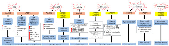
Figure 1.
Arabidopsis thaliana
PLDα1 was induced upon cold stress, causing a decrease in the ratio of PC to PA leading to membrane instability and freezing sensitivity [9]. In contrast, transgenic Arabidopsis Class I AtACBP6-OEs were conferred freezing tolerance via the PLDδ-mediated pathway in rosettes and the ABA-mediated pathway in flowers, resulting in changes in lipids, sugars and stress-related genes [8][52]. During drought, transgenic Arabidopsis Class II AtACBP2-OEs exhibited elevated
AtAREB1
AtRBOHF expression which led to ROS production, subsequent stomatal closure and reduced water loss [11]. Proper stem cuticle development conferred by Class I AtACBP6, Class III AtACBP3 or Class IV AtACBP4 protects wild-type Arabidopsis from water loss [61]. Under high salinity,
AtACBP1
AtAREB1 expression were upregulated in wild-type seeds [44]. The overexpression of AtACBP1 in transgenic Arabidopsis triggers nuclear translocation of AtAREB1, leading to the induction of stress marker genes (
RD22
RD29B
PKS5
RAB18), thereby promoting stronger ABA responses during seed germination and seedling establishment [44]. When wild-type Arabidopsis undergoes hypoxia, the RAP2.12 transcription factor bound to AtACBP1 or AtACBP2, translocates to the nucleus and activates hypoxia-responsive gene transcription, conferring hypoxic protection [43][57][64][69]. Another hypoxic tolerance pathway involves the interaction of unsaturated VLC ceramide and the CTR1 protein with subsequent nuclear translocation of EIN2, resulting in the activation of CTR1-mediated ethylene signalling [67]. AtACBP1 is involved in phytoremediation and its overexpression in transgenic Arabidopsis confers Pb(II) tolerance [65]. AtACBP2 can interact with AtFP6 or LYSOPL2, mediating heavy metal transport and phospholipid repair respectively, and hence transgenic Arabidopsis AtACBP2-OEs were resistant to Cd(II) and Cd(II)-induced oxidative stress [36][6][47]. On wounding, the up-regulation of
AtACBP3
AtACBP6 expression in the wild type suggested their involvement in JA-mediated local and systemic wound responses [54][55]. Orange and yellow boxes indicate transgenic Arabidopsis AtACBP-OEs and wild-type Arabidopsis AtACBPs respectively, used in studies on abiotic stress. Blue boxes represent the signalling pathways. White boxes indicate the molecular events that occur along the signalling pathway. Red and blue arrows indicate increase and a decrease, respectively. Black arrows denote the flow of events. ABA, abscisic acid; ACBP, acyl-CoA-binding protein; AREB1, ABA-RESPONSIVE ELEMENT BINDING PROTEIN1; FP6, FARNESYLATED PROTEIN6; COR, COLD-RESPONSIVE; CTR1, CONSTITUTIVE TRIPLE RESPONSE1; EIN2, ETHYLENE-INSENSITIVE2; JA, jasmonic acid; LYSOPL2, LYSOPHOSPHOLIPASE2; MGDG, monogalactosyldiacylglycerol; PA, phosphatidic acid; PC, phosphatidylcholine; PKS5, PROTEIN KINASE SOS2-LIKE5; PLD, PHOSPHOLIPASE D; RAB18, RESPONSIVE TO ABA18; RAP2.12, RELATED TO APETALA2.12; RBOHF, RESPIRATORY BURST OXIDASE HOMOLOG F; RD, RESPONSIVE TO DESSICATION; ROS, reactive oxygen species; VLC, very-long-chain.
2.1. Cold Stress
Figure 1) [8][52]. Northern-blot and Western-blot analyses showed that the expression of
AtACBP6 and its protein in the wild type was induced at 48 h after 4°C cold treatment [8]. The
atacbp6 mutant showed increased sensitivity to freezing temperature (−8°C) in contrast to the AtACBP6-OE plants [8]. Lipid profiles of rosettes upon freezing treatment of AtACBP6-OE transgenic Arabidopsis recorded decreases in PC and increases in PA, over the wild-type plants [8]. Furthermore, in vitro filter-binding assays revealed that rAtACBP6 binds PC, but not PA or lysoPC, suggesting a role for AtACBP6 in phospholipid metabolism in Arabidopsis [8]. On the other hand, in transgenic Arabidopsis AtACBP6-OE flowers, PC and monogalactosyldiacylglycerol (MGDG) levels were elevated while PA decreased [52]. In AtACBP6-OE rosettes,
PHOSPHOLIPASE Dδ
PLDδ
COLD-RESPONSIVE
COR)-related gene induction [8], while flowers showed increased expression of
COR
C-repeat binding factors
CBFs
INDUCER OF CBF EXPRESSION1
ICE1
MYB15), PC-related genes, MGDG-related genes and ABA-related genes [52]. These results suggest a differential mechanism of freezing tolerance conferred by AtACBP6 in rosettes and flowers, possibly mediated by soluble sugar and proline accumulation and the ABA signalling pathway, respectively (
Figure 1) [9]. AtACBP1-OE transgenic Arabidopsis plants were more cold-sensitive, accompanied by PC reduction and PA elevation, while
atacbp1 plants were better protected from freezing arising from an increase in PC and a reduction in PA [9]. Although AtACBP1 and AtACBP6 belong to the same protein family, they play distinctive roles in cold tolerance. In vitro binding of rAtACBP1 to PA indicated possible enhanced PA interaction in AtACBP1-OE plants [7][9]. PLDα1, an important enzyme that catalyses the conversion of PC to PA, showed a higher gene expression in AtACBP1-OE plants than in
atacbp1 [9]. In contrast,
PLDδ
atacbp1 [9]. As AtACBP1 is localised to the ER and plasma membrane, it may maintain a membrane-associated PA pool through PA binding, thereby regulating the expression of
PLDα1
PLDδ [9].
VvACBP was upregulated in leaves upon cold and heat shock stresses in comparison to the nontreated control [21]. In maize, the expression levels of
ZmACBP2
ZmACBP3
ZmACBP5
ZmACBP6
ZmACBP1
ZmACBP4
ZmACBP7
ZmACBP8
ZmACBP9 mRNA levels declined after cold treatment [24]. These changes in expression levels depicted the potential roles of ZmACBPs in cold stress response which remain to be further elucidated. RNA-seq data analysis of the expression of soybean
GmACBPs
GmACBP11
GmACBPs displayed a lack of significant changes of expression in comparison to the nontreated control [25].
2.2. Drought Stress
Drought stress has received massive attention as it threatens worldwide crop production. ABA is a plant hormone that plays vital roles in many physiological processes including responses to abiotic stresses such as drought and salinity [70]. Under water deficiency, plants produce adaptive responses through the expression of various genes upon elevation of ABA [71][72]. Many signal transducers have been reported to participate in ABA signalling, including PA, diacylglycerol (DAG), phosphoinositides, reactive oxygen species (ROS), cyclic adenosine 5′-diphosphate ribose, sphingosine 1-phosphate and calcium [73][74][75][76][77][78][79][80][81][82].
AtACBP2 expression was induced by ABA and drought treatment in wild-type Arabidopsis seedlings [11]. On top of that, transgenic Arabidopsis AtACBP2-OEs showed better drought tolerance than the wild type, whereas the
atacbp2 mutant plants were more sensitive after drought treatment [11]. ABA-signalling genes including
AREB1
RESPIRATORY BURST OXIDASE HOMOLOG F
AtRBOHF
AtRBOHD
ABA DEFICIENT2
ABA2) increased only after ABA treatment. These results support the role of AtACBP2 in ABA signalling and hence in drought tolerance, as characterized by stomatal closure and reduced water loss [11].
Figure 1) [61]. Transmission electron microscopy (TEM) showed that the leaves of the
atacbp3
atacbp4
atacbp6 mutants each had an abnormal and more permeable cuticle in comparison to the wild type, resulting in water loss after drought stress [61]. Furthermore, marked changes of cuticular wax and cutin monomer profiles in
atacbp3
atacbp4
atacbp6 single mutant plants depicted that AtACBPs play an important role in cuticle formation as well as in drought tolerance [61]. In soybean, expression profiles of roots were analysed by RNA-seq following dehydration stress [83]. Data mining of
GmACBP expression by Azlan et al. [25] revealed that Class II (
GmACBP3
GmACBP4
GmACBP7
GmACBP9
2.3. Salinity Stress
High salt in soil is detrimental to plant growth and development, and this in turn severely affects the crop yield worldwide. Salt stress can induce other stresses including osmotic stress, ionic stress and oxidative stress [84][85]. Osmotic stress arises from the reduction of water potential due to high amount of salt at the root surface, leading to a reduction in water uptake by the plant [86]. Ionic stress occurs as there is excessive uptake of sodium (Na
+
+) ions by plant roots, which eventually accumulate in leaves [87]. Besides, ROS production also increases upon exposure to salt stress, causing oxidative stress in plants [88][89][90][91][92][93].
Salt sensing and signalling are complex. One of the early salt-signalling components are phospholipids, including polyphosphoinositides and PA [94][95]. PI signalling triggers the biosynthesis of phosphoinositides and JA-related proteins upon salt stress and can rapidly remodel soybean lipid composition for stress adaption [96]. Under salt stress, Na
+
+ influx promotes PLDα1 enzyme activity, causing a rise in PA levels [97]. Acting as a signal relay, PA activates MITOGEN-ACTIVATED PROTEIN KINASE6 (MPK6) which then phosphorylates SOS1, a potential intracellular Na
+ sensor [98][99][100]. Several
pld mutants exhibit enhanced sensitivity to salt stress [101].
ChACBP1
Chlorella sp.) JB6, was induced under various abiotic stresses including salinity, oxidation, heavy metals and cold stresses [13]. Given the binding of rChACBP1 protein to PC and the improved tolerance of yeast and Arabidopsis overexpressing
ChACBP1 to abiotic stresses, these responses may be mediated through phospholipid metabolism [13]. Following NaCl or mannitol treatment of Arabidopsis seeds, the expression of
AtACBP1
AtAREB1 were upregulated over the water-treated control [44]. The overexpression of AtACBP1 rendered higher sensitivity of transgenic Arabidopsis to NaCl or mannitol treatment during seed germination and seedling establishment over the wild type, whereas the
atacbp1
Figure 1) [44]. In transgenic Arabidopsis DsRed-AtAREB1/AtACBP1-OEs, the overexpression of AtACBP1 led to nuclear translocation of DsRed-AtAREB1 [44]. Salt and osmotic stress marker genes (
RD22
RD29B
PKS5
RAB18) were also induced in AtACBP1-OEs [44]. These results suggested that enhanced AtAREB1 production in AtACBP1-OEs promotes stronger ABA responses during seed to seedling transition when AtAREB1 is released from AtACBP1 to enter the nucleus (
Figure 1) [44]. A recent study revealed that the overexpression of OsACBP4 and AtACBP2 conferred salt resistance in both transgenic rice and Arabidopsis [15]. Four salinity-responsive elements in the
OsACBP4 5′-flanking region were confirmed to interact with nuclear proteins from salt-treated rice [15]. On top of that, the up-regulation of genes encoding acyl-CoA synthase under salt stress and the binding of rOsACBP4 to long-chain acyl-CoA esters suggested that OsACBP4 may regulate salinity responses via lipid metabolism [15].
A recent study by Zhu et al. [24] showed that Class I
ZmACBP1
ZmACBP3 gene expression was induced after NaCl or mannitol treatment. Transgenic Arabidopsis overexpressing ZmACBP1 and ZmACBP3 exhibited better growth and longer roots in comparison to the vector control [24]. The expression levels of the lipid metabolic genes (
FAD2
DGAT
PLA2
PLC3
ACX
COR47
AREB1
RAB
ABI1
RD29A
RD29B) under NaCl or mannitol significantly increased in ZmACBP3-OEs compared to the wild type [24]. These results suggested that
ZmACBP3 overexpression may enhance stress tolerance through changes in lipid metabolism which led to the induction of stress-responsive genes [24]. In soybean response to NaCl stress, in silico analysis of
GmACBP
GmACBP3
GmACBP7
GmACBP9
GmACBP2
GmACBP10 [25]. As only Arabidopsis and rice Class II ACBPs have been reported in the NaCl response, the greater increase of Class III
GmACBP7
GmACBP3 expression implied different roles for GmACBPs in soybean [25].
2.4. Hypoxic Stress
Plants need oxygen for respiration. Hypoxia happens when plants encounter oxygen deprivation, usually arising from flooding and soil waterlogging. Plants regulate their oxygen-sensing ability by transcription factors belonging to group VII of the ETHYLENE-RESPONSE FACTORS (ERF-VIIs) which are protected against proteasomal degradation only under hypoxia [43]. The stabilized ERF-VIIs can translocate to the nucleus and bind the HYPOXIA-RESPONSIVE PROMOTOR ELEMENT (HRPE) to drive the transcription of anaerobic genes [102]. ERF-VII transcription factor, AtEBP interacts with AtACBP2 via the ankyrin repeats although AtEBP is colocalised to the nucleus, whereas AtACBP2 is found on the plasma membrane [42]. Under aerobic conditions, RAP2.12 interacts with AtACBP1 and AtACBP2 at the plasma membrane, preventing its translocation to the nucleus and protecting it from
N-end rule degradation [43]. When hypoxia arises, RAP2.12 is transported to the nucleus to activate the transcription of hypoxia-responsive genes (
Figure 1) [69]. Polyunsaturated 18:3-CoA was proven to regulate the release of RAP2.12 from the plasma membrane upon hypoxia [57]. Upon submergence, wild-type Arabidopsis significantly accumulated polyunsaturated 18:3-CoA [57]. Confocal microscopy and immunoblot analysis showed that 18:3-CoA promoted stronger stabilization of RAP2.12-GFP, HYPOXIA RESPONSIVE ERF 1 (HRE1)-GFP and RAP2.3-GFP fusions [57]. In vitro pull-down assays revealed that both 18:0- and 18:3-CoAs suppress the interaction of AtACBP1 and ERF-VII, suggesting that 18:3-CoA can modulate the dissociation of the AtACBP1-ERF-VII complex when hypoxia arises [57]. Moreover, 18:3-CoA treatment of
atacbp1 AtACBP2-RNAi lines indicates that AtACBP1 and AtACBP2 are important for the 18:3-CoA-induced stabilization of RAP2.12 and induction of hypoxia-responsive genes [57]. In addition, cellular energy depletion following hypoxia increased 18:1-CoA levels, triggering the dissociation of AtACBP1-bound RAP2.12 and its subsequent nuclear translocation for the activation of hypoxic gene transcription [64].
Other than Class II AtACBPs, AtACBP3 also plays a role in hypoxic response in Arabidopsis through binding of VLC acyl-CoA esters and regulation of fatty acid metabolism such as unsaturated VLC ceramides [66]. The interaction of unsaturated VLC ceramide with the CONSTITUTIVE TRIPLE RESPONSE1 (CTR1) protein promoted nuclear translocation of ETHYLENE-INSENSITIVE2 (EIN2), triggering CTR1-mediated ethylene signalling for hypoxic protection in Arabidopsis (
Figure 1) [67]. Besides ceramides and acyl-CoAs, other lipids including phospholipids, galactolipids, oxylipins, wax and cutin are important in plant hypoxic responses [103]. Upon submergence, total PC, PE and phosphatidylglycerol (PG) content declined but phosphatidylserine (PS), PA, PI, oxidized lipid, ceramide and hydroxyceramide levels increased significantly [66][67]. Moreover, significant increase of oxidized galactolipids [MGDG and digalactosyldiacylglycerol (DGDG)] and phospholipids (PC, PE and PG), arabidopsides and malondialdehyde (MDA), implied that an oxidative burst occurs during hypoxia or posthypoxic reoxygenation, leading to significant lipid peroxidation [57][67][104]. In addition, transcriptomic analyses have shown changes in the expression of genes encoding proteins essential for the ceramide and sphingolipid LCB biosynthesis [67], lipid transfer, and wax and cutin transport during submergence [105]. Moreover, JA biosynthesis genes were enhanced upon postsubmergence reoxygenation, implicating that oxylipins may modulate the posthypoxic reoxygenation response in plants [104].
2.5. Heavy Metal and Oxidative Stresses
Heavy metals such as lead [Pb(II)], cadmium [Cd(II)] and zinc [Zn(II)] are major pollutants threatening the environment and living organisms. Therefore, several studies have been performed to investigate the role of AtACBPs in response to heavy metal stresses [36][65]. Using metal-chelate affinity chromatography and fluorescence analysis using dansyl aziridine-labelled proteins, rAtACBP1 was reported to bind Pb(II) [65]. The overexpression of
AtACBP1
atacbp1
AtACBP1-overexpressing plants suggested a possible role of AtACBP1 in Pb(II) phytoremediation [65]. Besides
AtACBP1
AtACBP4 was also induced by Pb(II) in both Arabidopsis shoots and roots [53]. When transgenic
Brassica juncea
AtACBP1
AtACBP4 were grown in Pb(II)-containing media, Pb(II) accumulated in the cytosol of root tips and the vascular tissues, further corroborating to the function of AtACBPs in phytoremediation [53].
AtACBP2
AtACBP2
2
2
Figure 1) [36]. In the plasma membrane, AtACBP2 interacts via its ankyrin repeats with AtFP6, which has a metal-binding motif [36]. In Arabidopsis roots,
AtFP6 expression was induced after Cd(II) treatment [36]. The overexpression of AtFP6 conferred better Cd(II) resistance than the wild type, possibly by mediating heavy metal transport in plants [36]. LYSOPL2, another protein interactor of AtACBP2, is an intermediate of phospholipid metabolism and detoxifies lysoPC [6].
LYSOPL2
2
2
LYSOPL2
2
2
Figure 1) [6]. Possibly, the efficiency of membrane repair could be improved by the formation of an AtLYSOPL2-AtACBP2 complex, facilitated by lysoPC binding to AtACBP2 [47].
2.6. Wounding
In plants, wounding results following biotic attack (herbivores, insects and pathogens), mechanical damage or weather-induced damage, which may culminate in the entry of pathogens and nutrient loss. Mechanical injury triggers the transduction of mobile signals in the plants, leading to localised responses at the wound sites (local response) and distal responses in the undamaged tissues (systemic response) [106]. Cell wall-derived oligogalacturonides (OGs) and a polypeptide systemin are well-characterized wounding signals [107]. Upon wounding, systemin interacts with a cell-surface receptor to trigger several signalling events, including the release of linolenic acid from plant cell membranes and its conversion to 12 oxo-phytodienoic acid (OPDA) and JA [106][108][109]. The accumulation of JA in wounded plants subsequently activates various defence genes encoding proteinase inhibitor, thionin and enzymes involved in secondary metabolism [110].
Both Class I AtACBP6 and Class III AtACBP3 are involved in the local and systemic wound responses in Arabidopsis [54][55]. AtACBP6 and AtACBP3 proteins are localised to the companion cells, sieve elements and phloem [54][55]. On wounding,
AtACBP6
AtACBP3 were induced in Arabidopsis [54][55]. In comparison to
atacbp3
AtACBP3
JASMONATE ZIM-DOMAIN10, VEGETATIVE STORAGE PROTEIN2
LIPOXYGENASE2, were upregulated more significantly in locally wounded and systemic wild-type leaves [55]. Besides, lower levels of MeJA and oxylipin-related FAs, including C18:2-FA and C18:3-FA, were observed in
atacbp3
AtACBP3-RNAi over wild-type phloem exudates [55]. ITC data showed that rAtACBP3 binds medium and long-chain acyl-CoA esters but not MeJA, suggesting that AtACBP3 maintains FA pool but does not transport MeJA in the phloem [55]. Taken together, the evidence indicated that AtACBP3, a phloem-mobile protein, possibly regulates JA-mediated local and systemic wound responses by its binding to acyl-CoA esters (
AtACBP3
AtACBP6
OsACBP5
OsACBP6,
ZmACBPs
ZmACBP1
ZmACBP2
ZmACBP5
ZmACBP6), were rapidly induced after wound treatment [10][24]. However, their specific roles in wound response remain to be elucidated.
3. Membrane Lipids and ACBPs in Pathogen Defense
Figure 2) [61][33][14][50][56].
AtACBP3
Pseudomonas syringae
tomato
Botrytis cinerea) and treatments using pathogen elicitors (arachidonic acid) and defence-related phytohormones [1-aminocyclopropane-1-carboxylic acid (ACC), MeJA and salicylic acid (SA)] [50]. An S-box (TTTAA) regulatory element identified at the
AtACBP3 5′-flanking region was verified by electrophoretic mobility shift assay (EMSA) to bind nuclear proteins from pathogen-infected Arabidopsis leaves [51]. In addition, overexpression of
AtACBP3
PR1
PR2
PR5
2
2
P. syringae
tomato
atacbp3 mutant [50]. To determine whether the upregulation of
PR
npr1
P. syringae treatment [50]. Results showed that the
PR
npr1
P. syringae infection, implying that the pathogen protection of AtACBP3-OEs is mediated by the NPR1 signalling pathway [50]. As AtACBP3-OEs were more susceptible to necrotrophic fungus
B. cinerea
atacbp3, AtACBP3 is believed to play a differential role in the plant defence response against necrotrophic and biotrophic pathogens [50]. Apart from abiotic stress, grape VvACBP which belongs to the same Class III as AtACBP3, also plays a role in pathogen defence (
Figure 2) [21]. The expression of
VvACBP
P. syringae
tomato
Colletotrichum higginsianum upon infection [21].
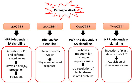
Figure 2.
Arabidopsis thaliana
PR1
PR2
PR5
2
2 production and eventually cell death [50]. AtACBP3 plays a distinct role in the plant defence response against necrotrophic and biotrophic pathogens as transgenic Arabidopsis AtACBP3-overexpressors (OEs) were protected against the biotrophic pathogen (
Pseudomonas syringae
tomato
Botrytis cinerea) [50]. In wild-type Arabidopsis, the expression of Class IV
AtACBP4
AtEBP
B. cinerea
AtACBP4
AtEBP are mediated by ethylene and/or JA signalling [48]. Rice Class III OsACBP5 protects transgenic Arabidopsis and rice plants against hemibiotrophs and biotrophs via NPR1-dependent SA signalling, and necrotrophs by JA signalling [14][56]. The
OsACBP5
OsACBP5 [14]. Proteomic studies showed that eleven biotic stress-related proteins were upregulated by
Rhizoctonia solani infection in transgenic Arabidopsis OsACBP5-OEs [56]. Grape Class III VvACBP conferred resistance to
P. syringae
Colletotrichum higginsianum
PDF1.2, the gene encoding plant defensin [21]. Orange boxes represent the named ACBPs involved in the pathogen response. White boxes indicate the molecular events that occur along the signalling pathway. Black arrows denote the flow of signalling events. ACBP, acyl-CoA-binding protein; EBP, ETHYLENE-RESPONSIVE BINDING PROTEIN; JA, jasmonic acid; NPR1, NONEXPRESSOR OF PR-1; PR, pathogenesis-related; SA, salicylic acid.
AtACBP4
AtEBP
B. cinerea
Figure 2) [48]. The interaction of AtACBP4 and AtEBP, as confirmed by yeast two-hybrid and coimmunoprecipitation, suggests that plant pathogen defence may be mediated by ethylene and/or JA signalling [48]. Another study revealed that
atacbp3
atacbp4
atacbp6
B. cinerea
C. higginsianum
P. syringae) pathogens [61]. Furthermore, AtACBP1 which is also important for stem cuticle formation, is suggested to confer resistance to
B. cinerea [33].
Rhizoctonia solani
Cercospora oryzae
Magnaporthe oryzae
Fusarium graminearum
Xanthomonas oryzae
Figure 2) [14]. Transgenic rice OsACBP5-OEs demonstrated stronger disease resistance against all pathogens tested [14]. In addition, enhanced resistance of OsACBP5-OEs against hemibiotrophs and biotrophs is mediated by SA signalling, while that against the necrotrophic pathogen
R. solani is regulated by JA signalling [14]. In the
OsACBP5
cis
R. solani
C. oryzae
M. oryzae
X. oryzae [14]. Furthermore, transgenic rice expressing the construct of the
OsACBP5
R. solani treatment compared to the promoter deletion lacking both W-boxes [14]. These results suggest that the W-boxes are important in the pathogen-responsiveness of
OsACBP5
Both Lipidex assays and ITC showed that rOsACBP5 binds to 18:3-CoA esters, suggesting that 18:3-FA, a precursor for JA biosynthesis, plays a role in basal defence against fungal pathogens [10][14]. Furthermore, proteomic analysis revealed that eleven biotic stress-related proteins were upregulated by
R. solani
R. solani [56].
Phakopsora pachyrhizi
GmACBPs comprising all four classes were detected in microarray data analysis [25]. The expression of Class I
GmACBP2
GmACBP4
GmACBP5
GmACBP6
GmACBP11 declined after 6 h post inoculation (hpi) of soybean with avirulent Hawaii 94-1 and virulent Taiwan 80-2 strains [25]. In addition,
Phytophthora sojae
GmACBP9
GmACBPs
GmACBP2
GmACBP5
GmACBP6
GmACBP7
GmACBP11 after 48 dpi [25]. Such differential changes in
GmACBPs
ACBPs
AtACBP3
VvACBP
OsACBP5