Recently, the intranasal route has emerged as a promising administration site for central nervous system therapeutics since it provides a direct connection to the central nervous system, avoiding the passage through the blood–brain barrier, consequently increasing drug cerebral bioavailability.
- stimuli-responsive hydrogel
- intranasal administration
- nose to brain
- neurodegenerative diseases
- Alzheimer’s disease
- Parkinson’s disease
- drug delivery
1. Introduction
Neurological disorders represent the first cause of disability and the second cause of death, around 17%, after cardiovascular diseases. Unfortunately, in the following years, the number of patients suffering from neurological disease is expected to rise dramatically due to the increase in average life expectancy, thus causing a heavy global burden [1][2][3].
Neurological disorders represent the first cause of disability and the second cause of death, around 17%, after cardiovascular diseases. Unfortunately, in the following years, the number of patients suffering from neurological disease is expected to rise dramatically due to the increase in average life expectancy, thus causing a heavy global burden [1,2,3].
The most frequent neurologic diseases are Alzheimer’s disease, Parkinson’s disease, multiple sclerosis, dementia, epilepsy, schizophrenia, stroke, and brain cancer. Although neurological disorders present different clinical manifestations and pathogenesis, they all mainly determine a progressive neuronal degeneration. Their etiology is complex and not completely known; however, genetic and environmental factors and aging are considered to have a key role [4][5][6][7].
The most frequent neurologic diseases are Alzheimer’s disease, Parkinson’s disease, multiple sclerosis, dementia, epilepsy, schizophrenia, stroke, and brain cancer. Although neurological disorders present different clinical manifestations and pathogenesis, they all mainly determine a progressive neuronal degeneration. Their etiology is complex and not completely known; however, genetic and environmental factors and aging are considered to have a key role [4,5,6,7].
Current therapeutic approaches against neurological disorders include oral, topical, or intravenous administration of drugs and more invasive techniques such as surgery, brain implants (i.e., deep brain stimulations), and so on. However, unfortunately, the currently available treatments are not associated with a repair and/or regeneration of the neural tissue and, consequently, resolution of the clinical situation but primarily act on alleviating symptoms and slowing the neurodegenerative process [8][9].
Current therapeutic approaches against neurological disorders include oral, topical, or intravenous administration of drugs and more invasive techniques such as surgery, brain implants (i.e., deep brain stimulations), and so on. However, unfortunately, the currently available treatments are not associated with a repair and/or regeneration of the neural tissue and, consequently, resolution of the clinical situation but primarily act on alleviating symptoms and slowing the neurodegenerative process [8,9].
The main limitation of central nervous system therapeutics is related to their delivery to the nervous system in therapeutic quantities. In this regard, the blood–brain barrier (BBB) plays a key role in preventing the passage of many substances, including drugs, to the brain [10][11].
The main limitation of central nervous system therapeutics is related to their delivery to the nervous system in therapeutic quantities. In this regard, the blood–brain barrier (BBB) plays a key role in preventing the passage of many substances, including drugs, to the brain [10,11].
The BBB is a complex vascular system consisting of endothelial cells interconnected by extended tight junctions without fenestrations, whose main function is to maintain the nervous system homeostasis. The endothelial cells are surrounded by pericytes and astrocytes that contribute to the structural and functional preservation of the BBB.
The BBB is the main structure responsible for the protection of the central nervous system from circulating xenobiotics, which could have harmful effects on the neural function, and of the supplying of required nutrients to the neural tissue [12][13].
The BBB is the main structure responsible for the protection of the central nervous system from circulating xenobiotics, which could have harmful effects on the neural function, and of the supplying of required nutrients to the neural tissue [12,13].
The main pathways for crossing the BBB are passive diffusion for small lipophilic compounds, active or passive transport for specific hydrophilic and/or ionized molecules (i.e., glucose, amino acids), and, lastly, for high-molecular-weight biological molecules, such as proteins and peptides, endocytosis [14][15]. The high selectivity of these crossing pathways massively prevents the entrance of circulating xenobiotics into the central nervous system, and for this reason, it has been estimated that around 98% of drugs is not able to cross the BBB [16].
The main pathways for crossing the BBB are passive diffusion for small lipophilic compounds, active or passive transport for specific hydrophilic and/or ionized molecules (i.e., glucose, amino acids), and, lastly, for high-molecular-weight biological molecules, such as proteins and peptides, endocytosis [14,15]. The high selectivity of these crossing pathways massively prevents the entrance of circulating xenobiotics into the central nervous system, and for this reason, it has been estimated that around 98% of drugs is not able to cross the BBB [16].
In order to overcome these limitations, the intranasal route has recently emerged as a promising administration site for central nervous system therapeutics [17][18]. In fact, it is a noninvasive route that provides a direct connection to the central nervous system, nose-to-brain route, via neural pathways, such as the olfactory and trigeminal ones, avoiding the passage through the BBB and, consequently, increasing drug concentration at the brain level [19][20] (
In order to overcome these limitations, the intranasal route has recently emerged as a promising administration site for central nervous system therapeutics [17,18]. In fact, it is a noninvasive route that provides a direct connection to the central nervous system, nose-to-brain route, via neural pathways, such as the olfactory and trigeminal ones, avoiding the passage through the BBB and, consequently, increasing drug concentration at the brain level [19,20] (
).
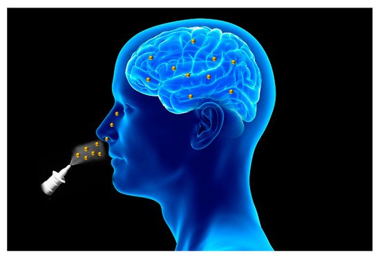
Figure 1.
Schematic representation of intranasal route for drug delivery to the brain.
Moreover, the intranasal route shows also the advantage, compared to the oral route, to prevent the gastrointestinal and first pass degradation, thus improving drug bioavailability [21].
2. Nasal Cavity: Anatomy and Physiology
The nose is the organ responsible for respiration and olfaction. Structurally, it is divided into the external nose and internal nose (nasal cavities).
The external nose is formed by bones and cartilages; it is placed in the center of the face and has the shape of a triangular pyramid. Small muscle groups controlled by the facial nerve that contribute to facial expression are connected to the external nose [22].
The nasal cavity extends around 12 cm in length from the external nose to the nasopharynx, and it is separated in two sections (left and right) by the nasal septum.
Structurally and functionally the nasal cavity can be divided into three regions: vestibular, olfactory, and respiratory [23].
The vestibular region is the smallest and outermost part of the nasal cavity lined firstly by stratified squamous epithelium followed by respiratory epithelium (pseudotratified columnar epithelium). It contains nasal hairs, called vibrissae, whose main function is to filter inhaled airborne particles, and sweat and sebaceous glands. This region can be considered irrelevant for drug absorption due to its structural features [7][24].
The vestibular region is the smallest and outermost part of the nasal cavity lined firstly by stratified squamous epithelium followed by respiratory epithelium (pseudotratified columnar epithelium). It contains nasal hairs, called vibrissae, whose main function is to filter inhaled airborne particles, and sweat and sebaceous glands. This region can be considered irrelevant for drug absorption due to its structural features [7,24].
The respiratory region has the largest surface area and represents around 80% of the nasal cavity; in fact, it covers three turbinates, bones that extend laterally from each nasal cavity [25].
It is lined by pseudostratified columnar epithelium and contains four cell types: ciliated, nonciliated, basal, and goblet cells.
The respiratory region is covered by a thick layer of mucus produced by nasal glands and basal cells that plays a key role in trapping inhaled particles, preventing their entrance into the respiratory system. Ciliated cells contribute to this defense mechanism, called mucociliary clearance, transporting mucus to the nasopharynx where it is expectorated or ingested [26].
This region is highly vascularized, since it is supplied by a branch of the maxillary artery and it is mainly innervated by the trigeminal nerve [27].
The nasal region can be considered the main site for systemic drug absorption due to its large surface area, high vascularization, and viscosity and can contribute to the nose-to-brain delivery of drugs via the trigeminal pathway [28].
The olfactory region is located in the superior part of the nasal cavity (which represents about 10% of the nasal cavity area), and it is responsible for the olfaction. It is lined by pseudostratified columnar epithelium and contains four different cell types: basal, sustentacular, trigeminal, and olfactory neural cells [29].
Sustentacular cells are prevalent in the olfactory region and surround the olfactory neural cells, providing structural and metabolic support [27]. Olfactory neural cells are bipolar neurons that extend their dendritic processes into the mucus layer, terminating as olfactory receptors, and project into the olfactory bulb, providing a direct portal between the nose and the central nervous system. In particular, their unmyelinated axons are covered by olfactory ensheathing cells and olfactory nerve fibroblasts that are in continuity with meninges and, consequently, with the subarachnoid space [30].
The basal lamina is located below the epithelium and contains blood and lymphatic vessels, nerve fibers, and Bowman’s glands responsible for the secretion of mucus that, in turn, solubilizes odor substances, cleans sensory receptors, and traps xenobiotics [8]. The olfactory region is mainly supplied by a branch of the olfactory artery.
2.1. Nose-To-Brain Delivery Pathways
The unique connection between the nasal cavity and the central nervous system makes the nasal mucosa a potential adsorption site for central nervous system therapeutics with minimal invasiveness, avoiding the passage via the blood–brain barrier and the gastrointestinal and first pass degradation, thus improving drug bioavailability. Currently, the exact mechanisms responsible for the nose-to-brain delivery of drugs are not well known; however, several evidences suggest that the trigeminal, olfactory, and systemic pathways are the main driving forces in delivering drugs via the nose-to-brain route. Presumably, although all these mechanisms contribute to the transport of drugs to the central nervous system, the properties of the drug and the formulation used may determine the prevalence of one of these pathways [31].2.1.1. The Olfactory Pathway
The olfactory pathway has been long considered to be the main pathway for the nose-to-brain delivery of drugs. In fact, the olfactory region contains olfactory neural cells that provide a direct portal between the nose and the central nervous system, terminating as olfactory receptors into the mucus layer and projecting into the olfactory bulb. Moreover, olfactory neural cells undergo a continuous turnover, making the nasal barrier an open environment due to the constant rearrangement of tight junctions [32][33].
Once they have crossed the olfactory epithelium and reached the lamina propria, drugs are transported to the central nervous system extracellularly through the perineural space, within olfactory ensheathing cells and olfactory nerve fibroblasts, by bulk flow mechanisms [35][36]; in this regard, probably, the conformational changes that also occur during the impulse propagation through the olfactory neural cells axons may contribute to the transport of drugs into the perineural space [37].
Olfactory neural cells have shown the ability to internalize some pathogens, such as herpes and polio viruses, large biological molecules, such as enzymes (i.e., horseradish peroxidase) and proteins (i.e., albumin and insulin), as well as gold nanoparticles, aluminum salts, and so on [38][39][40][41].
Pinocytosis is considered the main mechanism of drugs internalization into olfactory neural cells due to the high specificity of endocytosis [24]. Once inside, endosomes are translocated along axons toward the olfactory bulb where the drug is released via exocytosis and distributed in other central nervous system sites thanks to the projections existing between the olfactory bulb and other brain regions, such as the olfactory tract, hypothalamus, amygdala, etc. [34][42].
Unlike the extracellular pathway, the intracellular mechanism is a really slow and inefficient process, requiring 24 h to reach the central nervous system [39][43].
Moreover, the neuronal uptake is a quite specific process not applicable for a broad spectrum of drugs. In light of this, the extracellular pathway seems to be the predominant mechanism of transport, since several published studies highlight a rapid delivery and distribution of intranasally administrated drugs into the central nervous system [44][45][46].
2.1.2. The Trigeminal Pathway
The trigeminal branches present two different central nervous system entry points at the level of the pons and the olfactory bulb, respectively [5][18][47][48].
The intra-axonal transport, via the trigeminal route, has been observed for several intranasally administrated agents such as lidocaine, insulin-like growth factor-I, interferon β, vascular endothelial grow factor, nanoparticles, etc. [49][50][51][52][53][54].
3. Stimuli-Responsive Hydrogels
Hydrogels are three-dimensional networks formed by crosslinked hydrophilic polymers capable of absorbing large amounts of water. Recently, they have been studied as potential delivery platforms for biomedical applications due to their biocompatibility, biodegradability, nonimmunogenicity, and tunable properties [55].
Hydrogels are three-dimensional networks formed by crosslinked hydrophilic polymers capable of absorbing large amounts of water. Recently, they have been studied as potential delivery platforms for biomedical applications due to their biocompatibility, biodegradability, nonimmunogenicity, and tunable properties [63].
Hydrogels can be divided into physical or chemical gels depending on the type of crosslinking that can be based on covalent bonds or weak physical interactions. In particular, physical crosslinking shows the advantage of obtaining hydrogels with tunable properties and responsivity to different external cues such as pH, temperature, and ionic modulation [56]. In this regard, in order to overcome the limitations of the intranasal route, stimuli-responsive hydrogels have emerged as an interesting strategy for the intranasal administration of drugs thanks to their ability to increase drug retention time and bioavailability and decrease mucociliary clearance [57]; in fact, they can be administered as liquid formulation, guaranteeing an accurate administration, and then, after instillation, they undergo sol–gel transition triggered by external physiological factors such as pH, temperature, ionic modulation, etc., leading to an increase of drug retention time and protection from enzymatic degradation [7].
Hydrogels can be divided into physical or chemical gels depending on the type of crosslinking that can be based on covalent bonds or weak physical interactions. In particular, physical crosslinking shows the advantage of obtaining hydrogels with tunable properties and responsivity to different external cues such as pH, temperature, and ionic modulation [64]. In this regard, in order to overcome the limitations of the intranasal route, stimuli-responsive hydrogels have emerged as an interesting strategy for the intranasal administration of drugs thanks to their ability to increase drug retention time and bioavailability and decrease mucociliary clearance [65]; in fact, they can be administered as liquid formulation, guaranteeing an accurate administration, and then, after instillation, they undergo sol–gel transition triggered by external physiological factors such as pH, temperature, ionic modulation, etc., leading to an increase of drug retention time and protection from enzymatic degradation [7].
Several mucoadhesive polymers such as chitosan, pluronic, carbopol, and cellulose derivatives exhibit sol–gel behavior in response to several cues and, consequently, can be used for stimuli-responsive hydrogels preparation in order to further improve retention time and drug absorption [58].
Several mucoadhesive polymers such as chitosan, pluronic, carbopol, and cellulose derivatives exhibit sol–gel behavior in response to several cues and, consequently, can be used for stimuli-responsive hydrogels preparation in order to further improve retention time and drug absorption [66].
Moreover, the use of hydrogels as drug delivery systems shows the advantage to obtain a high drug-loading efficiency and sustained release, thus reducing the dose frequency and improving patient compliance and, thanks to their viscosity, a prolonged retention time [59].
Moreover, the use of hydrogels as drug delivery systems shows the advantage to obtain a high drug-loading efficiency and sustained release, thus reducing the dose frequency and improving patient compliance and, thanks to their viscosity, a prolonged retention time [67].
Stimuli-responsive hydrogels can be divided in several classes depending on the external cues, and among them, thermos-, pH-, and ion-responsive gels are the more efficient delivery platforms for intranasal administration [60].
Stimuli-responsive hydrogels can be divided in several classes depending on the external cues, and among them, thermos-, pH-, and ion-responsive gels are the more efficient delivery platforms for intranasal administration [68].
Thermoresponsive gels show sol–gel transition in response to a specific temperature change. Ideally, the temperature range should be at physiological values around 25–37 °C in order to guarantee an easy and accurate administration and avoid early drug loss [7].
Among thermoresponsive polymers, poloxamer, in particular poloxamer 407 and 188, is the most frequently used gelling agent for the preparation of in situ gels thanks to its mucoadhesiveness and sol–gel transition at physiological temperature values. It is a triblock polymer composed of one unit of polyoxypropylene and two units of polyoxyethylene that undergo sol–gel transition driven by supramolecular entanglements of micelle consequent to the dehydration of the polyoxypropylene units [61]. However, poloxomer-based hydrogels show low mechanical strength and viscosity in physiological conditions that limit their use, and, consequently, in order to overcome this problem, usually poloxamer is mixed with other polymers such as carbopol, chitosan, cellulose derivatives, etc. [62][63][64].
Among thermoresponsive polymers, poloxamer, in particular poloxamer 407 and 188, is the most frequently used gelling agent for the preparation of in situ gels thanks to its mucoadhesiveness and sol–gel transition at physiological temperature values. It is a triblock polymer composed of one unit of polyoxypropylene and two units of polyoxyethylene that undergo sol–gel transition driven by supramolecular entanglements of micelle consequent to the dehydration of the polyoxypropylene units [69]. However, poloxomer-based hydrogels show low mechanical strength and viscosity in physiological conditions that limit their use, and, consequently, in order to overcome this problem, usually poloxamer is mixed with other polymers such as carbopol, chitosan, cellulose derivatives, etc. [70,71,72].
In this context, Ahmed et al. designed a poloxamer-/chitosan-based thermoresponsive gel loaded with agomelantine and evaluated its suitability and efficacy for the delivery of antidepressant drugs via the nose-to-brain route [65]. Firstly, the hydrogel was prepared by physically mixing chitosan (0.5%
In this context, Ahmed et al. designed a poloxamer-/chitosan-based thermoresponsive gel loaded with agomelantine and evaluated its suitability and efficacy for the delivery of antidepressant drugs via the nose-to-brain route [73]. Firstly, the hydrogel was prepared by physically mixing chitosan (0.5%
w
/
v
), an agomelantine-based nanoemulsion and poloxamer 407 (20%
w
/
v
) (
Figure 2). Chitosan was added in order to increase the mucoadhesiveness of the formulation; in fact, the calculated mucoadhesive strengths were 3747.75 dynes/cm
3). Chitosan was added in order to increase the mucoadhesiveness of the formulation; in fact, the calculated mucoadhesive strengths were 3747.75 dynes/cm
2
and 6246.27 dynes/cm
2
for the formulation without and with chitosan, respectively, and, moreover, the obtained hydrogel showed high strength and viscosity, advantageous features that further enhance drug retention time and adsorption. In fact, the pharmacokinetics studies highlighted a ~three-fold higher drug bioavailability in the brain, when administered intranasally compared to the intravenous administration of the drug solution.
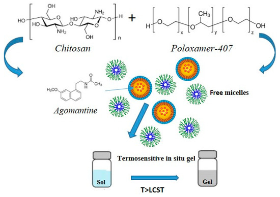
Figure 23.
In addition to poloxamer, other polymers such as chitosan usually mixed with polyols, cellulose derivatives like ethyl hydroxy-ethyl cellulose, and xyloglucan are used as gelling agents for the preparation of thermoresponsive hydrogels [66].
In addition to poloxamer, other polymers such as chitosan usually mixed with polyols, cellulose derivatives like ethyl hydroxy-ethyl cellulose, and xyloglucan are used as gelling agents for the preparation of thermoresponsive hydrogels [74].
3.1. Stimuli-Responsive Hydrogel for the Treatment of Alzheimer’s Disease
Alzheimer’s disease is the most common form of dementia, determining a progressive cognitive decline, involving first memory and subsequently other cognitive domains such as visuospatial, language, and thinking functions [67]. Physiopathologically, it characterized by the presence of β -amyloid plaques and neurofibrillary tangles of hyperphosphorylated forms of tau protein in the brain [68]. Moreover, several evidences suggest that also neuroinflammation and microglial activation play a key role in the pathogenesis of Alzheimer’s disease [69]. At the moment, unfortunately, there are no approved anti-Alzheimer effective drugs associated with a repair or regeneration of the neural tissue and/or able to block the neurodegenerative process, but rather current therapeutic approaches act mainly on reducing symptoms and slowing the neurodegeneration and include the oral administration of acetylcholinesterase inhibitors such as donezepil, rivastigmine, tacrine, galantamine, and NDMA receptors antagonists such as memantine. However, these drugs show low bioavailability at the brain level due to the BBB and gastrointestinal and first-pass degradation.
In order to overcome this problem and increase drug bioavailability, an interesting strategy has been recently proposed by Abouhussein et al. who developed thermoresponsive hydrogels for the nose-to-brain delivery of rivastigmine [70]. Several hydrogels were prepared using two different poloxamer types (407 and 188) and four mucoadhesive agents such as chitosan, hydroxypropyl methyl cellulose, carbopol 934, and sodium carboxy methyl cellulose via cold method, and their rheological behavior, mechanical strength, mucoadhesiveness, and release profile were evaluated. The formulation based on poloxamer 407 and carbopol 934 showed the best combination in terms of sol–gel transition temperature, mechanical properties, and mucoadhesiveness and, consequently, ex vivo permeation and in vivo pharmacokinetics studies were performed on it. Ex vivo results highlighted a much higher nasal permeation of rivastigmine incorporated into the gel compared to the drug solution (84% vs. 28%, respectively), attributable to the mucoadhesiveness of pluronic and carbopol that enhance drug retention time and decrease mucociliary clearance. Moreover, pharmacokinetic studies showed a greater rivastigmine cerebral distribution for the stimuli-responsive hydrogel compared to the intranasal and intravenous administration of the drug solution, with C
max
Figure 3).
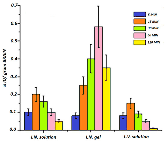
Figure 34. Distribution of rivastigmine in the brain after the intranasal administration of drug solution and rivastigmine-loaded pluronic-/carbopol-based gel and the intravenous administration of the drug solution. Adapted with modification from reference [70].
In addition to the conventional anti-Alzheimer drugs, since neuroinflammation and microglial activation are thought to play a key role in the pathogenesis of Alzheimer’s disease, some natural antioxidant compounds such as curcumin, resveratrol, and saponins have emerged as potential anti-Alzheimer agents. However, these agents show low bioavailability and cerebral distribution, thus limiting their use and efficacy [71][72][73].
In this regard, recently, Chen et al. designed a thermo- and pH-sensitive hydrogel for the nose-to-brain delivery of timosaponin BII and evaluated its potential use and efficacy for the prevention of Alzheimer’s disease [74]. The gelling agents poloxamer 407 and gellan gum were used for the preparation of the hydrogel via the cold method in order to obtain a dual-sensitive (thermo and pH) system. In vivo studies were performed on murine models of neuroinflammation and amyloidosis induced by lipopolysaccharide and highlighted that timosaponin BII improved memory and language functions, partially reducing the cognitive decline induced by LPS and inhibited iNOS expression with consequent reduction of the levels of some proinflammatory mediators such as TNF-α, IL-1β, and, therefore, neuroinflammation.
3.2. Stimuli-Responsive Hydrogels for the Treatment of Parkinson’s Disease
Parkinson’s disease is the second most common neurological disease characterized by bradykinesia, rigidity, and rest tremor, thus leading to a progressive motorial function decline. Physiopathologically, it is characterized by a progressive neuronal degradation in the substantia nigra, and, consequently, shortage of dopamine and accumulation of Lewy’s body at the brain level [75][76].
Current available treatments include levodopa, dopaminergic agonists such as ropinirole and pramipexole, monoamine oxidase-B inhibitors such as selegiline and rasagiline, anticholinergic agents, and amantadine [77].
Due to the motorial function decline induced by Parkinson’s disease, dysphagia can often occur in patients thus making difficult the oral administration of drugs, most common route for anti-Parkinson agents [78]. Moreover, several anti-Parkinson drugs show low oral bioavailability, such as rasagiline, a monoamine oxidase-B inhibitor with oral bioavailability of around 35% in humans, due to high gastrointestinal and first-pass degradation [79].
In this regard, in order to overcome these problems and increase rasagiline bioavailability and patient compliance, Ravi et al. designed thermosensitive gels and evaluated their potential use as intranasal formulations for rasagiline nose-to-brain delivery [80]. Several in situ gels were prepared by using two different poloxamer (407 and 188) types and two mucoadhesive agents such as carbopol 934P and chitosan via cold method, and their rheological behavior, drug release profile, effect on mucociliary clearance, and mucoadheseviness were evaluated. The optimal intranasal formulations resulted to be P407 15%
w
v
w
v
w
v
max
Figure 4).
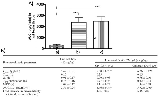
Figure 45.
a
b
c
p
A
max
max
max
e
1/2
(0–∞)
p
B). Adapted with permission form Taylor and Francis [80].
Among dopaminergic agonists used as anti-Parkinson agents, we find rotigotine, marketed as a transdermal patch called Neuropro due to a low oral bioavailability consequent to a massive hepatic degradation. However, this transdermal formulation showed some crystallization problems that lead to its withdrawal by FDA [81].
In this regard, interestingly, in order to develop a proper formulation for the nose-to-brain delivery of rotigotine, Wang et al. designed delivery platforms based on a thermosensitive gel incorporating rotigotine-loaded polymeric micelles [82]. Several thermosensitive formulations were prepared by using two types of Poloxamer (407 and 188) in different ratios via physical mixing into aqueous solution with rotigotine-loaded polymeric micelles obtained via solvent evaporation method. The optimal intranasal formulation resulted to be 22%
w
v
w
v
3.3. Stimuli-Responsive Hydrogels for the Treatment of Depression
Major depressive disorder is one of the most common psychiatric condition characterized by altered brain functions such as memory, attention, feelings, ability to think and concentrate, fatigue, sleep, and eating disorders, thus negatively impacting patients’ quality of life and representing one of the most common causes of disability [83].
Its etiology is complex and multifactorial, and, currently, the exact mechanisms responsible for its pathogenesis are not clearly known. However, several models have been proposed for major depressive disorder etiology: neuroinflammation, by reducing the levels of monoamines and increasing the levels of tryptophan catabolites, overactivity of the hypothalamic-pituitary-adrenal axis, and reduction of neurogenesis and neuroplasticity [84][85].
At present, the main pharmacological treatments act primarily on the monoaminergic system by increasing the synaptic levels of monoamines such as serotonin, norepinephrine, and dopamine and include selective serotonin reuptake inhibitors, that act mainly on the serotonin transporter (SERT) such as fluoxetine, paroxetine, and citalopram, serotonin-norepinephrine reuptake inhibitors, that act on both SERT and norepinephrine transporter (NET) such as duloxetine and venlafaxine, and tricyclic antidepressants such as imipramine and amitriptyline [86]. Among them, the selective serotonin reuptake inhibitors represent the most used antidepressants class thanks to their higher tolerability compared to the others; however, unfortunately, most of them show low oral bioavailability due to an extensive first-pass degradation [87]. In this regard, Thakkar et al. developed intranasal formulations for paroxetine nose-to-brain delivery [88]. Several in situ gels were prepared by using gellan gum as gelling agent, low-molecular-weight hydroxypropyl methyl cellulose as mucoadhesive agent, and hydroxypropyl-β-cyclodextrin as solubilizer and permeation enhancer, and their viscosity, strength, mucoadhesiveness, and loading efficiency were evaluated. The optimal intranasal formulation resulted to be 0.3%
w
v
w
v
w
v
max
max
A noteworthy further example of intranasal stimuli-responsive hydrogel for the nose-to-brain delivery of antidepressant drugs has been reported by Avachat et al. [89].
3.4. Stimuli-Responsive Hydrogels for the Treatment of Schizophrenia
Schizophrenia is a psychiatric disorder characterized by altered brain function such as memory, feelings and mental processing, hallucinations, delusions, and social withdrawal [90]. Its etiology is multifactorial, but genetic and environmental factors seem to have a key role [91]. Currently, the mechanisms responsible for the pathophysiology of schizophrenia remain still unclear; however, strong evidences suggest that the dopaminergic and the glutamic neurotransmission may be involved in the development of negative and positive symptoms, respectively.
The main pharmacological treatments act mainly on the dopaminergic circuit by blocking the D2 receptor and include clozapine, risperidone, haloperidol, paliperidone, etc. Antipsychotic drugs are mainly administrated orally and primarily used in acute episodes of schizophrenia alleviating positive symptoms [92].
In addition to conventional treatments, in the last years, a new class of antipsychotic peptide drugs is emerging as a promising therapeutic approach against schizophrenia showing a higher safety profile with reduced side effects compared to the conventional therapies. However, unfortunately, one of the main problems associated with this class of antipsychotics is related to their route of administration, since they cannot be administered orally due to an extensive enzymatic degradation. In this context, the intranasal route turns out to be a potential administration site thanks to its noninvasiveness, possibility of the nose-to-brain delivery, and avoidance of GI degradation; in this regard, a noteworthy example of antipsychotic peptide drug nose-to-brain delivery system has been recently proposed by Majcher et al. [93]. In this work, an innovative intranasal formulation based on oxidized starch nanoparticles and carboxymethyl chitosan has been designed and evaluated for the delivery of a preclinical antipsychotic peptide that acts as allosteric modulator of D2 receptor. In particular, promising data came from in vivo studies that highlighted a full alleviation of schizophrenia-related negative symptoms for up to 3 days after the intranasal administration of the carboxymethyl chitosan and starch nanoparticles-based gel compared to the drug solution alone, administrated nasally or intraperitoneally, indicating that the intranasal formulation increased drug nasal permeation and cerebral bioavailability and efficacy.
3.5. Stimuli-Responsive Hydrogels for the Treatment of Brain Injury
Stroke is the second cause of death worldwide and one of the major causes of disability. It is caused by impaired perfusion and blockage of cerebral blood vessels that lead to hypoxia and, consequently, cell death and cerebral damage. Stroke can be ischemic or hemorrhagic: ischemic stroke is caused by blood vessels occlusion and usually results in thrombotic and embolic conditions, while hemorrhagic stroke is caused by blood vessels rupture, and it is associated with higher mortality.
At present, therapeutic approaches are limited and involve mainly reperfusion treatments such as intravenous thrombolysis [94]. However, there are many natural and synthetic agents that have gained attention due to their neuroprotective effect; in this regard, Xie et al. developed an intranasal formulation combining polyamidoamine dendrimers with an in situ gellan-bum-based gel for the nose-to-brain delivery of paenol, a neuroprotective agent that was found to reduce cerebral stroke in murine models. Firstly, polyamidoamine dendrimers were prepared and loaded with paenol obtaining an encapsulation efficiency of ~54%, and, subsequently, incorporated in an ion-sensitive gel obtained by using gellan gum 0.45%
At present, therapeutic approaches are limited and involve mainly reperfusion treatments such as intravenous thrombolysis [107]. However, there are many natural and synthetic agents that have gained attention due to their neuroprotective effect; in this regard, Xie et al. developed an intranasal formulation combining polyamidoamine dendrimers with an in situ gellan-bum-based gel for the nose-to-brain delivery of paenol, a neuroprotective agent that was found to reduce cerebral stroke in murine models. Firstly, polyamidoamine dendrimers were prepared and loaded with paenol obtaining an encapsulation efficiency of ~54%, and, subsequently, incorporated in an ion-sensitive gel obtained by using gellan gum 0.45%
w
/
v
and hydroxypropyl cellulose 0.3%
w
/
v via cold method. The developed formulation showed advantageous features such as low critical ion concentration, facilitating the sol–gel transition under physiological condition, adequate viscosity and strength guaranteeing easy of spraying, and sustained release profile with ~80% of the drug released within 12 h. Moreover, fluorescence studies investigating the nose-to-brain delivery mechanism were performed and highlighted a greater cerebral bioavailability of paenol after the intranasal administration of the gel compared to the drug solution [95].
via cold method. The developed formulation showed advantageous features such as low critical ion concentration, facilitating the sol–gel transition under physiological condition, adequate viscosity and strength guaranteeing easy of spraying, and sustained release profile with ~80% of the drug released within 12 h. Moreover, fluorescence studies investigating the nose-to-brain delivery mechanism were performed and highlighted a greater cerebral bioavailability of paenol after the intranasal administration of the gel compared to the drug solution [108].