Metal oxide nanoparticles (NPs) have received a great deal of attention as potential theranostic agents. Despite extensive work on a wide variety of metal oxide NPs, few chemically active metal oxide NPs have received Food and Drug Administration (FDA) clearance. The clinical translation of metal oxide NP activity, which often looks so promising in preclinical studies, has not progressed as rapidly as one might expect. The lack of FDA approval for metal oxide NPs appears to be a consequence of the complex transformation of NP chemistry as any given NP passes through multiple extra- and intracellular environments and interacts with a variety of proteins and transport processes that may degrade or transform the chemical properties of the metal oxide NP. Moreover, the translational models frequently used to study these materials do not represent the final therapeutic environment well, and studies in reduced preparations have, all too frequently, predicted fundamentally different physico-chemical properties from the biological activity observed in intact organisms. Understanding the evolving pharmacology of metal oxide NPs as they interact with biological systems is critical to establish translational test systems that effectively predict future theranostic activity.
- cell trafficking
- endocytosis
1. Introduction
There is tremendous interest in metal oxide nanoparticles (NPs) for use in therapeutic applications such as diagnostic tools and drugs in which the nanoparticles are either the active agent or passive, drug delivery nanocarriers. To-date, there are over thirty different metal oxide formulations being studied that may have biological effects [1], but few have garnered FDA clearance. While nanomaterials have demonstrated potential therapeutic benefit in many biomedical applications, clinical translation of individual formulation has not progressed as rapidly as one would expect given the plethora of preclinical studies [2][3]. We believe that the slow progression to approved drugs may result, in part, from the types of translational models used to study these materials, and the emerging evidence that the activity of nanomaterials in cell-free conditions and reduced preparations can be fundamentally different from the biological properties of the nanomaterial when studied in either cell culture conditions or, more importantly, in intact organisms. A better understanding of the evolving pharmacology of metal oxide nanoparticles (NPs) as they interact with biological systems is critical to establish translational test systems that can effectively predict future drug potential.
2. The Origin of Biological Activity in the Structure of Nanoparticles
All nanoparticles, regardless of elemental composition or shape, have extremely high surface area:volume ratios that confer chemical reactivity not observed in particles with larger dimensions (i.e., > 100 nm) [4]. The solubility of nanomaterials in biological fluids is dictated by surface composition, surface charge, and the hydrophobicity/hydrophilicity profile. The surface electrostatic interactions between particles determine their propensity to aggregate and adsorb proteins to their surface. The chemical and biological reactivity as well as biodistribution of the nanomaterials are derived from these fundamental properties.
2.1. Redox Reactivity of Metal Oxides
Metal oxides NPs have an ability to participate in myriad biologically important redox reactions and mimic a wide range of enzymes including catalases, oxidases, dismutases, peroxidases, ATPases and phosphatases [5][6][7][8]. The native enzymes expressing similar redox activity play manifold and crucial roles in redox-dependent signaling cascades, and metal oxide NPs can disrupt or restore redox balance in cells through these reactions and signaling processes [8].Not all metal oxides exhibit the same enzymatic activities, and mimetic activities can be ‘biased’ by the local environment surrounding the particle. There are three key factors that determine the interaction between the biological milieu and the redox activity at the nano-bio interface: the half-cell potential of the elements comprising the particle, the organization of the surface atoms of the nanoparticle, and the oxidation state of the ions on and within the nanoparticle. Reduction in the oxidation number of the metal (i.e., the accounting of the number of electrons a metal possess or lacks) occurs when the crystal loses an oxygen atom and forms a vacancy in the NP. Thermodynamically, any oxide is potentially reducible [9], and the distinction between reducible and non-reducible metal oxides depends on the ease with which oxygen vacancies can be formed [9]. In non-reducible metal oxides, the thermodynamic cost of formation of oxygen vacancies is high, and redox activity is absent [10]. In reducible metal oxides, oxygen vacancy formation is thermodynamically more favorable and occurs at lattice surfaces and edges where the coordination number of the surface atoms (i.e., the total number of bonds to the atom) is less than inside the crystalline structure of the oxide. The edge is also where lattice strain is highest [11]; all of which facilitates the formation of oxygen vacancies [9][12]. Thus, the highest enzyme-mimetic activity occurs at the surface of the nanoparticle [9][13]. Many transition-metal oxides, such as TiO
2. The Origin of Biological Activity in the Structure of Nanoparticles
2.1. Redox Reactivity of Metal Oxides
2
2
3
2, are reducible because the energetic barriers to oxygen vacancy formation are low, and vacancies can occur spontaneously across the surface of the crystal.The chemical mechanisms underlying redox activity can be divided into either electrophilic or nucleophilic reactions. Extra-facial, adsorbed oxygen is responsible for most of the electrophilic reactions, whereas interfacial oxygen, where lattice oxygen vacancies are created, underlie the nucleophilic reactions [14]. In general, nucleophilic oxygen (e.g., the oxide ion) is capable of carrying out selective oxidations while it seems that electrophilic oxygen species, which are deficient in electrons (e.g., the superoxide radical), appear to be more promiscuous and are largely responsible for non-selective oxidation [15]. There is usually a ‘preferred’ or stable oxidation state in each NP, and surface defects created by spontaneous loss of oxygen result in different valence states (i.e., Ce
4+
3+). The redox state of the metal oxide can flip-flop repeatedly between valance states, which provide durable, recycling, catalytic activity.The mechanisms of cyclic, regenerative redox reactions have been studied in cerium oxide NPs because of the relatively low barrier for the transition between Ce
4+  Ce
Ce
Ce3+. Cerium oxide demonstrates both superoxide dismutase and catalase mimetic activity [16][17][18]. In the reaction scheme shown below, the hydroxyl radical is the ‘seed’ for the balanced set of redox processes. Given the high oxidation potential of
•
2−
2
2, likely contribute to oxidative stress and damage of DNA, proteins, and lipids [19].Formation of oxygen vacancies within the ceria nanoparticle lattice structure is central to this regenerative mimetic activity. The sequence of proposed reactions to explain the mimetic activity of cerium oxide is shown below [20][21]:

4+
3+
3+; both superoxide and hydrogen peroxide are consumed; the reaction is pH dependent; last, as pointed out by Reed et al. [21], the system of reactions is self-limiting (in the absence of a source of oxidizing agents like hydroxyl, there is little redox activity) and self-balancing. Other metal oxide NPs may have other preferred reactants so that the concentrations of a variety of oxidants, but especially the superoxide anion, may be reduced or regulated through multiple enzyme mimetic processes occurring simultaneously, even within the same NP.The oxygen vacancies in metal oxide NPs are transient and mobile across the surface of the metal oxide crystal, and the oxygen vacancies occur predominantly at the surface lattice boundaries or ‘edges,’ especially in smaller particles (~5 nm). The vacancies may then migrate internally where the coordination number with the metallic ions may be increased (Figure 1). The ability of the metal oxide to undergo reduction (vacancy formation), and the subsequent reincorporation of oxygen into the crystalline structure allows cyclically regenerative redox reactions, the durability of these reactions in vivo depends on particle retention in tissue and maintenance of the crystalline structure; dissolution of the crystal terminates redox reactivity.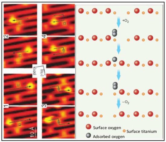 Figure 1. Scanning tunneling microscopic (STM) images (left) and molecular schematics (right) demonstrate the interactions of molecular oxygen adsorbed to the surface of titanium oxide at the site of oxygen vacancies within the crystal structure of the metal oxide. Each adjacent STM image shows the surface structure of the metal oxide before and after the interaction with molecular oxygen and the migration of the oxygen vacancy. Used with permission from Pinto et al. [9].In addition to reducing concentrations of oxidizing agents, most metal oxides can elicit free radical-mediated toxicity via the formation of hydroxyl through Fenton-type reactions [22][23]. Within reactive sites generated at oxygen vacancies, electron donor or acceptor regions interact with molecular O
2
2•
3
4 magnetic nanoparticles, for example, exhibited intrinsic peroxidase-like activity under acidic conditions and a catalase-like activity at neutral pH [24]. Moreover, both hematite nano-Fe
2
3
2
3 induced hydroxyl radical formation in more acidic environments through Fenton reactions. The specific reaction that predominates (i.e., oxidation or reduction) will depend on the valence state of the crystal, which is modified by the pH of the cellular compartment in which the particle resides (as shown above for cerium oxide). Redox cell damage may also occur if dissociation of the metal ions (i.e., Ag NPs and Quantum Dots) elicits cellular enzyme deactivation, membrane disruption, altered electron transfer, reduced mitochondrial membrane potentials, or changes in gene expression; all of which may increase the accumulation of cellular oxidants [25][26][27][28][29][30][31].The potential benefits of metal oxide nanoparticles for medical applications have emerged from their robust antioxidant properties [32][33][34]. Most studies fail to parse the impact of the local environment on nanoparticle reactivity and concentrate on the net effect of the nanoparticle as either pro- or anti-oxidant. This creates the (mistaken) impression that the metal oxide exhibits only one type of redox reactivity when in reality metal oxide NPs may have flexible redox reactivity that can be biased toward oxidation or reduction depending on the valence state and the milieu of the nanoparticle (pH, protein corona, cell-free media, serum, cell culture media, etc.).
2.2. Intracellular pH Environments and Metal Oxide NP Activity
Fan et al. [32] synthesized Pt-Ft nanoparticles using an apoferritin protein shell/scaffold as a nanoreactor to control the synthesis of size-tunable Pt nanostructures. One to two nm Pt–Ft NPs synthesized in this way possessed both catalase and peroxidase activities. However, these superparamagnetic iron particles (SPIONs) demonstrated peroxidase activity in acidic solutions, but lost this activity in more neutral solutions and instead expressed catalase-like activity through a series of coupled reactions [32]. The antioxidant properties of CeOx NPs dominate at physiological pH, whereas these particles exhibit high oxidase activity at acidic pH [35], likely related to a net shift in the valence of material to Ce
2.2. Intracellular pH Environments and Metal Oxide NP Activity
4+ [36]. Moreover, SOD activity is enhanced at lower pH relative to catalase activity, resulting in the accumulation of peroxide [37]. In more neutral conditions, CeOx NPs display both SOD and catalase activity [16]. Silver NPs were similarly sensitive to pH: the level of hydroxyl radical formation through a Fenton-like mechanism was dependent on pH-hydroxyl radical formation occurred at pH 4.6 or lower, but at more neutral pH, no significant formation of hydroxyl radicals occurred [38]. Thus, the tuning or biasing of the enzymatic mimesis of metal oxide NPs is modulated by intracellular pH, which can vary both by cellular localization (i.e., cytosol versus lysosome; see Figure 2) or whether the cells are immortalized or not [32][39].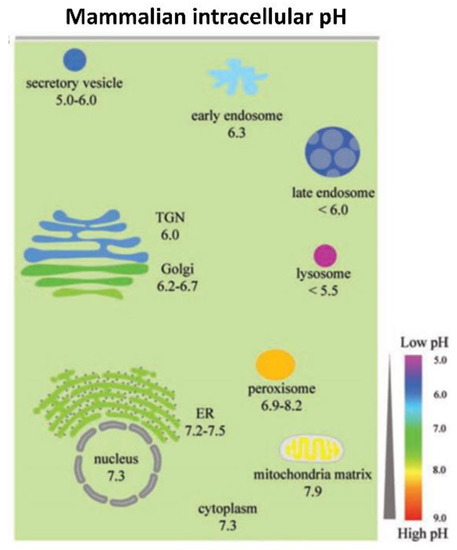 Figure 2. The range of pH values in intracellular compartments is shown schematically. The extent and type of chemical activity of metal oxide NPs may vary significantly, even in a single cell, across a wide range of pH values in different organelles. ER, endoplasmic reticulum and TGN, trans-Golgi network. From Shen et al. [40] with permission.
2.3. Model System Effects
The biological effects of nanoparticles depend not just on the properties of the material in standardized conditions, but also on the biological system in which the nanoparticles are active [41][42][43][44][45][46]. There is increasing evidence that immortalized cells (i.e., differentiated cancer cells) have unique redox profiles that are different from their native, healthy counterparts [47][48]. Selective cytoprotection has been reported following administration of nanoceria in normal, healthy cells, but not in cancer cells [49][50]. Often, cancer cells rely more on glycolysis for energy production, and consequently they maintain more acidic intracellular pH values [51]. Where additional protons are present (i.e., lactate accumulation or localization in acidic organelles), Ce
2.3. Model System Effects
3+
+
2•−
4+
2
2, leading to net oxidation [37][52]. Moreover, in a comparison of immortalized colorectal cells (HCT 116) and human embryonic kidney (HEK 293) cells, CeOx NPs increased the ROS load and subsequently induced apoptosis in colon cancer cells but not in the embryonic kidney cells, suggesting that differences in either cellular localization or baseline pH existed in these cell types [53]. The accumulation of CeOx NPs in this study was not evaluated, so it is possible that the amount of material taken up by these two cell types could have differed and impacted ROS formation. In a study of three different MnOx NPs (MnO
2
3
2
3
4
3
4
3
2
2. While the MnOx NPs were all cytotoxic, they protected cells when the cells were challenged with peroxide—suggesting that catalase mimetic activity was protective [54]. Unlike many other metal oxides, the MnOx NPs were devoid of peroxidase or hydroxyl radical scavenging activity in cell-free assays, but when studied in cells, the MnOx NPs were located in the cytosol, which has a higher pH than most other organelles in the cell, and the local pH may have biased the enzyme mimetic activities of the different valences and allowed the particles to provide cytoprotective activity when the cells were challenged with peroxide. Consistent with these findings, MnOx nanoparticles increased catalase and SOD activities, while they also decreased glutathione levels in cell culture [55]. The decreases in cell viability caused by MnOx NPs were associated with mitochondrial dysfunction and apoptosis, presumably secondary to the reduction in glutathione levels. Glutathione is critical to maintain mitochondrial function and cell viability, and loss of sufficient glutathione levels in mitochondria increased oxidative stress [56]. Most often, MnOx NPs are cytotoxic in immortalized cell cultures, but the outcome of administration of these materials in whole animals is variable, and some studies show that they are safe (Xiao et al., 2013) but not others [57]. Hence, these nanoparticles may be protective in certain redox states and certain cell types but not others.The variable redox effects of metal oxide NPs, which may be either pro-or antioxidant, have been vexing. Beyond the effects of the cells studied and the impact of pH in these test systems, redox activity of NPs may be related to the manner of synthesis (valence ratio), the size of the particles, the complement of adsorbed proteins, and the cellular localization of the material. The redox activity of metal oxide NPs is not easily predicted since local environments may vary so much. Moreover, findings in cell-free systems are not fully recapitulated in more representative biological environments like cell culture or intact animals. The biological impact of these materials seems to be tied to the baseline redox status of the cells being studied, which adds yet another source of variability when trying to characterize the likely therapeutic effect of nanoparticles. While many disease states elevate oxidative stress in tissues, not all tissues will have the same redox changes driven by the disease state. Thus, even within a single organism, the redox activity of a nanoparticle may differ organ by organ or even organelle by organelle. Since the delivery of metal oxides occurs passively, these materials distribute widely throughout the body including healthy cells, and healthy cells may be negatively impacted by NPs while the benefit of these materials as antioxidants may be observed only in cells that have a high oxidative load [47][48]. Understanding how these factors modify redox reactivity will be critical to the future development of therapeutic nanoparticles [58][59].