1. Genetic Findings from Two Families with Myotonia Congenita (MC)
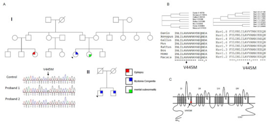
Two probands from two individual Taiwanese–Chinese families were selected. They had developed stiffness with difficulty relaxing their limb and truncal muscles. No periodic weakness occurred, and their muscle bulk was conspicuously large as compared with the age-matched controls. According to the family pedigree (Family I, upper panel of Figure 1A), the index case developed NDM, his sister had mental subnormality, and one of his cousins had a history of epilepsy. The nuclear family (Family II, lower panel of Figure 1A) with two affected brothers developed similar features of NDM. The candidate gene screening for a
Two probands from two individual Taiwanese–Chinese families were selected. They had developed stiffness with difficulty relaxing their limb and truncal muscles. No periodic weakness occurred, and their muscle bulk was conspicuously large as compared with the age-matched controls. According to the family pedigree (Family I, upper panel of A), the index case developed NDM, his sister had mental subnormality, and one of his cousins had a history of epilepsy. The nuclear family (Family II, lower panel of A) with two affected brothers developed similar features of NDM. The candidate gene screening for a
mutation did not find any sequence variant; however, further sequencing of
SCN4A gene identified a missense variant, c.1333G>A, that results in the p.V445M change in both index cases. The DNA from the parents of the proband in Family I was not available. Although the parents of Family II were dead, both the index case and his brother harbored the mutation. The amino acid Val445 was found to be not only conserved across the species in evolution but also across the alpha-subunits of the human voltage-gated sodium channel protein family (Figure 1B). Topologically, the p.V445M mutation is located at the sixth transmembrane α–helix of the first domain (DI/S6, Figure 1C). The mutation has been reported to be pathogenic in previous studies [1][2].
gene identified a missense variant, c.1333G>A, that results in the p.V445M change in both index cases. The DNA from the parents of the proband in Family I was not available. Although the parents of Family II were dead, both the index case and his brother harbored the mutation. The amino acid Val445 was found to be not only conserved across the species in evolution but also across the alpha-subunits of the human voltage-gated sodium channel protein family (B). Topologically, the p.V445M mutation is located at the sixth transmembrane α–helix of the first domain (DI/S6, C). The mutation has been reported to be pathogenic in previous studies [19,20].
Sequence chromatography of the mutation and multiple sequence alignments for Na
1.4 channels. (
) The pedigrees from two individual families (Family I and II) with the index cases are shown in the upper and lower panels, respectively. Both index cases suffered from muscle myotonia with muscle hypertrophy. In Family I, the sister of the index case suffered from mental subnormality and one of her relative had seizure history. Both the index case and his brother from Family II suffered from myotonic myopathy. The sequence chromatography (left side of lower panel) shows a variant, c.G1333A at exon 9 of the
gene, which causes a p.V445M mutation. Both probands have the sequence variant. The variant is not found in the relatives of the index case of Family I. In Family II, both the proband and his brother share the same sequence variant. (
) The amino acid V445 is highly conserved among species during evolution and in the protein family of voltage-gated sodium channels. (
) The topology of the Na
v1.4 channel shows that the mutation is located at the sixth transmembrane segment of Domain I (DI/S6).
1.4 channel shows that the mutation is located at the sixth transmembrane segment of Domain I (DI/S6).
2. A Smaller Sustained Na+ Current in the WT Channel than in the p.V445M Mutant Channel
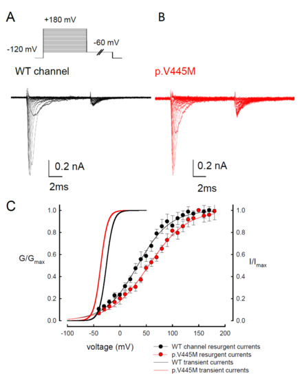
We compared the electrophysiological characteristics of the basic gating control of the WT and p.V445M mutant Na
We compared the electrophysiological characteristics of the basic gating control of the WT and p.V445M mutant Na
1.4 channels. A shows the sweeps after the depolarization via either an escalating (from −140 to +40 mV, left panel of A) or constant (+10 mV, right panel of A) depolarizing current. The activation and inactivation curves of the WT and p.V445M mutant channels were fitted with the following Boltzmann function: 1 / [1 + exp((V
− V) /
)], where V is the membrane potential and
is the slope factor. From the equation, V
and
were −25.79 ± 1.5 mV and 6.67 ± 0.43 for the activation curve and −63.99 ± 2.17 mV and −8.93 ± 0.33 for the fast steady-state inactivation curve in the WT channel, respectively and −36.53 ± 1.4 mV and 6.61 ± 0.23 for the activation curve and −73.81 ± 1.2 mV and −9.11 ± 0.21 for the inactivation curve in the p.V445M mutant channel, respectively (
= 9;
< 0.05; B). Both the activation and fast steady-state inactivation curves in the p.V445M mutant channel were hyperpolarization-shifted compared with those in the WT channel. The slopes of both gating curves of the Na
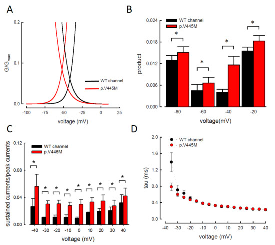
1.4 channels remained grossly unchanged (B). We further analyzed the window currents, which were defined by the area under the activation and fast steady-state of the inactivation curves. The window currents obviously increased in the mutant channel as compared with the WT channel (A), although hyperpolarization shifting was observed at both the activation and inactivation curves. B shows the products of the activation and inactivation curves, which indicate that the window currents or sustained Na
currents were significantly larger in the mutant channels than in the WT channels at different membrane potentials (i.e., −20, −40, −60 and −80 mV). Moreover, the ratio between the sustained and peak currents in the p.V445M mutant channel was significantly larger than that of the WT channel (C). The increase of a sustained Na
current associated with a large window current may partially account for the phenotype of hyperexcitability. Theoretically, increased sustained Na
currents could imply a decreased inactivation rate and/or increase of a reverse rate. These findings suggest that the p.V445M mutation plays a role as a gating modifier of the Na
1.4 channel. To investigate whether the kinetics of fast-inactivation were changes by p.V445M mutant channel, we measure the time constants of fast-inactivation (D). The inactivation time constants were measured by fitting the decaying phases of transient sodium currents (Na
T) from 80% of maximal to the end with a mono-exponential function. The inactivation rate of p.V445M was faster than that of WT channel at -35 mV but it was not significantly different at more depolarized potentials.
) from 80% of maximal to the end with a mono-exponential function. The inactivation rate of p.V445M was faster than that of WT channel at -35 mV but it was not significantly different at more depolarized potentials.
Activation and inactivation curves of wild-type (WT) and p.V445M mutant channels in the CHO-K1 cells. (
) Sample sweeps of the activation (left panel) and inactivation (right panel) curves of the WT and p.V445M mutant channels with 0.1 mM Na
β4 peptide. For the current-voltage (I-V) plot, each patch cell configuration was held at −120 mV, and step test pulses were from −140 to +40 mV for 100 ms in each 5 mV escalation and plotted against test pulse voltage where each with 5 potential step. For the inactivation curve of Na
currents, the maximal currents at +10 mV were documented after 100 ms prepulses at various voltages. (
) The activation and fast steady-state inactivation curves of the WT and p.V445M mutant channels are fitted with a Boltzmann function according to the following formula: 1 / [1 + exp((V
− V) /
)], where V is the membrane potential, V
and
are −25.79 ± 1.5 mV and 6.67 ± 0.43 for the activation curve and −63.99 ± 2.17 mV and −8.93 ± 0.33 for the inactivation curve in the WT Na
1.4 channel; −36.53 ± 1.4 mV and 6.61 ± 0.23 for the activation curve and −73.81 ± 1.2 mV and −9.11 ± 0.21 for the inactivation curve in the p.V445M mutant channel, respectively (
n = 9 for each measurement).
= 9 for each measurement).
Larger window currents in the p.V445M mutant channel in the Chinese hamster ovary (CHO-K1) cells (
A) An enlarger view of Figure 2B between −100 and +5 mV. (
) An enlarger view of B between −100 and +5 mV. (
) The product of G/G
and I/I
at each voltage from B is plotted against the different voltages in the WT and p.V445M mutant channels (*,
< 0.05 compared to the WT). (
C) The ratio between the sustained (the average currents between 90 and 95 ms of the pulse) and peak (maximal) transient currents at +40 to −40 mV (sample sweeps in Supplementary data in Figure S2) is significantly smaller in the WT channels in comparison with the p.V445M mutant channels (*,
) The ratio between the sustained (the average currents between 90 and 95 ms of the pulse) and peak (maximal) transient currents at +40 to −40 mV (sample sweeps in Supplementary data Figure S2) is significantly smaller in the WT channels in comparison with the p.V445M mutant channels (*, <0.05 between the WT and p.V445M mutant channel). (
) Fast-inactivation kinetics of WT and p.V445M mutant channels. The inactivation time constants were obtained by fitting the decay phase of transient sodium currents (Na
T) shown in Figure 2A with a mono-exponential function. Data are expressed as means ± SEM.
) shown in A with a mono-exponential function. Data are expressed as means ± SEM.
3. The p.V445M Mutant Channel Increases Resurgent Na+ Currents
Resurgent Na
currents can be observed in voltage-gated Na
channels containing a Na
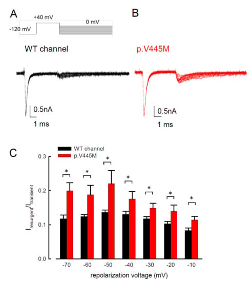
vβ4 peptide at the repolarization stage [3]. Figure 4 shows the resurgent Na
β4 peptide at the repolarization stage [21]. shows the resurgent Na currents elicited at the repolarization (0 to −100 mV) following a prepulse depolarization at +40 mV in the p.V445M mutant and WT Na
1.4 channels. The sweeps show that the magnitude of the resurgent Na
currents in the p.V445M mutant channel was larger (B) compared with that of the WT channel (A). Upon evaluating the ratio between the resurgent and transient currents, the mutant channel had a significantly high ratio at the repolarizing voltages, from −70 mV up to −10 mV (C). These results demonstrate that the p.V445M mutation located at Domain I/segment VI (DI/S6) may play a role in the genesis of resurgent Na
currents that is mediated by the inter-transformation between inactive and open (activation) states of the channel (refer to Discussion). The changes in the gating conformation caused by p.V445M mutant channels facilitate repetitive firing, which, in turn, can induce a drastic increase in muscular stiffness and clinical symptoms of SCM. We also evaluated whether the activation or inactivation curves can be modified by the peptide of Na
β4, which we added during the genesis of resurgent current. Both activation and inactivation curves of the WT Na
1.4 channel are evidently hyperpolarized shift on the membrane potential axis with the presence of 0.1 mM Na
vβ4 peptides in comparison those without the peptides (Supplementary data in Figure S1). However, the slope of the curves is unchanged. The window current remains unchanged either in the presence or absence of Na
β4 peptides in comparison those without the peptides (Supplementary data Figure S1). However, the slope of the curves is unchanged. The window current remains unchanged either in the presence or absence of Na β4 peptide.
Smaller resurgent Na
currents in the WT channel than that in the p.V445M mutant channel in the CHO-K1 cells (
) The protocol to generate resurgent Na
currents is as follows: the patched cells were held at −120 mV, and the resurgent Na
currents of the WT channel were evoked by pulses following a depolarization prepulse of +40 mV for ~10 ms in the presence the Na
β4 peptide (upper panel). Sample sweeps for the WT channel show the occurrence of resurgent Na
currents at ~4 ms post repolarization in the presence of 100 μM Na
β4 peptide (lower panel). (
) Sample sweeps for the p.V445M mutant channel in the presence 0.1-mM Na
β4 peptide following the same protocol as in part
(
) The ratio between resurgent and peak transient currents is significantly smaller in the WT than in the p.V445M mutant channel between −70 and −10 mV. (*,
< 0.05).
4. Evidence of Two Open States for Transient and Resurgent Na+ Currents in the WT Nav1.4 Channel
We observed the changes of resurgent Na
We observed the changes of resurgent Na
currents with a trend of escalating depolarization prepulses between −60 and +180 mV. Depolarization via prepulses within 10 ms could produce a similar activation curve of resurgent Na
currents in both the WT and p.V445M mutant channels (A,B). The 10 ms of depolarization induced resurgent Na
currents, indicating that an open (activation) state of the Na
1.4 channel can be obtained. The activation curves of the transient Na
currents and those of the resurgent Na
currents of the WT and mutant channels were replotted. The activation curve of the transient sodium current showed a hyperpolarizing shift in the mutant channel (C), which is in line with our previous study (B). However, the activation curve of the resurgent Na
currents moved to a more depolarized membrane voltage range in the mutant channel compared with the WT channel (C). After fitting with Boltzmann functions, the activation curves of the resurgent Na
currents from the WT channel showed that the cumulative results were +33.64 ± 1.7 mV for V
and 30.45 ± 1.45 for
, while for the p.V445M mutant channel, the results were 54.04 ± 1.43 mV for V
(
= 6;
< 0.05) and 33.84 ± 1.12 for
(
= 6;
< 0.05) (C). In both the WT and p.V445M mutant channels, the activation curve for the transient Na
current was in a much lower range of membrane potential than that of the resurgent Na
currents (C). The slope of the activation curve of the resurgent Na
current was much less steep. These findings suggest that there may be two open states (O
and O
2) for the channel during activation (refer to Discussion).
) for the channel during activation (refer to Discussion).
Activation curve of resurgent Na
currents in the CHO-K1 cells (
) The protocol to generate resurgent Na
currents are as follows: the cell was held at −120 mV for ~30 ms, and then stepped to various depolarization between −60 and +180 mV for 5 ms in 10 mV increments. Resurgent Na
currents were then evoked by a pulse of −60 mV for 150 ms (upper panel). Sample sweeps were obtained in the presence of 0.1 mM Na
β4 peptide for the WT channel (lower panel). (
) Sample sweeps for the p.V445M mutant channel in the presence 0.1 mM Na
β4 peptide. (
) The activation curves of transient and resurgent Na
currents for the WT and p.V445M mutant channels are replotted for comparison. The activation curve of resurgent Na
currents of the WT channel (the block point fitting line) for each cell are obtained by fittings with Boltzmann functions, and the cumulative results for V
are +33.64 ± 1.7 mV and
are 30.45 ± 1.45 for ~10 ms prepulses, respectively (
= 8). The resurgent activation curve of the p.V445M mutant channel (the red-point-fitting line) for each cell are also obtained by fitting Boltzmann functions and the cumulative results for V
and
are +54.04 ± 1.43 mV (
= 6) and 33.84 ± 1.12 (
= 6), respectively .
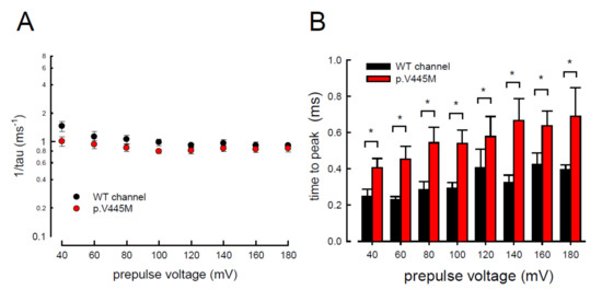
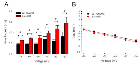
5. Changes of the Decay Rate and Time to Peak of Resurgent Na+ Currents in Mutant Channels
In addition to measuring the magnitude of the resurgent Na
In addition to measuring the magnitude of the resurgent Na
currents, we also examined the kinetic changes in the resurgent Na
currents induced by the p.V445M mutation. As shown in A, the decay kinetics of the resurgent Na
currents were not significantly changed in the p.V445M mutant channel within a wide range of depolarizing prepulses (from +40 to +180 mV). However, the time to peak of the resurgent Na
currents during repolarization was significantly longer in the mutant channel than in the WT channel (B). The changes of these parameters under a series of repolarizing potentials from −70 up to −20 mV were also evaluated. Compared with the WT channel, the time to peak was significantly slower in the mutant channel during repolarization (A). Moreover, the decay kinetics (1/tau) of the p.V445M mutant channel matched those of the WT channel without an obvious change (1/tau
= 0.22 × exp(−0.64V/25) ms
for the WT channel and 1/tau
= 0.18 × exp(−0.69V/25) ms
for the p.V445M mutant channel; B). These findings suggest that two different individual open (activation) states are responsible for the transient and resurgent Na
currents. The decay rate from the open (activation) state of the resurgent Na
currents in the p.V445M mutant channel was the same as that observed in the WT channel. However, compared with those in the WT channel, the resurgent Na
currents in the p.V445M mutant channel were conspicuously slow, indicating that the mutation results in a protracted period of open state of the Na
v1.4 channel, either after strong depolarizing prepulses or under different repolarization potentials.
1.4 channel, either after strong depolarizing prepulses or under different repolarization potentials.
Accelerated decay of resurgent Na
currents by hyperpolarization between the WT and p.V445M mutant channels in the CHO-K1 cells (
) The inverses of decay time constants (1/tau) of the resurgent Na
currents are obtained at −60 mV after the depolarizing prepulses between +40 and +180 mV in 20 mV increments for ~10 ms in the WT and p.V445M mutant channels (
= 8). (
) The time to peak of resurgent Na
currents in the WT channel is always faster than that in the p.V445M mutant channel at the prepulse voltages tested (
= 8 *;
< 0.05).
Slower time to peak of the resurgent Na
currents in the p.V445M mutant channel in the CHO-K1 cells (
) The time to the resurgent current peak was measured with the same protocol in (
= 6). The time to the resurgent Na
current peak is significantly shortened in the WT than in the p.V445M mutant channel between −20 and −70 mV (*;
< 0.05). (
) The reciprocal of the time constants (1/tau) for the decay phase of resurgent Na
currents was plotted against the voltage in semi-logarithmic scales for the WT and p.V445M mutant channels (
= 8). The lines are linear regression fits of the form: 1/tau
= 0.22 × exp(−0.64V/25) ms
for the WT channel and 0.18 × exp(−0.69V / 25) ms
−1 for the p.V445M mutant channel, respectively.
for the p.V445M mutant channel, respectively.
6. Discussion
In summary, we identified the missense mutation p.V445M of the
In summary, we identified the missense mutation p.V445M of the
gene from two patients with SCM who were recruited from two individual families. To determine the electrophysiological function of this mutation, a transfected CHO-K1 cell model was employed for the whole-cell patch-clamp recording. Evaluation of the transient Na
current showed a hyperpolarizing shift in both the activation and inactivation curves of the mutant channel as compared with the WT channel. The window and sustained Na
currents of the mutant channel were larger than those of the WT. Upon transfection of both Na
1.4 and a Na
β4 peptide, resurgent Na
currents could be obtained 3~5 ms after a prepulse of depolarization. Compared with that of the WT channel, the magnitude of the resurgent Na
currents were conspicuously larger in the mutant channel. Moreover, the time to peak of the resurgent Na
+ currents in the mutant channel was slower than that of the WT channel, although no difference was found between them in the decay kinetics.
currents in the mutant channel was slower than that of the WT channel, although no difference was found between them in the decay kinetics.
Myotonia results from an involuntary muscle contraction that persists for several seconds after the cessation of voluntary effect [4]. The hyperexcitability of muscles with a burst of muscle action potentials leads to an after-contraction that can last for several seconds. The spontaneous, painless discharges show a waxing and waning pattern upon EMG study. A sustained burst of discharges can be elicited by a brief voluntary contraction, and the firing frequency is usually 20~80 Hz, which means the interval of the depolarizing potentials is between 12.5 and 50 ms [5]. The repetitive, high-frequency discharges in myotonia suggest that resurgent Na
Myotonia results from an involuntary muscle contraction that persists for several seconds after the cessation of voluntary effect [22]. The hyperexcitability of muscles with a burst of muscle action potentials leads to an after-contraction that can last for several seconds. The spontaneous, painless discharges show a waxing and waning pattern upon EMG study. A sustained burst of discharges can be elicited by a brief voluntary contraction, and the firing frequency is usually 20~80 Hz, which means the interval of the depolarizing potentials is between 12.5 and 50 ms [23]. The repetitive, high-frequency discharges in myotonia suggest that resurgent Na currents may be a critical factor involving the phenotype. Channels are usually refractory to be activated after inactivation until the membrane potential has been sufficiently hyperpolarized. Nevertheless, channels can reopen, allowing a surge of inward current during repolarization, i.e., resurgent Na
currents. Raman and Bean first described resurgent Na
+ currents in cerebellar Purkinje neurons [6]. In the cerebellum, the resurgent Na
currents in cerebellar Purkinje neurons [16]. In the cerebellum, the resurgent Na currents following relief of an ultra-fast open-channel block by an endogenous blocking particle, probably the C-terminal portion of the Na
vβ4 subunit [7], may contribute to high-frequency firing [6][8]. The Na
β4 subunit [24], may contribute to high-frequency firing [16,25]. The Na 1.6 channel is the major carrier of resurgent Na
+ currents in cerebellar [9] and DRG [10] neurons. In the whole-cell configuration of transfected CHO-K1 cells expressing the Na
currents in cerebellar [26] and DRG [27] neurons. In the whole-cell configuration of transfected CHO-K1 cells expressing the Na 1.7 channel, resurgent Na
+ currents after a long depolarizing prepulse have also been identified [11]. Resurgent Na
currents after a long depolarizing prepulse have also been identified [13]. Resurgent Na currents from the mutant Na
v1.7 channel may play an important role in the episodic attacks of severe neuropathic pain found in both erythromelalgia and paroxysmal extreme pain disorders [12]. Enhancement of resurgent Na
1.7 channel may play an important role in the episodic attacks of severe neuropathic pain found in both erythromelalgia and paroxysmal extreme pain disorders [28]. Enhancement of resurgent Na + currents has even been proposed to be highly correlated with the severity of neuropathic pain [12]. In the present study, a hyperpolarizing shift was found in both activation and inactivation curves between the mutant and WT channels. Although the window currents increased in the mutant channel, the hyperexcitability with repetitive recurrent discharges could not be fully explained by the changes in the transient Na
currents has even been proposed to be highly correlated with the severity of neuropathic pain [28]. In the present study, a hyperpolarizing shift was found in both activation and inactivation curves between the mutant and WT channels. Although the window currents increased in the mutant channel, the hyperexcitability with repetitive recurrent discharges could not be fully explained by the changes in the transient Na current alone. Here, we described the resurgent Na
currents in the Na
1.4 channel. Compared with the WT channel, the larger magnitude and slowing of the time to peak of the resurgent Na
currents in the mutant channel may be one of the crucial factors of the functional disturbances of SCM. It has been proposed that impaired inactivation is a major determinant of resurgent Na
currents in Na
v1.7 channels [12]. Future research should investigate whether mediation of the inactivation process governs the functional consequences resulting from a mutation in the Na
1.7 channels [28]. Future research should investigate whether mediation of the inactivation process governs the functional consequences resulting from a mutation in the Na v1.4 channel, although prolonged recovery of slow inactivation has been observed in the p.V445M mutant channel [2].
1.4 channel, although prolonged recovery of slow inactivation has been observed in the p.V445M mutant channel [20].
In our study, resurgent currents of Nav1.4 channel are discernible only in the presence of the Na
In our study, resurgent currents of Nav1.4 channel are discernible only in the presence of the Na
β4 peptides (Figure S1). According to the prevailing model, this is ascribable to the competition between the Na vβ4 peptides and the inactivating peptide for the open channel pore [13][14][15][16]. However, quite other biophysical characteristics, including (1) the concomitant increase of resurgent and sustained currents with the p.V445M mutant channels (Figure 3A–C and Figure 4), (2) the very much different activation curves for the transient and resurgent currents (Figure 5C) all strongly argue against that the Na
β4 peptides and the inactivating peptide for the open channel pore [14,15,16,17]. However, quite other biophysical characteristics, including (1) the concomitant increase of resurgent and sustained currents with the p.V445M mutant channels (A–C and ), (2) the very much different activation curves for the transient and resurgent currents (C) all strongly argue against that the Na β4 peptide competes with the inactivating molecule for the same single open state, (3) the slower time to peak but unchanged decay kinetics of the resurgent currents with p.V445M mutant channels also contradicts that Na
β4 peptide competes with the inactivating molecule for the same single open state to generate the resurgent currents. We therefore propose that there may be two distinct open states (with two corresponding inactivated states) of the Na
1.4 channel (), each responsible for the transient (O
) and resurgent (O
) currents, respectively. Significant occupancy of O
is possible only in the presence of the Na
vβ4 peptides, which is thus chiefly a gating modifier inducing new gating conformations rather than a pore blocker competing with the inactivating peptides.
β4 peptides, which is thus chiefly a gating modifier inducing new gating conformations rather than a pore blocker competing with the inactivating peptides.
The activation curves of the resurgent Na
The activation curves of the resurgent Na
currents in both the WT and p.V445M mutant channels markedly moved to a more positive voltage range, with a much less steep slope than those of the transient currents (C). We proposed that there may be two open states of the channel and that the gating modification illustrates the electrophysiological changes of the genesis of resurgent Na
+ currents [11]. This study showed that either after a depolarizing prepulse (Figure 6B) or under a variable extent of repolarization (Figure 7A), the time to peak of the resurgent Na
currents [13]. This study showed that either after a depolarizing prepulse (B) or under a variable extent of repolarization (A), the time to peak of the resurgent Na currents in the mutant channel was extended significantly compared with that in the WT channel; however, the decay kinetics (1/tau) remained similar between the channels. The p.V445M mutation significantly accelerated the entry and protracted the duration of the new open (activation) state, leading to an increase in the magnitude of the resurgent Na
+ currents. The reopening of the fast inactivated channels was probably accompanied by conformational changes of the domain I/S6 (DI/S6), where V445M is located.
currents. The reopening of the fast inactivated channels was probably accompanied by conformational changes of the domain I/S6 (DI/S6), where V445M is located.
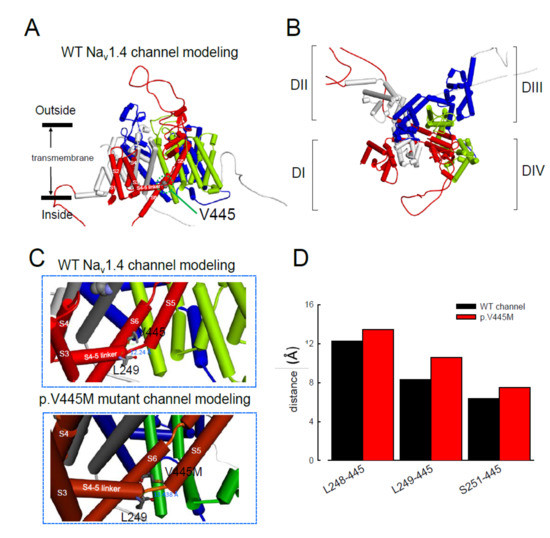
The computer homology model of the WT Na
The computer homology model of the WT Na
1.4 channel evaluated the topological structure change caused by the p.V445M mutation (A,B). The model estimated that the change of Val445 to Met may increase the distance between V445 and the residues at the S4–S5 linker (DI/S4-5 linker) (e.g., L248, L249 and S251; ). While the S4–S5 linker plays an imperative role in governing the activation/inactivation of resurgent Na
+ currents [11], such a distance change could contribute to the attenuation of inactivation and uncoupling between activation and inactivation. In the previous study, genesis of resurgent Na
currents [13], such a distance change could contribute to the attenuation of inactivation and uncoupling between activation and inactivation. In the previous study, genesis of resurgent Na currents in the Na
v1.7 channel was found to be possibly due to the existence of two open states in activation [11]. Through modulating the transitional kinetics between these two states, the S4–S5 linker in Domain III seems to play an important role in activation–inactivation coupling. Mutations involving the linker and relevant nearby regions may, therefore, enhance the sustained and resurgent Na
1.7 channel was found to be possibly due to the existence of two open states in activation [13]. Through modulating the transitional kinetics between these two states, the S4–S5 linker in Domain III seems to play an important role in activation–inactivation coupling. Mutations involving the linker and relevant nearby regions may, therefore, enhance the sustained and resurgent Na currents, leading to marked hyperexcitability and repetitive firing in the corresponding tissues. Herein, we postulated that the change of distance between V445 and the residues of the S4–S5 linker may be one of the critical factors for the coupling effect. Aberrations in modulation of the coupling facilitate the generation of resurgent Na
currents and increase both the magnitude and time to peak, augmenting the repetitive firing. The mutation causes slowing of the time to peak of the resurgent Na
currents elicited in the Na
1.4 channel, which increases the firing frequency of the muscle membrane. The action potential propagates to the T-tubule, which induces the opening of the voltage-gated calcium channel from the sarcoplasmic reticulum. The released calcium ion binds to troponin C, which induces the cross bridge to contract. The resurgent Na
currents introduce repetitive high-frequency firing, facilitating the binding leading to muscle stiffness. The essential tremors and chronic seizures found in the family members with a p.G1537S mutation in the
SCN4A gene [17] may also have been attributable to the disturbance of the resurgent Na
gene [29] may also have been attributable to the disturbance of the resurgent Na currents in the Na
1.4 channel.
Homology modeling of the WT and mutant human Na
1.4 channels. (
) Side view of the homology model shows the transmembrane helixes of the four domains. The four domains I, II, III and IV are colored red, white, blue and green. The side chain of V445 in the S6 linker of Domain I (DI) is indicated in the CPK model. (
) A closer view of the WT Na
1.4 channel from the external side of the pore. (
) A diagram of Domain I of the homology model of the WT Na
1.4 and p.V445M mutant channels is shown in the schematic presentation. The side chains of V445 in the S6 segment and L248, L249 and S251 in the S4–5 linker of Domain I are shown with sticks of various colors. A closer view of the area is shown in the panel, demonstrating inter-residue distances of ~8.3196, ~12.24 and ~6.348 Å between V445 and L248, V445M and L249 and V445 and S251, respectively, in the WT Na
1.4 channel, while the distances between p.V445M and L248, p.V445M and S249 and p.V445M and S251 are ~7.54, ~13.438 and ~10.551 Å, respectively, in the p.V445M mutant channel. (
D) The relative distances between the residue V445 and L248, L249 and S251 in the homology modeling is shown. Note the conspicuously decreased distances in all cases in the WT channel compared with those in the p.V445M mutant channel.
) The relative distances between the residue V445 and L248, L249 and S251 in the homology modeling is shown. Note the conspicuously decreased distances in all cases in the WT channel compared with those in the p.V445M mutant channel.
In addition to SCM, mutations in Na
In addition to SCM, mutations in Na
1.4 are also responsible for PMC. Clinically, mutations involving the Arg1448 of the
SCN4A gene are frequently found in patients with PMC [18]. The resurgent Na
gene are frequently found in patients with PMC [30]. The resurgent Na currents in the Na
1.4 channel were first described using rat DRG neurons transfected with Na
1.4r-R1448P. Jarecki et al. reported that the resurgent Na
+ currents could be obtained and that the average relative amplitude was 4.8± 0.7% [19], much lower than our findings (~10% in the WT and approximately 15–20% in mutant channels, Figure 4C). Furthermore, the Arg1448 locates at DIV/S4, which is the outermost extracellular charged residue in the sodium channel voltage sensor that modulates the activation/inactivation coupling of the channel. The study showed that the Na
currents could be obtained and that the average relative amplitude was 4.8± 0.7% [18], much lower than our findings (~10% in the WT and approximately 15–20% in mutant channels, C). Furthermore, the Arg1448 locates at DIV/S4, which is the outermost extracellular charged residue in the sodium channel voltage sensor that modulates the activation/inactivation coupling of the channel. The study showed that the Na 1.4r-R1448P channels slow the inactivation and generate resurgent Na
currents, suggesting that the mutation slows the rate of open-channel fast inactivation and may be able to induce resurgent Na
currents. In another study, large resurgent Na
currents could be observed in nearly 62% of Na
1.8-null DRG neurons using Biolistic transfected with Na
1.6r during the repolarization stage; however, no resurgent Na
currents were identified in neurons transfected with a TTX-R version of the rat skeletal muscle sodium channel Na
1.4, although they generated a larger peak sodium current amplitude than the Na
v1.6 channels [10]. The discrepancy may have been due to the different expression systems and the modification of the channel to become TTX-resistant since resurgent Na
1.6 channels [27]. The discrepancy may have been due to the different expression systems and the modification of the channel to become TTX-resistant since resurgent Na + currents are difficult to obtain using DRG neurons or WT skeletal muscles [10]. In a recent report, a p.G1306E mutation of SCN4A gene caused NDM and Brugada syndrome after treated with flecainide. The different isoforms of Nav1.4 expressed in the skeletal and cardiac muscles may result in such a rare phenotype [20].
currents are difficult to obtain using DRG neurons or WT skeletal muscles [27]. In a recent report, a p.G1306E mutation of SCN4A gene caused NDM and Brugada syndrome after treated with flecainide. The different isoforms of Nav1.4 expressed in the skeletal and cardiac muscles may result in such a rare phenotype [31].
In conclusion, mutations in
In conclusion, mutations in
are responsible for SCM. The hyperpolarizing shifting of the activation and inactivation curves, which results in an increase of window and sustained Na
currents, may contribute to the hyperexcitability of neural-muscle tissues. The Na
1.4 channel can generate resurgent Na
currents, which may further increase repetitive firing of the action potential in skeletal muscle, one of the hallmarks of SCM in EMG. In addition to myotonia, patients with SCM frequently experience episodes of muscle weakness. Overall,
mutations resulting in defects of slow inactivation and delay of time to peak in resurgent Na
+ currents are likely factors contributing to constant myotonia and weakness in SCM patients.
currents are likely factors contributing to constant myotonia and weakness in SCM patients.
- Rosenfeld, J.; Sloan-Brown, K.; George, A.L., Jr. A novel muscle sodium channel mutation causes painful congenital myotonia. Ann. Neurol. 1997, 42, 811–814. [Google Scholar] [CrossRef]
- Takahashi, M.P.; Cannon, S.C. Enhanced slow inactivation by V445M: A sodium channel mutation associated with myotonia. Biophys. J. 1999, 76, 861–868. [Google Scholar] [CrossRef]
- Huang, C.W.; Lai, H.J.; Huang, P.Y.; Lee, M.J.; Kuo, C.C. Anomalous enhancement of resurgent Na(+) currents at high temperatures by SCN9A mutations underlies the episodic heat-enhanced pain in inherited erythromelalgia. Sci. Rep. 2019, 9, 12251. [Google Scholar] [CrossRef] [PubMed]
- Rudel, R.; Lehmann-Horn, F. Membrane changes in cells from myotonia patients. Physiol. Rev. 1985, 65, 310–356. [Google Scholar] [CrossRef] [PubMed]
- Miller, T.M. Differential diagnosis of myotonic disorders. Muscle Nerve 2008, 37, 293–299. [Google Scholar] [CrossRef] [PubMed]
- Grieco, T.M.; Malhotra, J.D.; Chen, C.; Isom, L.L.; Raman, I.M. Open-channel block by the cytoplasmic tail of sodium channel beta4 as a mechanism for resurgent sodium current. Neuron 2005, 45, 233–244. [Google Scholar] [CrossRef]
- Khaliq, Z.M.; Gouwens, N.W.; Raman, I.M. The contribution of resurgent sodium current to high-frequency firing in Purkinje neurons: An experimental and modeling study. J. Neurosci. 2003, 23, 4899–4912. [Google Scholar] [CrossRef]
- Afshari, F.S.; Ptak, K.; Khaliq, Z.M.; Grieco, T.M.; Slater, N.T.; McCrimmon, D.R.; Raman, I.M. Resurgent Na currents in four classes of neurons of the cerebellum. J. Neurophysiol. 2004, 92, 2831–2843. [Google Scholar] [CrossRef]
- Cummins, T.R.; Dib-Hajj, S.D.; Herzog, R.I.; Waxman, S.G. Nav1.6 channels generate resurgent sodium currents in spinal sensory neurons. FEBS Lett. 2005, 579, 2166–2170. [Google Scholar] [CrossRef]
- Theile, J.W.; Jarecki, B.W.; Piekarz, A.D.; Cummins, T.R. Nav1.7 mutations associated with paroxysmal extreme pain disorder, but not erythromelalgia, enhance Navbeta4 peptide-mediated resurgent sodium currents. J. Physiol. 2011, 589, 597–608. [Google Scholar] [CrossRef]
- Bergareche, A.; Bednarz, M.; Sanchez, E.; Krebs, C.E.; Ruiz-Martinez, J.; De La Riva, P.; Makarov, V.; Gorostidi, A.; Jurkat-Rott, K.; Marti-Masso, J.F.; et al. SCN4A pore mutation pathogenetically contributes to autosomal dominant essential tremor and may increase susceptibility to epilepsy. Hum. Mol. Genet. 2015, 24, 7111–7120. [Google Scholar] [CrossRef]
- Featherstone, D.E.; Fujimoto, E.; Ruben, P.C. A defect in skeletal muscle sodium channel deactivation exacerbates hyperexcitability in human paramyotonia congenita. J. Physiol. 1998, 506, 627–638. [Google Scholar] [CrossRef]
- Cavalli, M.; Fossati, B.; Vitale, R.; Brigonzi, E.; Ricigliano, V.A.G.; Saraceno, L.; Cardani, R.; Pappone, C.; Meola, G. Flecainide-Induced Brugada Syndrome in a Patient With Skeletal Muscle Sodium Channelopathy: A Case Report With Critical Therapeutical Implications and Review of the Literature. Front. Neurol. 2018, 9, 385. [Google Scholar] [CrossRef] [PubMed]