
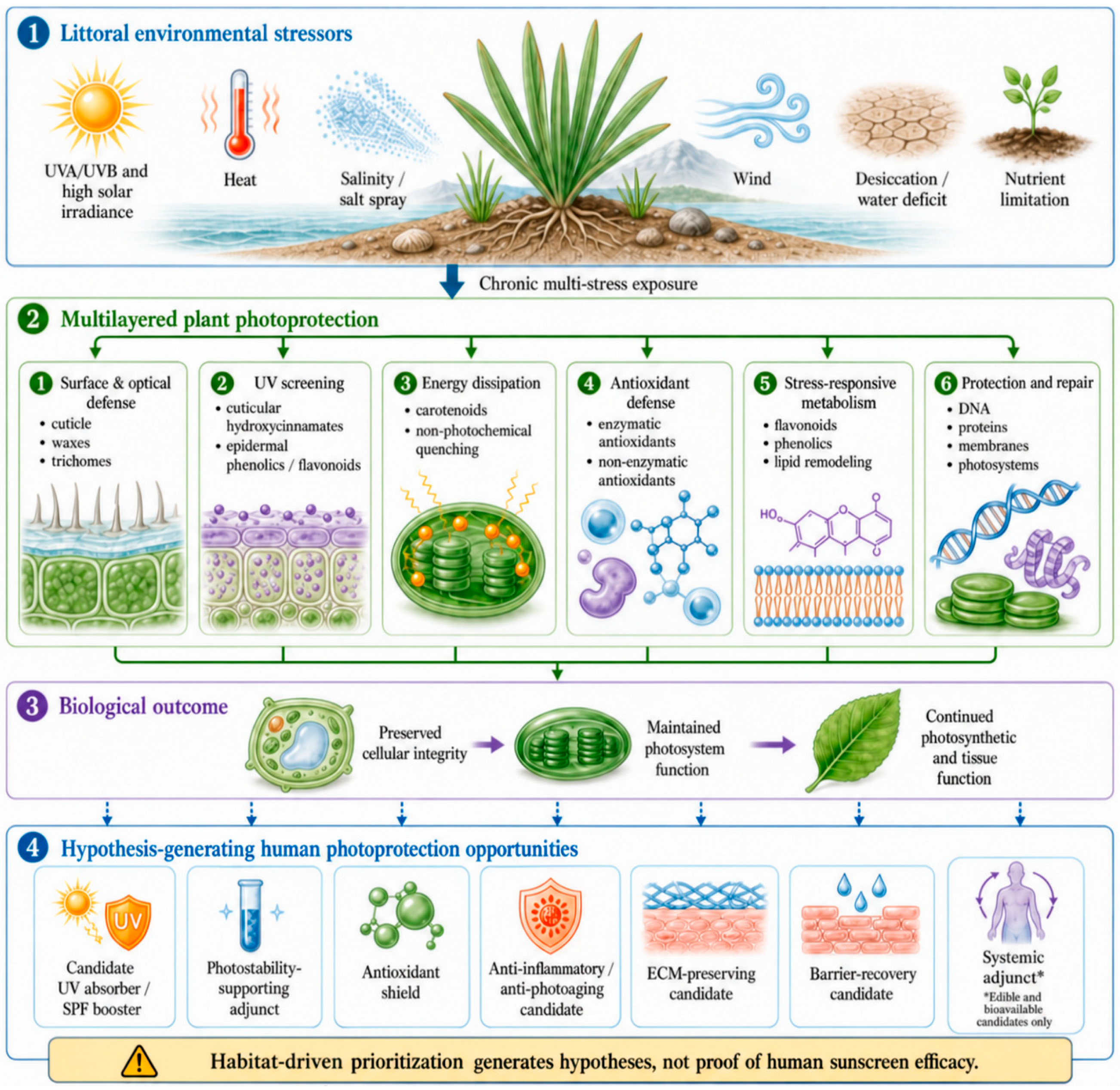
Coastal vascular plants survive chronic exposure to ultraviolet radiation, salinity, salt spray, heat, wind, and water limitation through coordinated structural, chemical, antioxidant, and repair mechanisms. Collectively, these adaptations constitute the “littoral plant photoprotectome” and offer an ecologically informed source of molecules and design principles for human skin photoprotection. This review maps the ecophysiological, phytochemical, optical, dermatological, formulation, safety, and translational evidence concerning littoral vascular plants. The review is a critical review based on a structured literature search, not a formal scoping or systematic review, and it classifies evidence from ecological rationale through optical screening and cell or tissue models to human data. Evidence was synthesized using the proposed Filter–Shield–Repair–Prime framework, which distinguishes direct attenuation of ultraviolet radiation from the biological suppression of oxidative and inflammatory injury, restoration of epidermal barrier and extracellular-matrix homeostasis, and systemic support before irradiation. Phenolic-rich cuticles, epidermally localized flavonoids, hydroxycinnamic acids, carotenoid-dependent energy dissipation, antioxidant networks, and stress-responsive secondary metabolism collectively contribute to plant photoprotection. Among the candidates examined, Calophyllum inophyllum presents the most integrated evidence, combining ultraviolet attenuation, chemically identified ultraviolet-absorbing constituents, and anti-inflammatory effects in UVB-exposed keratinocytes. Controlled human evidence for littoral candidates remains scarce, comprised of a clinical sun protection factor/UVA protection grade (SPF/PA) test of neat tamanu oil (SPF 6.3 ± 0.4; PA 2.9 ± 0.6), a placebo-controlled split-face study of a Salicornia europaea cream that improved skin-texture parameters in 29 volunteers under natural sun exposure, and a pilot barrier-recovery study of Crithmum maritimum biomass, whereas most other efficacy signals derive from cell and tissue models that cannot by themselves predict clinical photoprotection. Scaevola taccada, Carpobrotus edulis, Salicornia europaea, Terminalia catappa, Nypa fruticans, Crithmum maritimum, and Limonium algarvense demonstrate complementary optical, antioxidant, anti-inflammatory, extracellular-matrix-preserving, or barrier-restoring activities, although their translational maturity remains heterogeneous. Antioxidant assays, solution-phase ultraviolet absorbance, and calculated sun protection factor values alone cannot establish sunscreen efficacy. Translation therefore requires standardized full-spectrum optical testing, photostability and phototoxicity assessment, mechanistic human skin models, finished-formulation evaluation, clinical validation, and sustainable sourcing. Quantitative effect sizes, exposure doses and controls are reported where available, and demonstrated effects, mechanistic hypotheses and translational applications are distinguished throughout. The proposed framework and LITTORAL Readiness Score provide a structured basis for classifying candidates and guiding the development of topical and systemic photoprotective interventions.
- littoral vascular plants
- coastal halophytes
- ultraviolet radiation
- photoprotection
- photoaging
- phytochemicals
- cosmeceuticals
- nutricosmetics
- bioinspired sunscreens
References
- Mouret, S.; Charveron, M.; Favier, A.; Cadet, J.; Douki, T. Differential repair of UVB-induced cyclobutane pyrimidine dimers in cultured human skin cells and whole human skin. DNA Repair 2008, 7, 704–712.
- Salminen, A.; Kaarniranta, K.; Kauppinen, A. Photoaging: UV radiation-induced inflammation and immunosuppression accelerate the aging process in the skin. Inflamm. Res. 2022, 71, 817–831.
- Wei, M.; He, X.; Liu, N.; Deng, H. Role of reactive oxygen species in ultraviolet-induced photodamage of the skin. Cell Div. 2024, 19, 1.
- Hobohm, C.; Schaminée, J.; van Rooijen, N. Environmental Challenges and Solutions; Springer International Publishing: Cham, Switzerland, 2021; pp. 279–310.
- Ievinsh, G. Creating a conceptual framework for analysis of vascular plant diversity in a coastal landscape: Functional aspects and ecosystem services for plants at the Baltic Sea. Environ. Exp. Biol. 2024, 22, 201–244.
- Du, J.; Hesp, P.A. Salt spray distribution and its impact on vegetation zonation on coastal dunes: A review. Estuaries Coast 2020, 43, 1885–1907.
- Sachdev, S.; Ansari, S.A.; Ansari, M.I.; Fujita, M.; Hasanuzzaman, M. Abiotic stress and reactive oxygen species: Generation, signaling, and defense mechanisms. Antioxidants 2021, 10, 277.
- Zandi, P.; Schnug, E. Reactive oxygen species, antioxidant responses and implications from a microbial modulation perspective. Biology 2022, 11, 155.
- Chen, B.; Wang, Y.; Zhu, Y.; Li, J.; Wan, L.; Zhu, C.; Huang, Q.; Elhamid, M.A.A.; Soaud, S.A.; Yan, K.; et al. Membrane stability under heat stress: Molecular signaling, lipid remodeling, and defense mechanisms in plants. Plant Cell Rep. 2026, 45, 32.
- Karabourniotis, G.; Liakopoulos, G.; Bresta, P.; Nikolopoulos, D. The optical properties of leaf structural elements and their contribution to photosynthetic performance and photoprotection. Plants 2021, 10, 1455.
- Chen, Z.; Dong, Y.; Huang, X. Plant responses to UV-B radiation: Signaling, acclimation and stress tolerance. Stress Biol. 2022, 2, 51.
- Ferreyra, M.L.F.; Serra, P.; Casati, P. Recent advances on the roles of flavonoids as plant protective molecules after UV and high light exposure. Physiol. Plant. 2021, 173, 736–749.
- González Moreno, A.; de Cózar, A.; Prieto, P.; Domínguez, E.; Heredia, A. Radiationless mechanism of UV deactivation by cuticle phenolics in plants. Nat. Commun. 2022, 13, 1786.
- Heredia, A.; Benítez, J.J.; González Moreno, A.; Domínguez, E. Revisiting plant cuticle biophysics. New Phytol. 2024, 244, 65–73.
- Madhavan, C.; Meera, S.P.; Kumar, A. Anatomical adaptations of mangroves to the intertidal environment and their dynamic responses to various stresses. Biol. Rev. Camb. Philos. Soc. 2025, 100, 1019–1046.
- Li, X.; Yang, S.; Zhang, J.; Wan, S. Photo-oxidative stress in plants: ROS signaling, damage propagation, and systems-level resilience. Antioxidants 2026, 15, 371.
- Dalton, W.J. Employing Ultrafast Laser Spectroscopy Techniques to Identify and Design Nature-Inspired Chromophores for Photoprotection Applications. Ph.D. Thesis, University of Warwick, Coventry, UK, 2025. Available online: https://wrap.warwick.ac.uk/193820/ (accessed on 23 July 2026).
- Abiola, T.T. Ultrafast Spectroscopy of Plant-Inspired UV Filters for Sunscreen and Photothermal Applications. Ph.D. Thesis, University of Warwick, Coventry, UK, 2022. Available online: https://wrap.warwick.ac.uk/176131/ (accessed on 27 July 2026).
- Lovelock, C.E.; Clough, B.F.; Woodrow, I.E. Distribution and accumulation of ultraviolet-radiation-absorbing compounds in leaves of tropical mangroves. Planta 1992, 188, 143–154.
- Wingfield, J.L.; Ruane, L.G.; Patterson, J.D. A three-dimensional spatial mapping approach to quantify fine-scale heterogeneity among leaves within canopies. Appl. Plant Sci. 2017, 5, 1700056.
- Poorter, H.; Niinemets, Ü.; Ntagkas, N.; Siebenkäs, A.; Mäenpää, M.; Matsubara, S.; Pons, T.L. A meta-analysis of plant responses to light intensity for 70 traits ranging from molecules to whole plant performance. New Phytol. 2019, 223, 1073–1105.
- Nikalje, G.C.; Variyar, P.S.; Joshi, M.V.; Nikam, T.D.; Suprasanna, P. Temporal and spatial changes in ion homeostasis, antioxidant defense and accumulation of flavonoids and glycolipid in a halophyte Sesuvium portulacastrum (L.) L. PLoS ONE 2018, 13, e0193394.
- Cao, D.; Zhang, W.; Yang, N.; Li, Z.; Zhang, C.; Wang, D.; Ye, G.; Chen, J.; Wei, X. Proteomic and metabolomic analyses uncover integrative mechanisms in Sesuvium portulacastrum tolerance to salt stress. Front. Plant Sci. 2023, 14, 1277762.
- Wang, Y.; Ma, W.; Fu, H.; Li, L.; Ruan, X.; Zhang, X. Effects of salinity stress on growth and physiological parameters and related gene expression in different ecotypes of Sesuvium portulacastrum on Hainan Island. Genes 2023, 14, 1336.
- Fitzner, M.; Schreiner, M.; Baldermann, S. Between eustress and distress: UVB induced changes in carotenoid accumulation in halophytic Salicornia europaea. J. Plant Physiol. 2023, 291, 154124.
- Pereira, C.G.; Rodrigues, M.J.; Nawrot-Hadzik, I.; Matkowski, A.; Custódio, L. Seasonal and geographic dynamics in bioproperties and phytochemical profile of Limonium algarvense Erben. Molecules 2024, 29, 481.
- Rodrigues, M.J.; Monteiro, I.; Castañeda-Loaiza, V.; Placines, C.; Conceição Oliveira, M.; Reis, A.C.M.; Caperta, A.D.; Soares, F.; Pousão-Ferreira, P.; Pereira, C.; et al. Growth performance, in vitro antioxidant properties and chemical composition of the halophyte Limonium algarvense Erben are strongly influenced by the irrigation salinity. Ind. Crops Prod. 2020, 143, 111930.