Acrylic bone cements (ABC) are widely used in orthopedics for joint fixation, antibiotic release, and bone defect filling, among others. Most of the commercial ABCs available today consist of two components, one solid, based mainly on poly(methyl methacrylate) (PMMA), and one liquid, based on methyl methacrylate (MMA), which are mixed and, through the polymerization reaction of the monomer, transformed into a hardened cement paste.
- Acrylic Bone Cements
- orthopedics
- joint fixation
- bone cement
- PMMA bone cement
1. Introduction
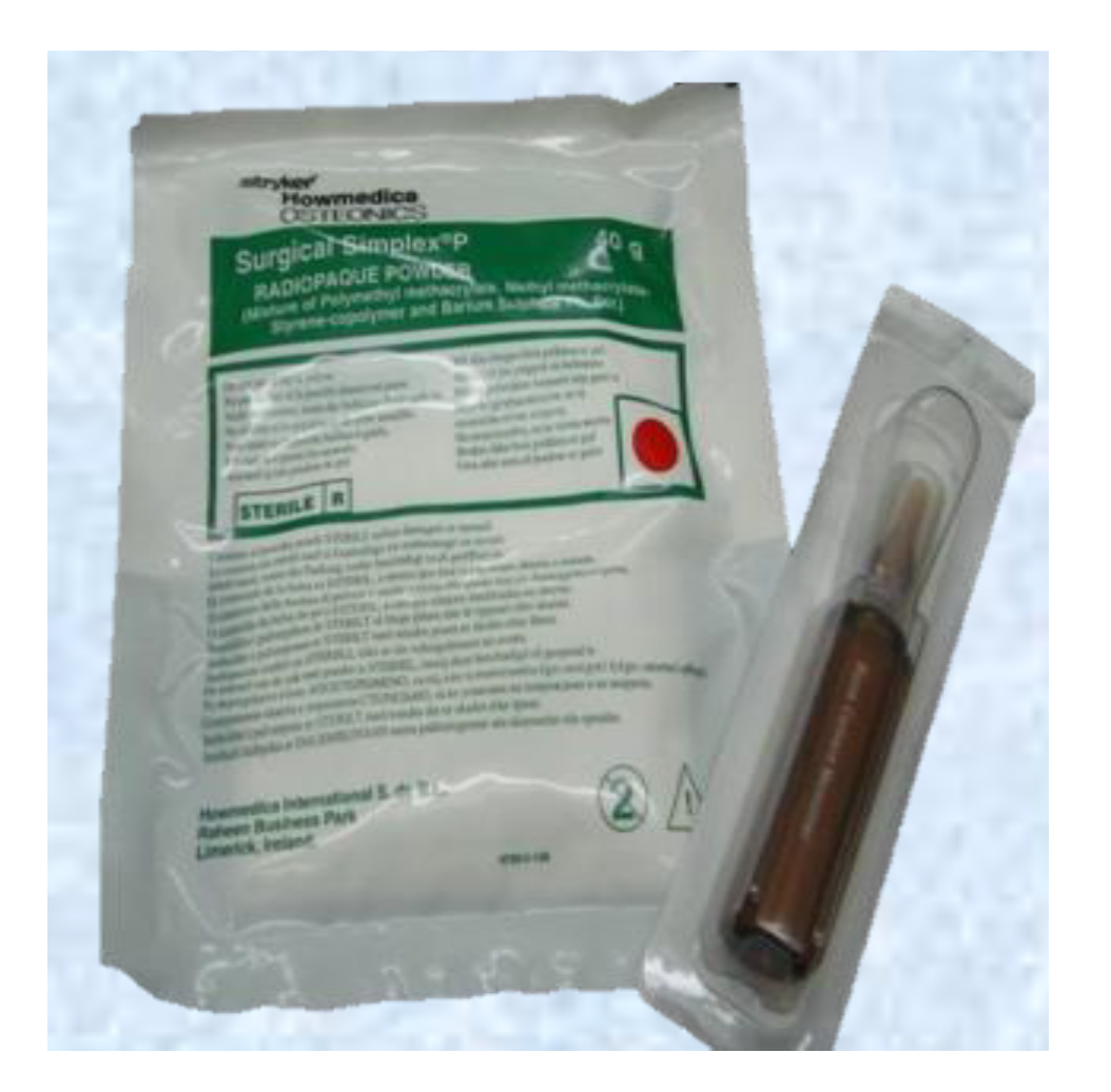
Figure 3Figure1 shows the presentation of Surgical Simplex P commercial cement.
PMMA cements are the most commonly used to bond and load transfer between the implant and the bone [14,15][1][2]. The main advantage of using it is the excellent primary fixation obtained between the implant and the bone and the patient’s faster recovery [16][3]. On the other hand, PMMA is a fragile material with a low resistance to fracture and low fatigue life [11][4].
Commercial ABCs do not differ drastically between brands. The main differences between them are the addition of PMMA copolymers or antibiotics into the solid phase, comonomers to the liquid phase, solid/liquid ratio variation, radiopaque agent variation, or additives chlorophyll Palacos® [11,17][4][5].
The polymerization process begins after mixing of the benzoyl peroxide (BPO) (in the solid) and N,N-Dimethyl-p-Toluidine (DMPT) (in the liquid) with the production of benzoyl radicals at room temperature [18,19][6][7]. The heat produced during polymerization is between 52 and 57 kJ per mole of MMA [3][8]. The maximum temperature reached during the polymerization reaction is known as Tmax [20][9], exceeding 100 °C. These high temperatures can cause cellular bone necrosis and contribute to aseptic loosening [10,21,22,23][10][11][12][13]. Polymerization temperatures experienced under in vivo conditions are much lower (between 40 and 47 °C) at the bone interface due to the reduced thickness of the bone cement layer used in TJR, the presence of blood circulation, and heat dissipation through the implant and surrounding tissue [19,24,25,26][7][14][15][16].
The leading cause of failure of cemented arthroplasties is aseptic loosening of the prosthesis, which usually occurs at the bone–cement interface and requires a second surgery to replace the whole system [27][17]. The loosening occurs according to:
- High polymerization temperature of the cements (between 67 and 124 °C) [28], which generates thermal necrosis of the bone [29], alteration of the local blood circulation, and predisposition to the formation of a fibrous membrane in the bone—cement interface [16,
, alteration of the local blood circulation, and predisposition to the formation of a fibrous membrane in the bone—cement interface [
30].High polymerization temperature of the cements (between 67 and 124 °C) [18], which generates thermal necrosis of the bone [19]3][20].
- Release of unreacted residual monomer or MMA, which generates chemical necrosis of the bone.
Release of unreacted residual monomer or MMA, which generates chemical necrosis of the bone.
- Contraction of the cement during polymerization.
Contraction of the cement during polymerization.
- A significant difference between the cement’s stiffness and the adjacent bone generates an inappropriate load transfer [27].
A significant difference between the cement’s stiffness and the adjacent bone generates an inappropriate load transfer [17].
- Interaction of the cement particles with the surrounding tissues, which produces the inflammatory responses of the periprosthetic tissue and increased bone destruction.
Interaction of the cement particles with the surrounding tissues, which produces the inflammatory responses of the periprosthetic tissue and increased bone destruction.
- Lack of osseointegration due to its inert nature [14].
Lack of osseointegration due to its inert nature [1].
All these disadvantages generate a lack of strong cement–bone interaction as the only adhesive force is the interdigitation of the cement with the bone, without any apparent chemical reaction. Therefore, fibrous tissue is encapsulated, causing instability, and movements in the bone—cement—prosthesis interface, which is considered the weak bonding zone. These micromovements can accelerate aseptic loosening, causing implant failure [14,31][1][21].
Since a substantial fixation of the ABCs to the bone depends primarily on mechanical anchorage [32][22], many investigations in two directions are ongoing to overcome loosening in the prosthesis. The first consists of generating bioactivity of the cement, which is a critical factor in achieving long-term stability of the implant [14][1], since chemical bonding is achieved between the bone and the cement, while the second consists of providing antimicrobial properties to the ABCs since infections are common in arthroplasty and also lead to septic loosening.
References
- Lissarrague, M.H.; Fascio, M.L.; Goyanes, S.; D’Accorso, N.B. Acrylic Bone Cements: The Role of Nanotechnology in Mechanical Properties. J. Biomed. Nanotechnol. 2014, 10, 3536–3557.
- Franco-Marquès, E.; Méndez, J.A.; Gironès, J.; Ginebra, M.P.; Pèlach, M.A. Evaluation of the influence of the addition of biodegradable polymer matrices in the formulation of self-curing polymer systems for biomedical purposes. Acta Biomater. 2009, 5, 2953–2962.
- Endogan, T.; Kiziltay, A.; Kose, G.T.; Comunoglu, N.; Beyzadeoglu, T.; Hasirci, N. Acrylic bone cements: Effects of the poly(methyl methacrylate) powder size and chitosan addition on their properties. J. Appl. Polym. Sci. 2014, 131, 39662.
- Deb, S.; Koller, G. Chapter 8. Acrylic bone cement: Genesis and evolution. In Orthopaedic Bone Cements; Deb, S., Ed.; Woodhead Publishing Limited: Cambridge, UK, 2008; pp. 167–182. ISBN 978-1-84569-517-0.
- Deb, S.; Koller, G. Chapter 14 Antibiotic-loaded bone cements. In Orthopaedic Bone Cements; Deb, S., Ed.; Woodhead Publishing Limited: Cambridge, UK, 2008; Volume 1, pp. 311–331. ISBN 978-1-84569-376-3.
- Madigan, S.; Towler, M.R.; Lewis, G. Optimisation of the composition of an acrylic bone cement: Application to relative amounts of the initiator and the activator/co-initiator in Surgical Simplex®P. J. Mater. Sci. Mater. Med. 2006, 17, 307–311.
- Kühn, K.-D. Bone Cements; Springer: Berlin/Heidelberg, Germany, 2000; ISBN 9783642641152.
- Jayaram, R.; O’Donnell, P.W.; Puleo, D.A. Systems for local, sustained release of zoledronic acid as a potential treatment for metastatic bone disease. Mater. Sci. Eng. C 2021, 118, 111395.
- International Standard ISO 5833: Implants for Surgery-Acrylic Resin Cements; International Standards Organization: Geneva, Switzerland, 2002; pp. 1–22.
- Dunne, N.; Ormsby, R.; Mitchell, C.A. Chapter 8. Carbon Nanotubes in Acrylic Bone Cement. In Biologically Responsive Biomaterials for Tissue Engineering; Antoniac, I., Ed.; Springer: New York, NY, USA, 2013; Volume 1, pp. 173–199. ISBN 978-1-4614-4327-8.
- Madigan, S.; Towler, M.R.; Lewis, G. Influence of two changes in the composition of an acrylic bone cement on some of its properties: The case of Surgical Simplex® P. J. Mater. Sci. 2006, 41, 5758–5759.
- Sharma, R.; Kapusetti, G.; Bhong, S.Y.; Roy, P.; Singh, S.K.; Singh, S.; Balavigneswaran, C.K.; Mahato, K.K.; Ray, B.; Maiti, P.; et al. Osteoconductive Amine-Functionalized Graphene-Poly(methyl methacrylate) Bone Cement Composite with Controlled Exothermic Polymerization. Bioconjug. Chem. 2017, 28, 2254–2265.
- Hasenwinkel, J.M.; Lautenschlager, E.P.; Wixson, R.L.; Gilbert, J.L. A novel high-viscosity, two-solution acrylic bone cement: Effect of chemical composition on properties. J. Biomed. Mater. Res. 1999, 47, 36–45.
- Toksvig-Larsen, S.; Franzen, H.; Ryd, L. Cement interface temperature in hip arthroplasty. Acta Orthop. Scand. 1991, 62, 102–105.
- Feith, R. Side-effects of acrylic cement. Acta Orthop. Scand. 1975, 46, 3–136.
- Webb, J.C.J.; Spencer, R.F. The role of polymethylmethacrylate bone cement in modern orthopaedic surgery. J. Bone Jt. Surg. Br. Vol. 2007, 89-B, 851–857.
- Boesel, L.F.; Cachinho, S.C.P.; Fernandes, M.H.V.; Reis, R.L. The in vitro bioactivity of two novel hydrophilic, partially degradable bone cements. Acta Biomater. 2007, 3, 175–182.
- Lewis, G. Properties of acrylic bone cement: State of the art review. J. Biomed. Mater. Res. 1997, 38, 155–182.
- Fernández, M.; Méndez, J.A.; Vázquez, B.; San Román, J.; Ginebra, M.P.; Gil, F.J.; Manero, J.M.; Planell, J.A. Acrylic-phosphate glasses composites as self-curing controlled delivery systems of antibiotics. J. Mater. Sci. Mater. Med. 2002, 13, 1251–1257.
- Lopes, P.P.; Ferreira, B.L.; Gomes, P.S.; Correia, R.N.; Fernandes, M.H.; Fernandes, M.H. V Silicate and borate glasses as composite fillers: A bioactivity and biocompatibility study. J. Mater. Sci. Mater. Med. 2011, 22, 1501–1510.
- Shinzato, S.; Nakamura, T.; Kokubo, T.; Kitamura, Y. A new bioactive bone cement: Effect of glass bead filler content on mechanical and biological properties. J. Biomed. Mater. Res. 2001, 54, 491–500.
- He, Q.; Chen, H.; Huang, L.; Dong, J.; Guo, D.; Mao, M.; Kong, L.; Li, Y.; Wu, Z.; Lei, W. Porous Surface Modified Bioactive Bone Cement for Enhanced Bone Bonding. PLoS ONE 2012, 7, e42525.