
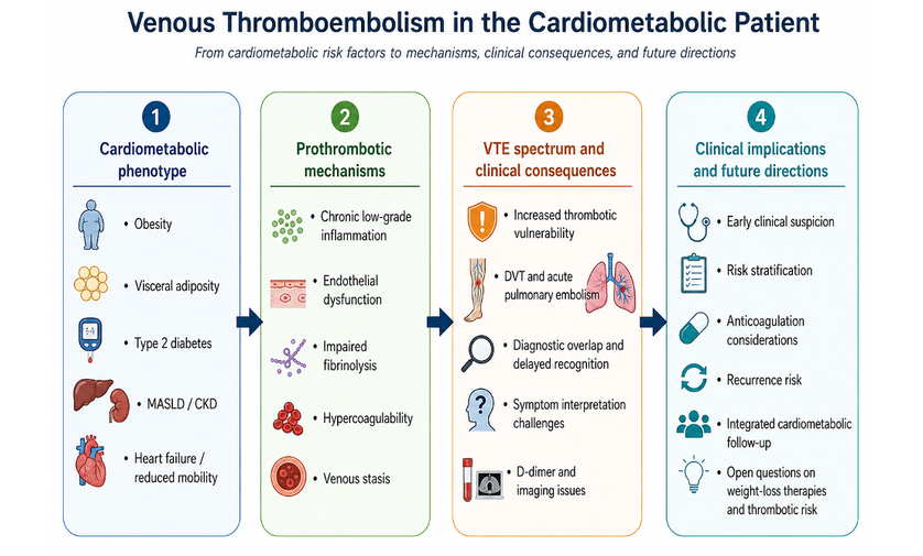
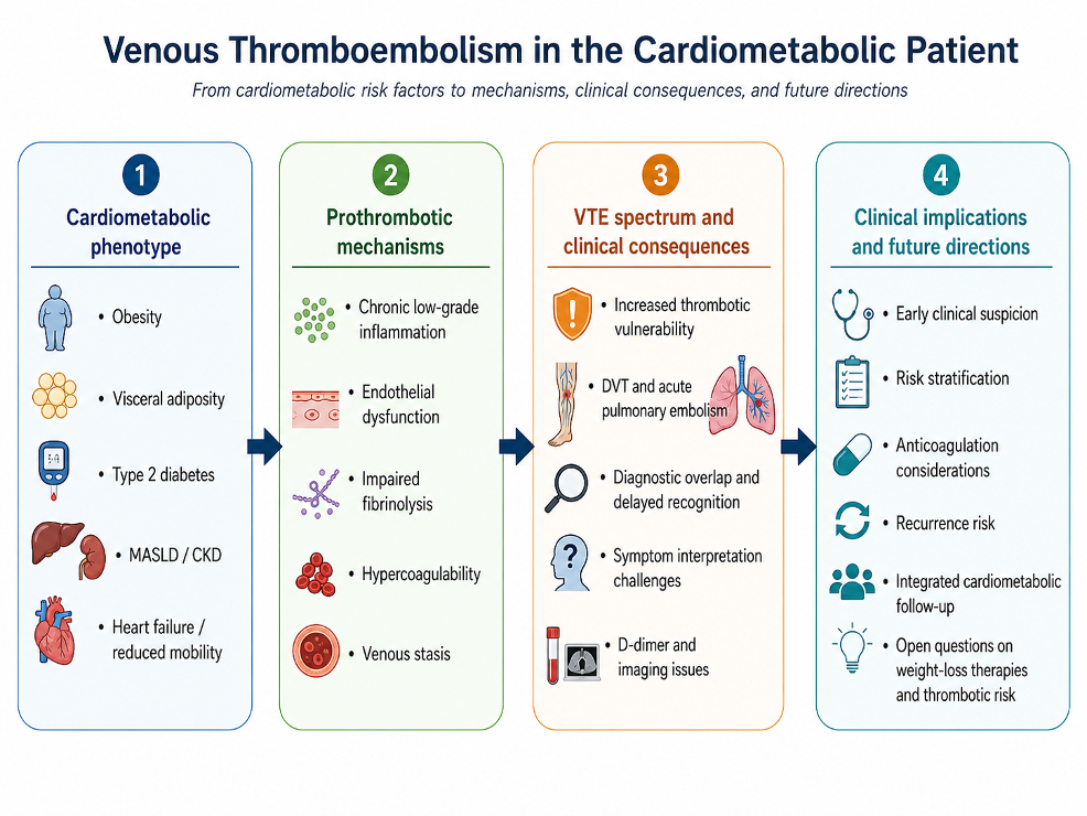
Venous thromboembolism (VTE), including deep vein thrombosis and pulmonary embolism, is increasingly recognized within the broader spectrum of cardiometabolic disease. Obesity, visceral adiposity, type 2 diabetes, chronic kidney disease, and metabolic dysfunction-associated steatotic liver disease may contribute to a prothrombotic milieu through inflammation, endothelial dysfunction, impaired fibrinolysis, hypercoagulability, and venous stasis. This entry summarizes the main pathophysiological links between cardiometabolic dysfunction and VTE, their clinical implications, and emerging areas of interest in risk stratification and management.
- venous thromboembolism
- pulmonary embolism
- obesity
- visceral adiposity
- type 2 diabetes
- MASLD
- hypercoagulability
1. IDefintroductition
Venous thromboembolism (VTE), comprising deep vein thrombosis and acute pulmonary embolism (PE), is traditionally classified as a disease entity distinct from atherothrombosis, with its own risk factors, its own diagnostic algorithms and its own therapeutic logic. In cardiometabolic medicine this separation has become progressively harder to defend. Patients with obesity, visceral adiposity, type 2 diabetes (T2D), chronic kidney disease and metabolic dysfunction-associated steatotic liver disease (MASLD) are routinely identified as high-risk for myocardial infarction, stroke and heart failure, yet the same biological terrain—chronic low-grade inflammation, endothelial dysfunction, impaired fibrinolysis, reduced mobility—also favours venous thrombosis [1]. The term venous thromboembolism in the cardiometabolic patient therefore designates not a distinct nosological entity but an interpretive frame: acute PE understood as a downstream expression of systemic metabolic and thrombo-inflammatory vulnerability rather than as an isolated vascular accident [1][2][3][1,2,3].
2. The Pprothrombotic Mmilieu
The modern cardiometabolic patient is defined by clustering rather than by a single diagnosis. Insulin resistance, dysglycaemia, dyslipidaemia, hypertension, renal dysfunction, hepatic steatosis and reduced exercise capacity coexist and reinforce one another. Viewed through the lens of venous thrombosis, this clustering is consequential because each arm of Virchow's triad is simultaneously affected [3][4][3,4].
Blood composition shifts toward hypercoagulability: adipose tissue and hepatic dysfunction increase circulating plasminogen activator inhibitor-1 (PAI-1), thrombin generation rises, tissue factor activity increases, and fibrinolytic capacity falls [4][5][4,5]. The vessel wall is affected through chronic inflammatory signalling, oxidative stress and endothelial activation, which convert the endothelium from an antithrombotic to a permissive surface [3,4]. Stasis, the third arm, is generated by mechanisms that are clinical rather than molecular: deconditioning, obesity-related limitation of mobility, venous insufficiency, recurrent hospitalisation and cardiorespiratory compromise [1][3][1,3].
A second, frequently underappreciated mechanism operates alongside the biological one. Cardiometabolic disease increases exposure to conventional thrombotic triggers—elective and bariatric surgery, orthopaedic procedures, immobilisation, acute medical admissions, polypharmacy. Excess adiposity therefore acts both as a direct biological driver and as a multiplier of situational risk, which helps explain why epidemiological signals persist across heterogeneous study designs [3][4][3,4].
3. Adiposity Bbeyond Body Mass Ibody mass index
Among cardiometabolic determinants, obesity carries the most robust epidemiological support. A dose-response meta-analysis of cohort studies including approximately four million participants demonstrated a significant linear relationship between body mass index (BMI) and the risk of both VTE and PE, indicating that excess adiposity is a graded contributor rather than a categorical one [6].
BMI, however, is a blunt instrument. It cannot distinguish metabolically quiescent excess weight from ectopic, centrally distributed fat, and this distinction matters because the inflammatory and insulin-resistant phenotype tracks with fat distribution rather than with total mass. Mechanistic work in morbid obesity showed that visceral adiposity is an independent determinant of hypercoagulability as measured by thrombin generation, implying that where fat is stored modifies coagulation independently of how much of it there is [7]. Imaging-based evidence extended this observation: visceral adipose tissue volume demonstrated a stronger association with VTE than BMI, suggesting that central adiposity captures thrombotic vulnerability more faithfully than anthropometric surrogates [8].
Adipokine data complete the picture. In the Multi-Ethnic Study of Atherosclerosis, lower adiponectin and higher leptin concentrations were associated with incident VTE, positioning adipose tissue as an endocrine organ relevant to venous thrombosis and not merely as a component of metabolic risk scoring [9].
The methodological implication is worth stating explicitly, because it recurs across cardiometabolic research: the strength and even the direction of an association may depend on which measure of adiposity is privileged. BMI provides the most reproducible epidemiological signal; visceral fat quantification and adipokine profiling provide the more biologically informative one. These are not competing answers to the same question but answers to different questions, and conflating them accounts for part of the apparent inconsistency in the literature.
4. Type 2 Ddiabetes as a Ggraded Mmodifier
The relationship between T2D and VTE is considerably more ambiguous than that of obesity, and this ambiguity is itself informative. Several meta-analyses reported an increased risk of VTE in people with diabetes [10][11][10,11], whereas others found that the association weakens or loses statistical significance after adjustment for confounders, particularly obesity [12].
Biological plausibility is not the limiting factor. Chronic hyperglycaemia, protein glycation, oxidative stress, endothelial injury, increased tissue factor expression and hypofibrinolysis all provide coherent pathways through which diabetes could promote venous thrombosis [5], and a nested case-control analysis has examined glycaemic control as a modifier of that risk [13]. The difficulty is epidemiological: patients with T2D who develop VTE frequently also have obesity, chronic kidney disease, restricted mobility, systemic inflammation or established cardiovascular disease, and these exposures are difficult to disentangle in observational data.
The defensible position is therefore not that diabetes is an independent cause of VTE, nor that it is irrelevant, but that it functions as a graded modifier of thrombotic vulnerability. Longer disease duration, poor glycaemic control and microvascular complications—diabetic kidney disease in particular—plausibly shift a patient toward a more unfavourable prothrombotic profile even where the pooled epidemiological signal remains inconsistent [12][13][14][12,13,14]. Framed this way, T2D identifies a patient in whom the threshold for thrombosis may be lower, the symptom burden more confusing and the consequences of an event more severe. Overclaiming a direct causal role weakens rather than strengthens the clinical argument.
5. MASLD as an Aamplifier
MASLD offers the most conceptually useful bridge between metabolic and haemostatic thinking, because the liver is the site where the two systems physically converge: it synthesises coagulation factors, natural anticoagulant proteins and the principal regulators of fibrinolysis. Steatotic liver disease is accordingly not a hepatic diagnosis appended to obesity and diabetes but a marker of multisystem metabolic dysfunction with direct haemostatic consequences [15].
Evidence indicates that patients with steatotic liver disease exhibit altered primary and secondary haemostasis, reduced fibrinolytic balance and a broader procoagulant profile [15,17]. Experimental and translational work in metabolic dysfunction-associated steatohepatitis supports enhanced venous thrombosis and hypercoagulability in both murine models and human samples [16], and increased hepatic production of procoagulant mediators, including PAI-1, provides a mechanistic explanation for amplification rather than mere co-occurrence [15][16][17][15,16,17].
The metabolic syndrome literature frames this within a longer trajectory. Early case-control work linked metabolic syndrome to unprovoked venous thrombosis [18], and a patient-level meta-analysis subsequently identified abdominal adiposity as the strongest single component while preserving relevance for the cluster as a whole [19][20][19,20]. Notably, metabolic syndrome has been associated with increased VTE recurrence after both acute PE and deep vein thrombosis, indicating that the metabolic terrain continues to matter after the index event [21][22][21,22].
6. Clinical Rrecognition and Mmanagement
Acute PE is difficult to recognise because its presentation—dyspnoea, chest discomfort, tachycardia, exercise intolerance, presyncope, hypoxaemia—is nonspecific [2]. In the cardiometabolic patient these symptoms are more likely to be misattributed, since obesity-related exertional breathlessness, heart failure, obstructive sleep apnoea, chronic lung disease, anaemia and deconditioning all supply plausible alternative explanations. The consequence is diagnostic delay rather than diagnostic complexity [1].
Biomarker interpretation is a second obstacle. D-dimer specificity falls in the presence of obesity, chronic inflammation and multimorbidity, increasing false-positive results and downstream imaging. Prospective data nonetheless indicate that age-adjusted D-dimer strategies remain safe in obese patients with suspected PE and improve efficiency relative to a fixed cut-off [23]. Computed tomography pulmonary angiography remains the reference modality but is technically more demanding in severe obesity, where body habitus and suboptimal contrast enhancement may degrade image quality; ventilation/perfusion imaging and lower-limb compression ultrasonography retain a role within a structured probability-based approach [2].
Prognostic tools such as the Pulmonary Embolism Severity Index require interpretation within a broader clinical assessment, since multimorbidity, impaired functional reserve and renal or respiratory limitation are incompletely captured by validated scores. Regarding anticoagulation, contemporary reviews and meta-analyses are broadly reassuring: direct oral anticoagulants show efficacy and safety at least comparable to warfarin in obese and morbidly obese patients, though high-quality randomised evidence at extremes of body weight remains limited [24][25][26][24,25,26]. Because diabetic kidney disease is common in this population, periodic reassessment of renal function is essential when selecting and dosing these agents [27].
7. Open Qquestions
Several questions remain unresolved and define the current research frontier.
The first concerns incretin-based therapy. Substantial and rapid weight loss achieved with GLP-1 receptor agonists and dual agonists alters adipose mass, adipokine profiles and, potentially, fibrinolytic balance, yet the net effect on venous thrombotic risk has not been prospectively quantified. The composition of the weight lost—the relative contribution of visceral fat versus lean mass—may determine whether the haemostatic benefit matches the anthropometric one. Cardiovascular outcome trials in this class have not adopted VTE as a prespecified endpoint, leaving the question structurally unanswered.
Second, pharmacokinetic interactions deserve attention. Delayed gastric emptying under incretin therapy may in principle affect the absorption of orally administered anticoagulants, and periprocedural suspension recommendations for these agents intersect with anticoagulation timing in ways that have not been formally studied.
Third, no validated risk-assessment model currently integrates cardiometabolic phenotype—visceral adiposity, hepatic steatosis, glycaemic control, renal function—into VTE prediction or into decisions about extended anticoagulation, despite evidence that the metabolic terrain influences recurrence [21][22][21,22]. Whether pharmacological or surgical resolution of MASLD modifies haemostatic parameters, and whether such modification translates into fewer clinical events, remains unknown.
[1][2][3][4][5][6][7][8][9][10][11][12][13][14][15][16][17][18][19][20][21][22][23][24][25][26][27]
References
- Labate AM, Villari P. Acute pulmonary embolism in the cardiometabolic patient. J Cardiol Cardiovasc Sci. 2026;7(2):17-21.
- Konstantinides SV, Meyer G, Becattini C, Bueno H, Geersing GJ, Harjola VP, Huisman MV, Humbert M, Jennings CS, Jiménez D, et al. 2019 ESC Guidelines for the diagnosis and management of acute pulmonary embolism developed in collaboration with the European Respiratory Society (ERS). Eur Heart J. 2020;41(4):543-603. doi:10.1093/eurheartj/ehz405.
- Pastori D, Cormaci VM, Marucci S, Franchino G, Del Sole F, Capozza A, Fallarino A, Corso C, Valeriani E, Menichelli D, et al. A comprehensive review of risk factors for venous thromboembolism: from epidemiology to pathophysiology. Int J Mol Sci. 2023;24(4):3169. doi:10.3390/ijms24043169
- Zawadzka PS, Imiela AM, Pruszczyk P. The interplay between obesity and venous thromboembolism: from molecular aspects to clinical issue. Int J Mol Sci. 2025;26(21):10292. doi:10.3390/ijms262110292.
- Panchagnula N, Lee LH, White RH. Hyperglycemia and venous thromboembolism. Res Pract Thromb Haemost. 2024;8:e70036.
- Rahmani J, Haghighian Roudsari A, Bawadi H, Thompson J, Khalooeifard R, Clark C, Ryan P, Ajami M, Rahimi Sakak F, Salehisahlabadi A, et al. Relationship between body mass index, risk of venous thromboembolism and pulmonary embolism: a systematic review and dose-response meta-analysis of cohort studies among four million participants. Thromb Res. 2020;192:64-72. doi:10.1016/j.thromres.2020.05.014.
- Chitongo PB, Roberts LN, Arya R. Visceral adiposity is an independent determinant of hypercoagulability as measured by thrombin generation in morbid obesity. TH Open. 2019;3(2):e146-e154.
- . Xiang R, Huang A, Stevens H, Singh M, Peter K, Takeuchi F, McFadyen JD. Visceral adipose tissue demonstrates a stronger association with venous thromboembolism than body mass index. J Thromb Haemost. 2025;23(9):2883-2889. doi:10.1016/j.jtha.2025.05.020.
- Broni EK, Ogunmoroti O, Quispe R, Sweeney T, Varma B, Fashanu OE, Lutsey PL, Allison M, Szklo M, Ndumele CE, et al. Adipokines and incident venous thromboembolism: the Multi-Ethnic Study of Atherosclerosis. J Thromb Haemost. 2023;21(2):303-310. doi:10.1016/j.jtha.2022.11.012.
- Bai J, Ding X, Du X, Zhao X, Wang Z, Ma Z. Diabetes is associated with increased risk of venous thromboembolism: a systematic review and meta-analysis. Thromb Res. 2015;135(1):90-95. doi:10.1016/j.thromres.2014.11.003.
- Ding C, Guo C, Du D, Gong X, Yuan Y. Association between diabetes and venous thromboembolism: a systematic review and meta-analysis. Medicine (Baltimore). 2023;102(42):e35329. doi:10.1097/MD.0000000000035329.
- Bell EJ, Folsom AR, Lutsey PL, Selvin E, Zakai NA, Cushman M, Alonso A. Diabetes mellitus and venous thromboembolism: a systematic review and meta-analysis. Diabetes Res Clin Pract. 2016;111:10-18. doi:10.1016/j.diabres.2015.10.019.
- Charlier SHR, Meier C, Jick SS, Meier CR, Becker C. Association between glycemic control and risk of venous thromboembolism in diabetic patients: a nested case-control study. Cardiovasc Diabetol. 2022;21(1):2. doi:10.1186/s12933-021-01432-1.
- Marx N, Federici M, Schütt K, Müller-Wieland D, Ajjan RA, Antunes MJ, Christodorescu RM, Crawford C, Di Angelantonio E, Eliasson B, et al. 2023 ESC Guidelines for the management of cardiovascular disease in patients with diabetes. Eur Heart J. 2023;44(39):4043-4140. doi:10.1093/eurheartj/ehad192.
- Spinosa M, Stine JG. Nonalcoholic fatty liver disease—evidence for a thrombophilic state? Curr Pharm Des. 2020;26(10):1036-1044. doi:10.2174/1381612826666200131101553
- Pandey N, Anand SK, Kaur H, Richard KSE, Chandaluri L, Butler ME, Zhang X, Pearson-Gallion B, Rohilla S, Das S, et al. Enhanced venous thrombosis and hypercoagulability in murine and human metabolic dysfunction-associated steatohepatitis. J Thromb Haemost. 2024;22(12):3572-3580. doi:10.1016/j.jtha.2024.08.023.
- González-Mendoza DE, Fernández-Nogueira F, Uribe M, Chávez-Tapia NC, Nuño-Lámbarri N. Hemostatic alterations in metabolic dysfunction-associated steatotic liver disease (MASLD) and their link to venous thromboembolism (VTE). Thromb Res. 2025;253:109395. doi:10.1016/j.thromres.2025.109395.
- Ageno W, Prandoni P, Romualdi E, Ghirarduzzi A, Dentali F, Pesavento R, Crowther M, Venco A. The metabolic syndrome and the risk of venous thrombosis: a case-control study. J Thromb Haemost. 2006;4(9):1914-1918. doi:10.1111/j.1538-7836.2006.02132.x.
- . Ageno W, Di Minno MND, Ay C, Jang MJ, Hansen JB, Steffen LM, Vayà A, Rattazzi M, Pabinger I, Oh D, et al. Association between the metabolic syndrome, its individual components, and unprovoked venous thromboembolism: results of a patient-level meta-analysis. Arterioscler Thromb Vasc Biol. 2014;34(11):2478-2485. doi:10.1161/ATVBAHA.114.304085.
- Di Minno MND, Tufano A, Guida A, Di Capua M, De Gregorio AM, Cerbone AM, Tarantino G, Di Minno G. Abnormally high prevalence of major components of the metabolic syndrome in subjects with early-onset idiopathic venous thromboembolism. Thromb Res. 2011;127(3):193-197.
- Stewart LK, Kline JA. Metabolic syndrome increases risk of venous thromboembolism recurrence after acute pulmonary embolism. Ann Am Thorac Soc. 2020;17(7):821-828. doi:10.1513/AnnalsATS.201907-518OC.
- Stewart LK, Kline JA. Metabolic syndrome increases risk of venous thromboembolism recurrence in patients with deep vein thrombosis. Blood Adv. 2020;4(1):127-135. doi:10.1182/bloodadvances.2019000561.
- Gaugler JO, Righini M, Robert-Ebadi H, Sanchez O, Roy PM, Verschuren F, Miranda S, Delluc A, Le Gal G, Tritschler T. Obesity as a predictor for pulmonary embolism and performance of the age-adjusted D-dimer strategy in obese patients with suspected pulmonary embolism. Thromb Haemost. 2024;124(1):49-57. doi:10.1055/s-0043-57018.
- Sorodoc V, Asaftei A, Ceasovschih A, Lionte C, Crisan S, Constantin M, Indrei L, Sorodoc L. Anticoagulation approach in morbid obesity: a comprehensive review on venous thromboembolism management. Front Pharmacol. 2024;15:1457280. doi:10.3389/fphar.2024.1457280.
- Talerico R, Pola R, Klok FA, Huisman MV. Direct-acting oral anticoagulants in patients at extremes of body weight: a review of pharmacological considerations and clinical implications. TH Open. 2024;8(1):e31-e41. doi:10.1055/s-0043-1776989.
- Karakasis P, Ktenopoulos N, Pamporis K, Sagris M, Soulaidopoulos S, Gerogianni M, Leontsinis I, Giannakoulas G, Tousoulis D, Fragakis N, et al. Efficacy and safety of direct oral anticoagulants versus warfarin in obese patients (BMI ≥30 kg/m²) with atrial fibrillation or venous thromboembolism: an updated systematic review and meta-analysis. J Clin Med. 2024;13(13):3784. doi:10.3390/jcm13133784.
- Elenjickal EJ, Mavrakanas T, Brodsky S, Bansal N. Anticoagulation in patients with chronic kidney disease. Semin Nephrol. 2024;44(1):151443.