Your browser does not fully support modern features. Please upgrade for a smoother experience.
Please note this is a comparison between Version 2 by Lindsay Dong and Version 1 by Nathan Oesch.
Human language and social cognition are two key disciplines that have traditionally been studied as separate domains. Nonetheless, an emerging view suggests an alternative perspective. Drawing on the theoretical underpinnings of the social brain hypothesis (thesis of the evolution of brain size and intelligence), the social complexity hypothesis (thesis of the evolution of communication), and empirical research from comparative animal behavior, human social behavior, language acquisition in children, social cognitive neuroscience, and the cognitive neuroscience of language, it is argued that social cognition and language are two significantly interconnected capacities of the human species.
- social brain
- language brain
- social brain hypothesis
- social complexity hypothesis for animal communication
- social bonding
1. Introduction
Human beings are an incredibly sophisticated species: technologically, scientifically, and cerebrally—why is this? The social brain hypothesis posits that the cognitive pressures of residing in dynamic animal societies, selected for increases in the volume of the primate brain, explain the atypically large brains of a number of anthropoid primates [1][2][3]. The initial data for this thesis came primarily from the discovery that neocortex size correlates with the size of social groupings for a variety of anthropoid primates, including humans.
A simple extension of the social brain hypothesis, known as the social complexity hypothesis for animal communication (more informally, the social bonding hypothesis), postulates that species with complex social groupings demand complex communication systems to manage the complex social dynamics involved and promote social bonds [5,6][4][5]. The original findings in support of this thesis came from a comparative investigation of several dozen species of anthropoid primates, where both the average size of the social group and average grooming duration (as a standard metric of social bonds) were correlated with the size of the vocal repertoires of these primates.
Intriguingly, further support for the social bonding evolutionary function of human language, arguably the most complex of all primate communication systems, arises from sociolinguistic analyses of human conversational behavior. In particular, studies of conversational semantics reveal that, both in traditional cultures and industrial societies, gossip concerns predominate in typical dialogue, encompassing nearly 70% of daily dialogue time [11,12][6][7]. Moreover, further analyses have shown that the spread of information, relevant to personal reputation via gossip, facilitates prosocial behavior by encouraging mindful acquaintance choice when circumstances necessitate collaboration [13][8].
In summary, the subsequent analysis examines the relationship between social cognition and language acquisition within the framework of the social brain hypothesis. This hypothesis posits that specific brain regions form a dedicated neural network for processing social information, playing a crucial role in both understanding and interacting with other individuals. Crucially, deficits within this social brain network may contribute to the language impairments observed in autism spectrum disorder (ASD). For instance, evidence will be highlighted linking mentalizing abilities—a core function of the social brain—to the development of complex syntactic structures in both typical and atypical language development.
2. The Social Brain and Social Cognitive Neuroscience
Despite the clear anthropological and evolutionary connection between social cognition, social behavior, and the social brain, described above, this framework has not yet been fully integrated into our current understanding of social cognitive neuroscience. In truth, the intricacies of the neurological computations that underpin group living in primates are substantial, including activities like coalition formation, tactical deception, organizing grooming cliques, social play, and social learning [1]. In humans, a complex network of brain regions underlies important social activities, including the recognition and cognitive processing of social signals, recognizing faces, evaluating mental states (i.e., mentalizing or theory of mind), perceiving emotions, sharing attention, determining friends from foes, evaluating others’ perceptions and beliefs, social learning, relationship formation, and social bonding [1][4][59]. In a preliminary, noteworthy model of the social brain in the 1990s, neuroscientist Leslie Brothers [18][10] highlighted the contributions of the amygdala, orbitofrontal cortex (OFC), superior temporal sulcus (STS), and fusiform gyrus (FFG) to social information processing. More recently, functional magnetic resonance imaging (fMRI) has provided additional recognition of an interconnected network of regions joining the parietal and temporal brain lobes to the prefrontal brain lobes [4][11].Broadly speaking, the OFC is implicated in social reinforcement and social reward processing [4][612]. More specifically, the STS region, particularly the right-hemisphere posterior STS (pSTS) area, processes biological motion signals, like the hand, eye, and salient motions of the body, to predict and interpret the intentions and behaviors of other agents [4][612]. In addition to this area, the right inferior temporal gyrus, fusiform gyrus, right parietal lobule, and middle temporal gyrus in each hemisphere are differentially activated by processing the direction of gaze [4][612]. Moreover, the default mode network (DMN)—comprised of the dorsal medial prefrontal cortex (mPFC), posterior cingulate cortex, precuneus, angular gyrus, and, occasionally, right temporoparietal junction (rTPJ)—which is known for activation when an individual is unfocused on the external world and the brain is at conscious rest, further appears to be active when an individual is thinking about the self, the past and future, and most intriguingly, evaluating the mental states of other people (i.e., mentalizing or theory of mind) [7]. Further, much work now reveals that the social brain hypothesis explains not only variation in the volume of the brain between various primates, but also individual differences in the volume of the brain in humans, in regard to several different features of human social networking and social cognition. In particular, the volume of gray matter in the OFC, ACC, ventromedial prefrontal cortex (vmPFC), amygdala, and STS are associated with individual differences in higher-order intentionality capacity (i.e., advanced mentalizing or theory of mind) and social network size [8][913][1014]. Lastly, recent studies of ‘mirror neurons’—neurons in the brain that activate when an organism acts, as well as when the same organism observes this same action done by another—have been postulated to be integral for mentalizing or theory of mind, language, empathy, comprehending the intentions and acts of agents, and imitative learning [10][11][12]. In other words, studies of the default mode neural network, especially, in studies of adult monkeys, suggest that observing an action and producing the same action oneself are neurally equivalent, and, at least in monkeys, this capacity appears to occupy a role in social comprehension and imitation [10][11][12]. Though mirror neurons of the brain have been observed directly in non-human primates—most notably, in macaques—in humans, brain activity merely consistent with mirror neurons has been found in the primary somatosensory cortex, inferior and superior parietal lobes, inferior frontal cortex, premotor cortex, and supplementary motor region [1315].
3. The Social Brain and Cognitive Neuroscience of Language
In a similar fashion, despite the clear anthropological and evolutionary connection between the social brain and social communication, as described above, this framework has not yet been fully integrated into our current understanding of the cognitive neuroscience of human language [1416][1517]. Perhaps most critically, a complex neurological system of communication—for regulating interactions and social bonding with important members of the group—appears to be crucial for many non-human primates, including human social relationships [1618]. In humans, a complex network of brain regions underlies the processing of language, including speech comprehension and production, and substantive integration with the social brain, including social-semantic working memory, and encompassing regulation from the neural network level to the neurotransmitter level, including social neurotransmitters such as oxytocin, endorphins, and dopamine [17][18][19][20][21][22][23]. In an influential and noteworthy model of the cognitive neuroscience of language, Pierre Paul Broca determined in 1861 that language processing areas are located primarily in the left cerebral hemisphere of the brain [2224]. In later years, much research, including neuroanatomical analyses by Geschwind and Galaburda, further suggested left hemisphere dominance in brain areas dedicated to language [2325][2426], including myelinated axons and larger pyramidal neurons in the left hemisphere, allowing for more rapid and efficient processing of linguistic information [2527][2628]. Nonetheless, more recent work has further shown that additional areas, including the putamen, caudate nucleus, and internal capsule appear to play additional roles in language processing [49][29], while very young children also show significant activity in the inferior frontal and superior temporal regions of the right cerebral hemisphere—homologs of traditional left cerebral hemisphere language areas—with an activation profile in the right cerebral hemisphere that appears to diminish with age [50][30]. Intriguingly, homologous brain regions of Broca’s area and Wernicke’s area have also been discovered in the brains of social, group-adapted, nonhuman primates, strongly suggesting a shared evolutionary or phylogenetic history [51,52][31][32]. Though their function in nonhuman primates is poorly understood, an evolutionary perspective would suggest that they are probably central to nonhuman primate vocalization processing, in ways similar to human language processing [53,54,55,56,57,58,59][33][34][35][36][37][38][39].4. The Social Brain and First Language Acquisition
4.1. Social Signals That Facilitate Early Language Acquisition
Social interaction skills, including play, reading, reference, or joint attention between an infant or child and parent or guardian to an outward thing, and the face-to-face interactions involved in speaking in natural language environments, crucially aid the early acquisition of language (see Figure 51; [1820][2740][2841][2942][3043][3144][3245]). In particular, infant-directed speech (IDS) and child-directed speech (CDS), or the face-to-face communication cues between an infant or child and parent or guardian, aid language acquisition by delivering relevant social signals (e.g., gestures, facial and emotional expressions, and directed eye-gaze), provoking infant attention, and emphasizing important pragmatic signals. Crucially, social interaction appears to impact the development of both speech perception and comprehension [3329], as well as speech production learning [3430][3531][3632].
Figure 51. Social interaction skills, including play, reading, joint attention, and the face-to-face interactions involved in infant-directed speech (IDS) in natural language environments crucially aid the early acquisition of language. IDS aids language acquisition by providing relevant social signals (e.g., gestures, facial and emotional expressions, and directed eye gaze) that provoke infant attention and emphasize important pragmatic signals. Adapted image from the public domain.
Several important developments accompany the capacity to understand reference and joint attention of an infant and parent to an outward thing or object [68][46]. By 9 months of age, youngsters start to participate in individual–object–individual triadic activities in which interest is devoted to objects using gaze, which provokes attention from other individuals, also known as joint attention [65,66][47][48]. This shared perception of communicative intentions is likely to be critical for the infant’s learning of language [65[47][48][49],66,85], as well as understanding others as intentional agents [65,86][47][50].
In addition, the quality and quantity of speech stimuli (e.g., vocabulary diversity, amount of word units, and mean length of utterance (MLU)) are further associated with infant vocabulary growth [87,88,89][51][52][53]. Unfortunately, while most language acquisition research has been conducted on families of high socioeconomic status (SES), infants raised in poorer communities with multiple challenges can affect caregiver interactions, leading to greater variability in language abilities [90,91][54][55]; although, see [92][56] for a recent alternative perspective.
4.2. Infant-/Child-Directed Speech and Face-to-Face Communication
Infant-directed speech (IDS) and child-directed speech (CDS) are intrinsically multimodal, and many nonverbal social signals are present during this sort of communication (e.g., gestures, facial and emotional expressions, and directed eye gaze; see Figure 51). Previous research has shown that directed eye gaze is a key form of nonverbal communication, as it facilitates language acquisition in several regards, including language processing, development of vocabulary, and perceptual mapping of form-to-object [3733][3857][3935]. For instance, gaze following and directed eye contact provoke arousal and attention by emphasizing important social stimuli and facilitating the infant’s or child’s social engagement [4036][4137]. Directed gaze as a tool for language learning is typically distinguished by an early developmental trajectory where infants display a proclivity for open eyes on upright faces, involving the specialization of areas of the cortex associated with gaze processing [97,108,109,110,111][58][59][60][61][62]. Newborns develop the capacity for gaze following beginning from 3–4 months, becoming a consistent communication signal from 6–8 months of age [108,110][63][61]. However, it is not until 9–12 months that directed gaze begins to become an important tool used for indicating reference, facilitating language acquisition by providing directed eye gaze signaling [64,112,113,114,115][64][65][66][67][68]. Quality and quantity of speech during interactions are also significant factors in language learning, especially the growth of vocabulary [4238][4369][4446][4570]. For instance, studies have shown the quantity of child-directed speech at 18 months predicts vocabulary growth at 2 years [4671]. Parental engagement, namely, vocal reactions to infant vocalizations with either words or vowels, quickly influences infant vocal productions, as newborns start to assimilate phonological sound patterns spoken by the parent, facilitating the acquisition of new vocalizations [4772]. Child–parent social interactions are further affected by a number of environmental factors, like socioeconomic status (SES). More specifically, SES affects both the quantity and quality of parental speech stimuli [4238][4873]; for instance, children of low-SES families tend to display more sluggish real-time effectiveness of linguistic processing and subsequent growth of vocabulary [4974].5. The Social Brain, Cognitive Neuroscience of Language, and First Language Acquisition
In light of the significance of child–parent interactions for acquiring language, neuroimaging research has recently begun to investigate how just this sort of communication may impact the developing brain. As previously discussed, studies have found that, during early maturation, the quantity of linguistic information, as calculated by infant exposure to the number of adult words, is strongly predictive of myelin in white matter association tracts related to adult language abilities—especially the left arcuate fasciculus (AF) and superior longitudinal fasciculus (SLF) in younger children at 30 months of age—as well as youngster’s developing linguistic abilities [5075]. On the other hand, the quality of linguistic information—word richness, dialogue experience, and mean length of utterance (MLU)—appears to be more crucial for older youth 4–6 years of age, who show greater white matter connectivity involving left AF and SLF [5176][5277] and greater cortical volume in the left inferior frontal gyrus (IFG) and supramarginal gyri [5378], as well as older children 5–9 years of age, who show increased cortical areas in the left perisylvian areas [5447]. Additional social cognitive neuroscience studies have revealed that the neural circuits underpinning the discrimination of a mother’s voice—also known as ‘motherese’, as an important component of social bonding—include voice-perception and auditory areas of the temporal lobe, reward circuit areas in the orbitofrontal cortex (OFC), nucleus accumbens (NAc), and ventromedial prefrontal cortex (vmPFC), affective processing areas, especially the amygdala, and areas related to visual face processing, especially the fusiform cortex, predict the communication and linguistic function capacities in older youth at 7–12 years of age [1719]. That said, functional magnetic resonance imaging (fMRI) work on social interactions in youngsters has, until recently, primarily centered on brain activation in infants or children in reaction to a one-way social signal. However, a newly utilized technique, known as ‘hyperscanning’, allows for concurrent data collection of brain activation from multiple individuals at once, concurrently taking part in social interaction [144][79]. More specifically, real-time social interactions between a child and parent can be correlated with the temporal alignment of their brainwaves during such interactions. In particular, as a consequence of non-verbal and verbal signaling during social interaction, neural synchronization can occur [145,146][80][81]. Further, in at least one recent neuroimaging study, of a live two-way social interaction involving differences in speech prosody, eye gaze, and joint attention between adults and infants 9–15 months of age, distinctive paired activation occurred in infant and adult brains as a function of their social importance.6. The Social Brain and Second Language Acquisition
A relatively more recent aggregation of work has further explored the influence of bilingualism on mentalizing or perspective-taking, as well as empathy, on language processing in young children. For instance, at least one recent study found bilingual-speaking youngsters to be more accurate than monolingual speakers in a task that required analyzing an observer’s perspective from different positions [5548]. Moreover, a recent meta-analysis appears to indicate these general findings are robust [5649]. Though it is not fully understood how bilingualism provides this advantage, it has been suggested that bilingualism perhaps allows for additional occasions to develop executive function, metalinguistic comprehension, and improved sensitivity to the nuances of typical sociolinguistic interactions [5548][5649].7. The Social Brain, Developmental Dysfunctions, and Psychopathologies
Over the last few decades, increasing numbers of psychologists and neuroscientists have come to understand that many psychopathologies and developmental disorders can be largely attributed to dysfunctions of the evolved social brain [5750]. In the majority of cases, such dysfunctions typically involve substantive deficiencies in social cognition, social communication, and linguistic abilities. In particular, autism spectrum disorder (ASD) is a heterogeneous disruption of social cognition, generally entailing various social deficits, such as dysfunctions in social communication (e.g., atypical facial expressions and vocal tone), social interactions (e.g., joint attention, eye gaze, and gesture), imitation and social norms, mentalizing, empathy, analogies (e.g., sarcasm and jokes), unfamiliar situations, imagination (e.g., make believe or play), and planning for or predicting future events [5882]. Intriguingly, due to the profound neurogenetic and neurodevelopmental causes, as well as serious dysfunctions in social cognition that define ASD, ASD presents the occasion for neuroscientists, anthropologists, and psychologists to investigate the biological genesis of social cognition and social behaviors inherent to human nature.8. The Social Brain and Autism Spectrum Disorder
Autism spectrum disorder (ASD) is a neurodevelopmental dysfunction characterized by chronic deficits in social interaction, non-verbal and verbal communication, and social cognition, including deficits in mentalizing or the ability to understand the mental states of another individual [5952][6053]. Intriguingly, the complex interrelated genetic, social, and neurodevelopmental pathways and deficits found in ASD, present perhaps one of the clearest and most compelling connections between the social brain, language function, social cognition, and social bonding [612]. As the name suggests, autism is situated on a spectrum, with some individuals whose verbal capacities exist along the typical spectrum of abilities, while others never learn to speak [6154]. Interestingly, in those with adequate language and cognitive capacities, such as those with Asperger’s syndrome and high-functioning autism (HFA), specifically social communicative capacities ostensibly remain impaired. In other words, communication is typically unidirectional and used instrumentally and non-socially instead of for socially related functions [6255]. Neurological studies on cortical development in language-related areas of the frontal and temporal lobes of the brain have been further correlated with linguistic impairments in ASD, including asymmetrical turnaround of the frontal lobes [6383][64][6558], superior and anterior shifting of the left cerebral hemisphere, superior temporal sulcus, and inferior frontal sulcus [6684], bilateral decreases of gray matter volume in the superior temporal sulcus [6785], and apparently overall reduced left hemispheric dominance. Intriguingly, though challenging to disentwine the respective contributions of social cognition deficits in autism to linguistic deficits in autism, several recent studies in both autistic and neurotypical adults and children appear to suggest that mentalizing, which is impaired in autistic individuals, may be integral for the cognitive and linguistic ability to build subordinate and recursive embedded clauses (e.g., ‘‘Mary thinks that Sandra believes the broom is in the closet’’) (see Figure 62; [6886][6963][7060]), suggesting another direct link between social cognition and language ability.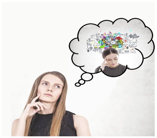
Figure 62. Several studies of both autistic and neurotypical adults and children appear to suggest that higher-order mentalizing (i.e., inferring the mental states of more than one individual) may be important for the syntactic ability to build subordinate and recursive embedded clauses (e.g., “Mary thinks that Sandra believes the broom is in the closet”), suggesting a direct link between social cognition and language ability. Adapted image from the public domain.
9. Early Biomarkers of Language-Related Abilities and Relevant Clinical Applications
Describing the early development of neurotypical and neuroatypical language neurobiology is critical for the early identification and potential treatment of clinical language disorders. Crucially, delays in language and speech in infants and children can negatively affect important social and academic skills such as attention, reading, writing, social interactions, and, of course, later educational outcomes [7161]. For instance, delays in language acquisition from 2–5 years are implicated in substandard reading comprehension in the classroom [7262][7365]. If such language delays persist after 5 years, related challenges often persist in the consequent maturation of attention, directed eye gaze, and socialization [7161][7466]. The majority of language delays are often noticed during parental observations or clinical check-ups when an important developmental landmark does not appear to be present, like syntactic challenges or speech onset delays. As a consequence of this rather crude ‘sit-and-wait’ approach, most youngsters are unfortunately not characterized as having had a disorder or delay of language until 2–3 years of age, which is often noted by the absence of combinatorial speech, or the capacity to formulate words into complete thoughts and sentences [7161][7587].
An alternative approach emphasizes the emergence of early indications, or biomarkers, of ultimate language capacities, early enough in development, to establish that any clinical interventions into speech and language delays and disorders might provide the greatest benefits. Perhaps surprisingly, there are currently no standardized or universally agreed-upon criteria in screening for language and speech deficiencies.
As might be expected, the diagnosis of language delays and disorders is usually grounded in comparable maturational landmarks observed in neurotypical language learning [175][88]. Children with language delays typically adhere to a normal maturational trajectory, albeit at more sluggish rates than would be expected [176][89], whereas children with language disorders tend to display regressions in language development (e.g., word loss from 14–21 months of age in ASD), serious and persistent delays in language learning (e.g., challenges with syntax in youngsters with specific language impairment (SLI), or impairments in at least two domains of development (e.g., such as motor function and language impairments in global developmental delay (GDD) [175,177,178][88][90][91]. As a general rule-of-thumb, language delays typically require clinical intervention when the development rate drops beneath 3/4 of the rate expected, for example, when a standard developmental landmark typically observed at 2 years of age fails to be met in a youngster at 30 months of age [179][92].
Nonetheless, speech and linguistic interventions should arguably begin even earlier in development. In fact, speech processing already begins in utero, in spite of the fact that the more observable first 24 months are distinguished by more obvious mappings of form-to-meaning at 5–7 months of age and proficiency at distinguishing native sounds from 6–12 months of age [7693].
References
- Dunbar, R.I.M.; Shultz, S. Evolution in the social brain. Science 2007, 317, 1344–1347.
- Semendeferi, K.; Armstrong, E.; Schleicher, A.; Zilles, K.; Van Hoesen, G.W. Prefrontal cortex in humans and apes: A comparative study of area. Am. J. Phys. Anthropol. 2001, 114, 224–241.
- Semendeferi, K.; Lu, A.; Schenker, N.; Damasio, H. Humans and great apes share a large frontal cortex. Nat. Neurosci. 2002, 5, 272–276.
- Freeberg, T.M.; Dunbar, R.I.M.; Ord, T.J. Social complexity as a proximate and ultimate factor in communicative complexity. Philos. Trans. R. Soc. B Biol. Sci. 2012, 367, 1785–1801.
- Oesch, N. Music and language in social interaction: Synchrony, antiphony and functional origins. Front. Psychol. 2019, 10, 1514. Oesch, N. Music and language in social interaction: Synchrony, antiphony and functional origins. Front. Psychol. 2019, 10, 1514.
- Dahmardeh, M.; Dunbar, R.I.M. What shall we talk about in Farsi?: Content of everyday conversations in Iran. Hum. Nat. 2017, 28, 423–433. Dahmardeh, M.; Dunbar, R.I.M. What shall we talk about in Farsi?: Content of everyday conversations in Iran. Hum. Nat. 2017, 28, 423–433.
- Dunbar, R.I.M.; Marriott, A.; Duncan, N.D. Human conversational behavior. Hum. Nat. 1997, 8, 231–246.
- Feinberg, M.; Willer, R.; Shultz, M. Gossip and ostracism promote cooperation in groups. Psychol. Sci. 2014, 25, 656–664.
- Adolphs, R. The social brain: Neural basis of social knowledge. Annu. Rev. Psychol. 2009, 60, 693–716. Oesch, N. Music and language in social interaction: Synchrony, antiphony and functional origins. Front. Psychol. 2019, 10, 1514.
- Dunbar, R.I.M. The social brain hypothesis and its implications for social evolution. Ann. Hum. Biol. 2009, 36, 562–572. Brothers, L. The social brain: A project for integrating primate behavior and neurophysiology in a new domain. Concepts Neurosci. 1990, 1, 27–51.
- Brothers, L. The social brain: A project for integrating primate behavior and neurophysiology in a new domain. Concepts Neurosci. 1990, 1, 27–51. Oesch, N. Social brain hypothesis. In International Encyclopedia of Anthropology; Callan, H., Ed.; John Wiley and Sons: New York, NY, USA, 2018; pp. 1–11.
- Oesch, N. Social brain hypothesis. In International Encyclopedia of Anthropology; Callan, H., Ed.; John Wiley and Sons: New York, NY, USA, 2018; pp. 1–11. Dahmardeh, M.; Dunbar, R.I.M. What shall we talk about in Farsi?: Content of everyday conversations in Iran. Hum. Nat. 2017, 28, 423–433.
- Johnson, M.H.; Griffin, R.; Csibra, G.; Halit, H.; Farroni, T.; de Haan, M.; Tucker, L.A.; Baron-Cohen, S.; Richards, J. The emergence of the social brain network: Evidence from typical and atypical development. Dev. Psychopathol. 2005, 17, 599–619. Adolphs, R. The social brain: Neural basis of social knowledge. Annu. Rev. Psychol. 2009, 60, 693–716.
- Buckner, R.L.; Andrews-Hanna, J.R.; Schacter, D.L. The brain’s default network: Anatomy, function, and relevance to disease. Ann. N. Y. Acad. Sci. 2008, 1124, 1–38. Dunbar, R.I.M. The social brain hypothesis and its implications for social evolution. Ann. Hum. Biol. 2009, 36, 562–572.
- Lewis, P.A.; Birch, A.; Hall, A.; Dunbar, R.I.M. Higher order intentionality tasks are cognitively more demanding. Soc. Cogn. Affect. Neurosci. 2017, 12, 1063–1071. Johnson, M.H.; Griffin, R.; Csibra, G.; Halit, H.; Farroni, T.; de Haan, M.; Tucker, L.A.; Baron-Cohen, S.; Richards, J. The emergence of the social brain network: Evidence from typical and atypical development. Dev. Psychopathol. 2005, 17, 599–619.
- Kwak, S.; Joo, W.; Youm, Y.; Chey, J. Social brain volume is associated with in-degree social network size among older adults. Proc. R. Soc. B 2018, 285, 20172708. Buckner, R.L.; Andrews-Hanna, J.R.; Schacter, D.L. The brain’s default network: Anatomy, function, and relevance to disease. Ann. N. Y. Acad. Sci. 2008, 1124, 1–38.
- Von Der Heide, R.; Vyas, G.; Olson, I.R. The social network-network: Size is predicted by brain structure and function in the amygdala and paralimbic regions. Soc. Cogn. Affect. Neurosci. 2014, 9, 1962–1972. Lewis, P.A.; Birch, A.; Hall, A.; Dunbar, R.I.M. Higher order intentionality tasks are cognitively more demanding. Soc. Cogn. Affect. Neurosci. 2017, 12, 1063–1071.
- Meltzoff, A.N.; Decety, J. What imitation tells us about social cognition: A rapprochement between developmental psychology and cognitive neuroscience. Philos. Trans. R. Soc. Lond. B Biol. Sci. 2003, 358, 491–500. Kwak, S.; Joo, W.; Youm, Y.; Chey, J. Social brain volume is associated with in-degree social network size among older adults. Proc. R. Soc. B 2018, 285, 20172708.
- Rizzolatti, G. The mirror neuron system and imitation. In Perspectives on Imitation: From Neuroscience to Social Science—I: Mechanisms of Imitation and Imitation in Animals; Hurley, S., Chater, N., Eds.; MIT Press: Cambridge, UK, 2005; pp. 55–76. Von Der Heide, R.; Vyas, G.; Olson, I.R. The social network-network: Size is predicted by brain structure and function in the amygdala and paralimbic regions. Soc. Cogn. Affect. Neurosci. 2014, 9, 1962–1972.
- Kilner, J.M.; Lemon, R.N. What we know currently about mirror neurons. Curr. Biol. 2013, 23, R1057–R1062. Meltzoff, A.N.; Decety, J. What imitation tells us about social cognition: A rapprochement between developmental psychology and cognitive neuroscience. Philos. Trans. R. Soc. Lond. B Biol. Sci. 2003, 358, 491–500.
- Adolphs, R. Cognitive neurosciences of human social behavior. Nat. Rev. Neurosci. 2003, 4, 165–178. Rizzolatti, G. The mirror neuron system and imitation. In Perspectives on Imitation: From Neuroscience to Social Science—I: Mechanisms of Imitation and Imitation in Animals; Hurley, S., Chater, N., Eds.; MIT Press: Cambridge, UK, 2005; pp. 55–76.
- Pulvermuller, F. Brain mechanisms linking language to action. Nat. Rev. Neurosci. 2005, 6, 574–582. Kilner, J.M.; Lemon, R.N. What we know currently about mirror neurons. Curr. Biol. 2013, 23, R1057–R1062.
- Abrams, D.A.; Chen, T.; Odriozola, P.; Cheng, K.M.; Baker, A.E.; Padmanabhan, A.; Ryali, S.; Kochalkaa, J.; Feinstein, C.; Menon, V. Neural circuits underlying mother’s voice perception predict social communication abilities in children. Proc. Natl. Acad. Sci. USA 2016, 113, 6295–6300. Adolphs, R. Cognitive neurosciences of human social behavior. Nat. Rev. Neurosci. 2003, 4, 165–178.
- Li, P.; Jeong, H. The social brain of language: Grounding second language learning in social interaction. Npj Sci. Learn. 2020, 5, 8. Pulvermuller, F. Brain mechanisms linking language to action. Nat. Rev. Neurosci. 2005, 6, 574–582.
- Rauchbauer, B.; Nazarian, B.; Bourhis, M.; Ochs, M.; Prévot, L.; Chaminade, T. Brain activity during reciprocal social interaction investigated using conversational robots as control condition. Philos. Trans. R. Soc. Lond. B Biol. Sci. 2019, 374, 20180033. Abrams, D.A.; Chen, T.; Odriozola, P.; Cheng, K.M.; Baker, A.E.; Padmanabhan, A.; Ryali, S.; Kochalkaa, J.; Feinstein, C.; Menon, V. Neural circuits underlying mother’s voice perception predict social communication abilities in children. Proc. Natl. Acad. Sci. USA 2016, 113, 6295–6300.
- Tamir, D.I.; Mitchell, J.P. Disclosing information about the self is intrinsically rewarding. Proc. Natl. Acad. Sci. USA 2012, 109, 8038–8043. Li, P.; Jeong, H. The social brain of language: Grounding second language learning in social interaction. Npj Sci. Learn. 2020, 5, 8.
- Zhang, G.; Xu, Y.; Wang, X.; Li, J.; Shi, W.; Bi, Y.; Lin, N. A social-semantic working-memory account for two canonical language areas. Nat. Hum. Behav. 2023, 7, 1980–1997. Rauchbauer, B.; Nazarian, B.; Bourhis, M.; Ochs, M.; Prévot, L.; Chaminade, T. Brain activity during reciprocal social interaction investigated using conversational robots as control condition. Philos. Trans. R. Soc. Lond. B Biol. Sci. 2019, 374, 20180033.
- Fancher, R.E. Pioneers of Psychology, 2nd ed.; W. W. Norton and Co.: New York, NY, USA, 1990; pp. 72–93. Tamir, D.I.; Mitchell, J.P. Disclosing information about the self is intrinsically rewarding. Proc. Natl. Acad. Sci. USA 2012, 109, 8038–8043.
- Galaburda, A.M.; LeMay, M.; Kemper, T.L.; Geschwind, N. Right-left asymmetrics in the brain. Science 1978, 199, 852–856. Lieberman, P. Human Language and Our Reptilian Brain: The Subcortical Bases of Speech, Syntax, and Thought; Harvard University Press: Cambridge, UK, 2000.
- Zatorre, R.J.; Belin, P. Spectral and temporal processing in human auditory cortex. Cereb. Cortex 2001, 11, 946–953. Oluladea, O.A.; Seydell-Greenwalda, A.; Chambersa, C.E.; Turkeltauba, P.E.; Dromericka, A.W.; Berlb, M.M.; Gaillard, W.D.; Newport, E.L. The neural basis of language development: Changes in lateralization over age. Proc. Natl. Acad. Sci. USA 2020, 117, 23477–23483.
- Galuske, R.A.; Schlote, W.; Bratzke, H.; Singer, W. Interhemispheric asymmetries of the modular structure in human in human temporal cortex. Science 2000, 289, 1946–1949. Schenker, N.M.; Hopkins, W.D.; Spocter, M.A.; Garrison, A.R.; Stimpson, C.D.; Erwin, J.M.; Hof, P.R.; Sherwood, C.C. Broca’s area homologue in chimpanzees (Pan troglodytes): Probabilistic mapping, asymmetry, and comparison to humans. Cereb. Cortex 2010, 20, 730–742.
- Hutsler, J.J. The specialized structure of human language cortex: Pyramidal cell size asymmetries within auditory and language-associated regions of the temporal lobes. Brain Lang. 2003, 86, 226–242. Striedter, G.F. Principles of Brain Evolution; Sinauer Associates: Sunderland, MA, USA, 2005.
- Lieberman, P. Human Language and Our Reptilian Brain: The Subcortical Bases of Speech, Syntax, and Thought; Harvard University Press: Cambridge, UK, 2000. Aboitiz, F. A brain for speech. Evolutionary continuity in primate and human auditory-vocal processing. Front. Neurosci. 2018, 12, 174.
- Oluladea, O.A.; Seydell-Greenwalda, A.; Chambersa, C.E.; Turkeltauba, P.E.; Dromericka, A.W.; Berlb, M.M.; Gaillard, W.D.; Newport, E.L. The neural basis of language development: Changes in lateralization over age. Proc. Natl. Acad. Sci. USA 2020, 117, 23477–23483. Ardesch, D.J.; Scholtens, L.H.; Longchuan, L.; Preuss, T.M.; Rilling, J.K.; van den Heuvel, M.P. Evolutionary expansion of connectivity between multimodal association areas in the human brain compared with chimpanzees. Proc. Natl. Acad. Sci. USA 2019, 116, 7101–7106.
- Schenker, N.M.; Hopkins, W.D.; Spocter, M.A.; Garrison, A.R.; Stimpson, C.D.; Erwin, J.M.; Hof, P.R.; Sherwood, C.C. Broca’s area homologue in chimpanzees (Pan troglodytes): Probabilistic mapping, asymmetry, and comparison to humans. Cereb. Cortex 2010, 20, 730–742. Hage, S.R.; Nieder, A. Dual neural network model for the evolution of speech and language. Trends Neurosci. 2016, 39, 813–829.
- Striedter, G.F. Principles of Brain Evolution; Sinauer Associates: Sunderland, MA, USA, 2005. Holstege, G.; Subramanian, H.H. Two different motor systems are needed to generate human speech. J. Comp. Neurol. 2016, 524, 1558–1577.
- Aboitiz, F. A brain for speech. Evolutionary continuity in primate and human auditory-vocal processing. Front. Neurosci. 2018, 12, 174. Kumar, V.; Croxson, P.L.; Simonyan, K. Structural organization of the laryngeal motor cortical network and its implication for evolution of speech production. J. Neurosci. 2016, 36, 4170–4181.
- Ardesch, D.J.; Scholtens, L.H.; Longchuan, L.; Preuss, T.M.; Rilling, J.K.; van den Heuvel, M.P. Evolutionary expansion of connectivity between multimodal association areas in the human brain compared with chimpanzees. Proc. Natl. Acad. Sci. USA 2019, 116, 7101–7106. Petkov, C.I.; Jarvis, E.D. Birds, primates, and spoken language origins: Behavioral phenotypes and neurobiological substrates. Front. Evol. Neurosci. 2012, 4, 12.
- Hage, S.R.; Nieder, A. Dual neural network model for the evolution of speech and language. Trends Neurosci. 2016, 39, 813–829. Schoenemann, P.T. Conceptual complexity and the brain: Understanding language origins. In Language Acquisition, Change and Emergence: Essays in Evolutionary Linguistics; Wang, W.S.-Y., Minett, J.W., Eds.; City University of Hong Kong Press: Hong Kong, China, 2005; pp. 47–94.
- Holstege, G.; Subramanian, H.H. Two different motor systems are needed to generate human speech. J. Comp. Neurol. 2016, 524, 1558–1577. Zhang, G.; Xu, Y.; Wang, X.; Li, J.; Shi, W.; Bi, Y.; Lin, N. A social-semantic working-memory account for two canonical language areas. Nat. Hum. Behav. 2023, 7, 1980–1997.
- Kumar, V.; Croxson, P.L.; Simonyan, K. Structural organization of the laryngeal motor cortical network and its implication for evolution of speech production. J. Neurosci. 2016, 36, 4170–4181. Fancher, R.E. Pioneers of Psychology, 2nd ed.; W. W. Norton and Co.: New York, NY, USA, 1990; pp. 72–93.
- Petkov, C.I.; Jarvis, E.D. Birds, primates, and spoken language origins: Behavioral phenotypes and neurobiological substrates. Front. Evol. Neurosci. 2012, 4, 12. Galaburda, A.M.; LeMay, M.; Kemper, T.L.; Geschwind, N. Right-left asymmetrics in the brain. Science 1978, 199, 852–856.
- Schoenemann, P.T. Conceptual complexity and the brain: Understanding language origins. In Language Acquisition, Change and Emergence: Essays in Evolutionary Linguistics; Wang, W.S.-Y., Minett, J.W., Eds.; City University of Hong Kong Press: Hong Kong, China, 2005; pp. 47–94. Zatorre, R.J.; Belin, P. Spectral and temporal processing in human auditory cortex. Cereb. Cortex 2001, 11, 946–953.
- Kuhl, P.K. Is speech learning ‘gated’ by the social brain? Dev. Sci. 2007, 10, 110–120. Galuske, R.A.; Schlote, W.; Bratzke, H.; Singer, W. Interhemispheric asymmetries of the modular structure in human in human temporal cortex. Science 2000, 289, 1946–1949.
- Ferjan Ramírez, N.; Lytle, S.R.; Fish, M.; Kuhl, P.K. Parent coaching at 6 and 10 months improves language outcomes at 14 months: A randomized controlled trial. Dev. Sci. 2019, 22, e12762. Hutsler, J.J. The specialized structure of human language cortex: Pyramidal cell size asymmetries within auditory and language-associated regions of the temporal lobes. Brain Lang. 2003, 86, 226–242.
- Kuhl, P.K. Brain mechanisms in early language acquisition. Neuron 2010, 67, 713–727. Kuhl, P.K. Is speech learning ‘gated’ by the social brain? Dev. Sci. 2007, 10, 110–120.
- Kuhl, P.K. Early language learning and the social brain. Cold Spring Harb. Symp. Quant. Biol. 2014, 79, 211–220. Tomasello, M. Constructing a Language; Harvard University Press: Cambridge, MA, USA, 2003.
- Schick, J.; Fryns, C.; Wegdell, F.; Laporte, M.; Zuberbühler, K.; van Schaik, C.P.; Townsend, S.W.; Stoll, S. The function and evolution of child-directed communication. PLoS Biol. 2022, 20, e3001630. Tomasello, M. The key is social cognition. In Language and Thought; Gentner, D., Kuczaj, S., Eds.; MIT Press: Cambridge, MA, USA, 2003; pp. 47–58.
- Sanchez-Alonso, S.; Aslin, R.N. Towards a model of language neurobiology in early development. Brain Lang. 2022, 224, 105047. Akhtar, N.; Tomasello, M. Intersubjectivity in early language learning and use. In Intersubjective Communication and Emotion in Early Ontogeny; Bråten, S., Ed.; Cambridge University Press: Cambridge, UK, 1998; pp. 316–335.
- Kuhl, P.K.; Tsao, F.-M.; Liu, H.-M. Foreign-language experience in infancy: Effects of short-term exposure and social interaction on phonetic learning. Proc. Natl. Acad. Sci. USA 2003, 100, 9096–9101. Meltzoff, A.N. Understanding the intentions of others: Re-enactment of intended acts by 18-month-old children. Dev. Psychol. 1995, 31, 838–850.
- Bloom, K. Social elicitation of infant vocal behavior. J. Exp. Child Psychol. 1975, 20, 51–58. Fernald, A.E.; Marchman, V.A. Causes and consequences of variability in early language learning. In Experience, Variation and Generalization: Learning a First Language; Arnon, I., Clark, E., Eds.; John Benjamins: New York, NY, USA, 2011; pp. 181–202.
- Bloom, K.; Esposito, A. Social conditioning and its proper control procedures. J. Exp. Child Psychol. 1975, 19, 209–222. Fernald, A.; Perfors, A.; Marchman, V.A. Picking up speed in understanding: Speech processing efficiency and vocabulary growth across the 2nd year. Dev. Psychol. 2006, 42, 98–116.
- Goldstein, M.; King, A.; West, M. Social interaction shapes babbling: Testing parallels between birdsong and speech. Proc. Natl. Acad. Sci. USA 2003, 100, 8030–8035. Weisleder, A.; Fernald, A. Talking to children matters: Early language experience strengthens processing and builds vocabulary. Psychol. Sci. 2013, 24, 2143–2152.
- Tomasello, M. Constructing a Language; Harvard University Press: Cambridge, MA, USA, 2003. Fernald, L.C.H.; Weber, A.; Galasso, E.; Ratsifandrihamanana, L. Socioeconomic gradients and child development in a very low income population: Evidence from Madagascar. Dev. Sci. 2011, 14, 832–847.
- Tomasello, M. The key is social cognition. In Language and Thought; Gentner, D., Kuczaj, S., Eds.; MIT Press: Cambridge, MA, USA, 2003; pp. 47–58. Fernald, A.; Marchman, V.A.; Weisleder, A. SES differences in language processing skill and vocabulary are evident at 18 months. Dev. Sci. 2013, 16, 234–248.
- Akhtar, N.; Tomasello, M. Intersubjectivity in early language learning and use. In Intersubjective Communication and Emotion in Early Ontogeny; Bråten, S., Ed.; Cambridge University Press: Cambridge, UK, 1998; pp. 316–335. Bergelson, E.; Soderstrom, M.; Schwarz, I.-C.; Rowland, C.F.; Ramírez-Esparza, N.; Hamrick, L.R.; Marklund, E.; Kalashnikova, M.; Guez, A.; Casillas, M.; et al. Everyday language input and production in 1001 children from 6 continents. Proc. Natl. Acad. Sci. USA 2024, 120, e2300671120.
- Meltzoff, A.N. Understanding the intentions of others: Re-enactment of intended acts by 18-month-old children. Dev. Psychol. 1995, 31, 838–850. Ardesch, D.J.; Scholtens, L.H.; Longchuan, L.; Preuss, T.M.; Rilling, J.K.; van den Heuvel, M.P. Evolutionary expansion of connectivity between multimodal association areas in the human brain compared with chimpanzees. Proc. Natl. Acad. Sci. USA 2019, 116, 7101–7106.
- Fernald, A.E.; Marchman, V.A. Causes and consequences of variability in early language learning. In Experience, Variation and Generalization: Learning a First Language; Arnon, I., Clark, E., Eds.; John Benjamins: New York, NY, USA, 2011; pp. 181–202. Carpenter, M.; Nagell, K.; Tomasello, M.; Butterworth, G.; Moore, C. Social cognition, joint attention, and communicative competence from 9 to 15 months of age. Monogr. Soc. Res. Child Dev. 1998, 63, i+iii+v–vi+1–174.
- Fernald, A.; Perfors, A.; Marchman, V.A. Picking up speed in understanding: Speech processing efficiency and vocabulary growth across the 2nd year. Dev. Psychol. 2006, 42, 98–116. D’Entremont, B.; Hains, S.M.J.; Muir, D.W. A demonstration of gaze following in 3- to 6-month-olds. Infant Behav. Dev. 1997, 20, 569–572.
- Weisleder, A.; Fernald, A. Talking to children matters: Early language experience strengthens processing and builds vocabulary. Psychol. Sci. 2013, 24, 2143–2152. Farroni, T.; Csibra, G.; Simion, F.; Johnson, M.H. Eye contact detection in humans from birth. Proc. Natl. Acad. Sci. USA 2002, 99, 9602–9605.
- Fernald, L.C.H.; Weber, A.; Galasso, E.; Ratsifandrihamanana, L. Socioeconomic gradients and child development in a very low income population: Evidence from Madagascar. Dev. Sci. 2011, 14, 832–847. Gredebäck, G.; Fikke, L.; Melinder, A. The development of joint visual attention: A longitudinal study of gaze following during interactions with mothers and strangers. Dev. Sci. 2010, 13, 839–848.
- Fernald, A.; Marchman, V.A.; Weisleder, A. SES differences in language processing skill and vocabulary are evident at 18 months. Dev. Sci. 2013, 16, 234–248. Perra, O.; Gattis, M. Attention engagement in early infancy. Infant Behav. Dev. 2012, 35, 635–644.
- Bergelson, E.; Soderstrom, M.; Schwarz, I.-C.; Rowland, C.F.; Ramírez-Esparza, N.; Hamrick, L.R.; Marklund, E.; Kalashnikova, M.; Guez, A.; Casillas, M.; et al. Everyday language input and production in 1001 children from 6 continents. Proc. Natl. Acad. Sci. USA 2024, 120, e2300671120. D’Entremont, B.; Hains, S.M.J.; Muir, D.W. A demonstration of gaze following in 3- to 6-month-olds. Infant Behav. Dev. 1997, 20, 569–572.
- Brooks, R.; Meltzoff, A.N. The development of gaze following and its relation to language. Dev. Sci. 2005, 8, 535–543.
- Carpenter, M.; Nagell, K.; Tomasello, M.; Butterworth, G.; Moore, C. Social cognition, joint attention, and communicative competence from 9 to 15 months of age. Monogr. Soc. Res. Child Dev. 1998, 63, i+iii+v–vi+1–174. Butler, S.C.; Caron, A.J.; Brooks, R. Infant understanding of the referential nature of looking. J. Cogn. Dev. 2000, 1, 359–377.
- Morales, M.; Mundy, P.; Delgado, C.E.F.; Yale, M.; Messinger, D.; Neal, R.; Schwartz, H.K. Responding to joint attention across the 6-through 24-month age period and early language acquisition. J. Appl. Dev. Psychol. 2000, 21, 283–298. Caron, A.J.; Butler, S.; Brooks, R. Gaze following at 12 and 14 months: Do the eyes matter? Br. J. Dev. Psychol. 2002, 20, 225–239.
- Çetinçelik, M.; Rowland, C.F.; Snijders, T.M. Do the eyes have it? A systematic review on the role of eye gaze in infant language development. Front. Psychol. 2021, 11, 589096. Johnson, S.C.; Ok, S.-J.; Luo, Y. The attribution of attention: 9-month-olds? Interpretation of gaze as goal-directed action. Dev. Sci. 2007, 10, 530–537.
- Yu, C.; Smith, L.B. Hand-eye coordination predicts joint attention. Child Dev. 2017, 88, 2060–2078. Woodward, A.L. Infants’ developing understanding of the link between looker and object. Dev. Sci. 2003, 6, 297–311.
- D’Entremont, B.; Hains, S.M.J.; Muir, D.W. A demonstration of gaze following in 3- to 6-month-olds. Infant Behav. Dev. 1997, 20, 569–572. Schoenemann, P.T. Conceptual complexity and the brain: Understanding language origins. In Language Acquisition, Change and Emergence: Essays in Evolutionary Linguistics; Wang, W.S.-Y., Minett, J.W., Eds.; City University of Hong Kong Press: Hong Kong, China, 2005; pp. 47–94.
- Farroni, T.; Csibra, G.; Simion, F.; Johnson, M.H. Eye contact detection in humans from birth. Proc. Natl. Acad. Sci. USA 2002, 99, 9602–9605. Ferjan Ramírez, N.; Lytle, S.R.; Fish, M.; Kuhl, P.K. Parent coaching at 6 and 10 months improves language outcomes at 14 months: A randomized controlled trial. Dev. Sci. 2019, 22, e12762.
- Gredebäck, G.; Fikke, L.; Melinder, A. The development of joint visual attention: A longitudinal study of gaze following during interactions with mothers and strangers. Dev. Sci. 2010, 13, 839–848. Kuhl, P.K. Brain mechanisms in early language acquisition. Neuron 2010, 67, 713–727.
- Perra, O.; Gattis, M. Attention engagement in early infancy. Infant Behav. Dev. 2012, 35, 635–644. Kuhl, P.K. Early language learning and the social brain. Cold Spring Harb. Symp. Quant. Biol. 2014, 79, 211–220.
- Butler, S.C.; Caron, A.J.; Brooks, R. Infant understanding of the referential nature of looking. J. Cogn. Dev. 2000, 1, 359–377. Schick, J.; Fryns, C.; Wegdell, F.; Laporte, M.; Zuberbühler, K.; van Schaik, C.P.; Townsend, S.W.; Stoll, S. The function and evolution of child-directed communication. PLoS Biol. 2022, 20, e3001630.
- Caron, A.J.; Butler, S.; Brooks, R. Gaze following at 12 and 14 months: Do the eyes matter? Br. J. Dev. Psychol. 2002, 20, 225–239. Sanchez-Alonso, S.; Aslin, R.N. Towards a model of language neurobiology in early development. Brain Lang. 2022, 224, 105047.
- Johnson, S.C.; Ok, S.-J.; Luo, Y. The attribution of attention: 9-month-olds? Interpretation of gaze as goal-directed action. Dev. Sci. 2007, 10, 530–537. Kuhl, P.K.; Tsao, F.-M.; Liu, H.-M. Foreign-language experience in infancy: Effects of short-term exposure and social interaction on phonetic learning. Proc. Natl. Acad. Sci. USA 2003, 100, 9096–9101.
- Woodward, A.L. Infants’ developing understanding of the link between looker and object. Dev. Sci. 2003, 6, 297–311. Bloom, K. Social elicitation of infant vocal behavior. J. Exp. Child Psychol. 1975, 20, 51–58.
- Hoff, E. How social contexts support and shape language development. Dev. Rev. 2006, 26, 55–88. Bloom, K.; Esposito, A. Social conditioning and its proper control procedures. J. Exp. Child Psychol. 1975, 19, 209–222.
- Hurtado, N.; Marchman, V.A.; Fernald, A. Does input influence uptake? Links between maternal talk, processing speed and vocabulary size in Spanish-learning children. Dev. Sci. 2008, 11, F31–F39. Goldstein, M.; King, A.; West, M. Social interaction shapes babbling: Testing parallels between birdsong and speech. Proc. Natl. Acad. Sci. USA 2003, 100, 8030–8035.
- Huttenlocher, J.; Waterfall, H.; Vasilyeva, M.; Vevea, J.; Hedges, L.V. Sources of variability in children’s language growth. Cogn. Psychol. 2010, 61, 343–365. Montague, P.R.; Berns, G.S.; Cohen, J.D.; McClure, S.M.; Pagnoni, G.; Dhamala, M.; Fisher, R.E. Hyperscanning: Simultaneous fMRI during linked social interactions. NeuroImage 2002, 16, 1159–1164.
- Rowe, M.L. A longitudinal investigation of the role of quantity and quality of child-directed speech in vocabulary development. Child Dev. 2012, 83, 1762–1774. Dumas, G.; Lachat, F.; Martinerie, J.; Nadel, J.; George, N. From social behaviour to brain synchronization: Review and perspectives in hyperscanning. IRBM 2011, 32, 48–53.
- Goldstein, M.H.; Schwade, J.A. Social feedback to infants’ babbling facilitates rapid phonological learning. Psychol. Sci. 2008, 19, 515–523. Hasson, U.; Ghazanfar, A.A.; Galantucci, B.; Garrod, S.; Keysers, C. Brain-to-brain coupling: A mechanism for creating and sharing a social world. Trends Cogn. Sci. 2012, 16, 114–121.
- Hoff, E. The specificity of environmental influence: Socioeconomic status affects early vocabulary development via maternal speech. Child Dev. 2003, 74, 1368–1378. Fernald, A.E.; Marchman, V.A. Causes and consequences of variability in early language learning. In Experience, Variation and Generalization: Learning a First Language; Arnon, I., Clark, E., Eds.; John Benjamins: New York, NY, USA, 2011; pp. 181–202.
- Fibla, L.; Forbes, S.H.; McCarthy, J.; Mee, K.; Magnotta, V.; Deoni, S.; Cameron, D.; Spencer, J.P. Language exposure and brain myelination in early development. J. Neurosci. 2023, 43, 4279–4290. Bergelson, E.; Soderstrom, M.; Schwarz, I.-C.; Rowland, C.F.; Ramírez-Esparza, N.; Hamrick, L.R.; Marklund, E.; Kalashnikova, M.; Guez, A.; Casillas, M.; et al. Everyday language input and production in 1001 children from 6 continents. Proc. Natl. Acad. Sci. USA 2024, 120, e2300671120.
- Romeo, R.R.; Leonard, J.A.; Robinson, S.T.; West, M.R.; Mackey, A.P.; Rowe, M.L.; Gabrieli, J.D.E. Beyond the 30-Million-word gap: Children’s conversational exposure is associated with language-related brain function. Psychol. Sci. 2018, 29, 700–710. Morales, M.; Mundy, P.; Delgado, C.E.F.; Yale, M.; Messinger, D.; Neal, R.; Schwartz, H.K. Responding to joint attention across the 6-through 24-month age period and early language acquisition. J. Appl. Dev. Psychol. 2000, 21, 283–298.
- Romeo, R.R.; Segaran, J.; Leonard, J.A.; Robinson, S.T.; West, M.R.; Mackey, A.P.; Yendiki, A.; Rowe, M.L.; Gabrieli, J.D. Language exposure relates to structural neural connectivity in childhood. J. Neurosci. 2018, 38, 7870–7877. Çetinçelik, M.; Rowland, C.F.; Snijders, T.M. Do the eyes have it? A systematic review on the role of eye gaze in infant language development. Front. Psychol. 2021, 11, 589096.
- Romeo, R.R.; Leonard, J.; Grotzinger, H.; Robinson, S.T.; Takada, M.E.; Mackey, A.; Scherer, E.; Rowe, M.; West, M.R.; Gabrieli, J. Neuroplasticity associated with changes in conversational turn-taking following a family-based intervention. Dev. Cogn. Neurosci. 2021, 49, 100967. Yu, C.; Smith, L.B. Hand-eye coordination predicts joint attention. Child Dev. 2017, 88, 2060–2078.
- Merz, E.C.; Maskus, E.A.; Melvin, S.A.; He, X.; Noble, K.G. Socioeconomic disparities in language input are associated with children’s language-related brain structure and reading skills. Child Dev. 2020, 91, 846–860. Johnson, S.C.; Ok, S.-J.; Luo, Y. The attribution of attention: 9-month-olds? Interpretation of gaze as goal-directed action. Dev. Sci. 2007, 10, 530–537.
- Montague, P.R.; Berns, G.S.; Cohen, J.D.; McClure, S.M.; Pagnoni, G.; Dhamala, M.; Fisher, R.E. Hyperscanning: Simultaneous fMRI during linked social interactions. NeuroImage 2002, 16, 1159–1164. Schum, R.L. Language screening in the pediatric office setting. Pediatr. Clin. N. Am. 2007, 54, 425–436.
- Dumas, G.; Lachat, F.; Martinerie, J.; Nadel, J.; George, N. From social behaviour to brain synchronization: Review and perspectives in hyperscanning. IRBM 2011, 32, 48–53. Rescorla, L. Age 17 language and reading outcomes in late-talking toddlers: Support for a dimensional perspective on language delay. J. Speech Lang. Hear. Res. 2009, 52, 16–30.
- Hasson, U.; Ghazanfar, A.A.; Galantucci, B.; Garrod, S.; Keysers, C. Brain-to-brain coupling: A mechanism for creating and sharing a social world. Trends Cogn. Sci. 2012, 16, 114–121. Tager-Flusberg, H.; Paul, R.; Lord, C. Language and communication in autism spectrum disorders. In Handbook of Autism and Pervasive Developmental Disorders: Diagnosis, Development, Neurobiology, and Behavior; Volkmar, F.R., Paul, R., Klin, A., Cohen, J.D., Eds.; John Wiley and Sons Inc.: New York, NY, USA, 2005; pp. 335–364.
- Greenberg, A.; Bellana, B.; Bialystok, E. Perspective-taking ability in bilingual children: Extending advantages in executive control to spatial reasoning. Cogn. Dev. 2013, 28, 41–50. Zengin-Akkuş, P.; Çelen-Yoldaş, T.; Kurtipek, G.; Özmert, E.N. Speech delay in toddlers: Are they only ‘late talkers’? Turk. J. Pediatr. 2018, 60, 165–172.
- Schroeder, S.R. Do bilinguals have an advantage in theory of mind? A meta-analysis. Front. Commun. 2018, 3, 36. Feldman, H.M. How young children learn language and speech. Pediatr. Rev. 2019, 40, 398–411.
- Brüne, M.; Ribbert, H.; Schiefenhövel, W. The Social Brain: Evolution and Pathology; John Wiley and Sons: West Sussex, UK, 2003. Woodward, A.L. Infants’ developing understanding of the link between looker and object. Dev. Sci. 2003, 6, 297–311.
- Guiora, A.Z.; Brannon, R.C.L.; Dull, C.Y. Empathy and second language learning. Lang. Learn. 1972, 22, 111–130.
- Groen, W.B.; Zwiers, M.P.; van der Gaag, R.-J.; Buitelaar, J.K. The phenotype and neural correlates of language in autism: An integrative review. Neurosci. Biobehav. Rev. 2008, 32, 1416–1425.
- Klin, A.; Pauls, D.; Schultz, R.; Volkmar, F. Three diagnostic approaches to Asperger syndrome: Implications for research. J. Autism Dev. Disord. 2005, 35, 221–234.
- Lord, C.; Paul, R. Language and communication in autism. In Handbook of Autism and Pervasive Developmental Disorders; Cohen, D., Volkmar, F.R., Eds.; John Wiley: New York, NY, USA, 1997.
- Fine, J.; Bartolucci, G.; Szatmari, P.; Ginsberg, G. Cohesive discourse in pervasive developmental disorders. J. Autism Dev. Disord. 1994, 24, 315–329.
- Abell, F.; Krams, M.; Ashburner, J.; Passingham, R.; Friston, K.; Frackowiak, R.; Happe, F.; Frith, C.; Frith, U. The neuroanatomy of autism: A voxel-based whole brain analysis of structural scans. Neuroreport 1999, 10, 1647–1651.
- DeFosse, L.; Hodge, S.M.; Makris, N.; Kennedy, D.N.; Caviness, V.S., Jr.; McGrath, L.; Steele, S.; Ziegler, D.A.; Herbert, M.R.; Frazier, J.A.; et al. Language-association cortex asymmetry in autism and specific language impairment. Ann. Neurol. 2004, 56, 757–766.
- Herbert, M.R.; Harris, G.J.; Adrien, K.T.; Ziegler, D.A.; Makris, N.; Kennedy, D.N.; Lange, N.T.; Chabris, C.F.; Bakardjiev, A.; Hodgson, J.; et al. Abnormal asymmetry in language association cortex in autism. Ann. Neurol. 2002, 52, 588–596.
- Levitt, J.G.; Blanton, R.E.; Smalley, S.; Thompson, P.M.; Guthrie, D.; McCracken, J.T.; Sadoun, T.; Heinichen, L.; Toga, A.W. Cortical sulcal maps in autism. Cereb. Cortex 2003, 13, 728–735.
- Boddaert, N.; Chabane, N.; Gervais, H.; Good, C.D.; Bourgeois, M.; Plumet, M.H.; Barthelemy, C.; Mouren, M.C.; Artiges, E.; Samson, Y.; et al. Superior temporal sulcus anatomical abnormalities in childhood autism: A voxel-based morphometry MRI study. Neuroimage 2004, 23, 364–369.
- Colle, L.; Baron-Cohen, S.; Hill, J. Do children with autism have a theory of mind? A non-verbal test of autism vs. specific language impairment. J. Autism Dev. Disord. 2007, 37, 716–723.
- Oesch, N.; Dunbar, R.I.M. The emergence of recursion in human language: Mentalising predicts recursive syntax task performance. J. Neurolinguist. 2017, 43, 95–106.
- Varley, R.; Siegal, M.; Want, S. Severe impairment in grammar does not preclude theory of mind. Neurocase 2001, 7, 489–493.
- McLaughlin, M.R. Speech and language delay in children. Am. Fam. Physician 2011, 83, 1183–1188.
- Catts, H.W.; Fey, M.E.; Tomblin, J.B.; Zhang, X. A longitudinal investigation of reading outcomes in children with language impairments. J. Speech Lang. Hear. Res. 2002, 45, 1142–1157.
- Durand, V.N.; Loe, I.M.; Yeatman, J.D.; Feldman, H.M. Effects of early language, speech, and cognition on later reading: A mediation analysis. Front. Psychol. 2013, 4, 586.
- Snowling, M.J.; Bishop, D.V.M.; Stothard, S.E.; Chipchase, B.; Kaplan, C. Psychosocial outcomes at 15 years of children with a preschool history of speech-language impairment. J. Child Psychol. Psychiatry 2006, 47, 759–765.
- Rosenbaum, S.; Simon, P. Speech and language disorders in children. In Speech and Language Disorders in Children: Implications for the Social Security Administration’s Supplemental Security Income Program; Rosenbaum, S., Simon, P., Eds.; National Academies Press: New York, NY, USA, 2016.
- Schum, R.L. Language screening in the pediatric office setting. Pediatr. Clin. N. Am. 2007, 54, 425–436.
- Rescorla, L. Age 17 language and reading outcomes in late-talking toddlers: Support for a dimensional perspective on language delay. J. Speech Lang. Hear. Res. 2009, 52, 16–30.
- Tager-Flusberg, H.; Paul, R.; Lord, C. Language and communication in autism spectrum disorders. In Handbook of Autism and Pervasive Developmental Disorders: Diagnosis, Development, Neurobiology, and Behavior; Volkmar, F.R., Paul, R., Klin, A., Cohen, J.D., Eds.; John Wiley and Sons Inc.: New York, NY, USA, 2005; pp. 335–364.
- Zengin-Akkuş, P.; Çelen-Yoldaş, T.; Kurtipek, G.; Özmert, E.N. Speech delay in toddlers: Are they only ‘late talkers’? Turk. J. Pediatr. 2018, 60, 165–172.
- Feldman, H.M. How young children learn language and speech. Pediatr. Rev. 2019, 40, 398–411.
- Moon, C.; Lagercrantz, H.; Kuhl, P.K. Language experienced in utero affects vowel perception after birth: A two-country study. Acta Paediatr. 2013, 102, 156–160.
- Oesch, N. Music and language in social interaction: Synchrony, antiphony and functional origins. Front. Psychol. 2019, 10, 1514.
- Dahmardeh, M.; Dunbar, R.I.M. What shall we talk about in Farsi?: Content of everyday conversations in Iran. Hum. Nat. 2017, 28, 423–433.
- Ardesch, D.J.; Scholtens, L.H.; Longchuan, L.; Preuss, T.M.; Rilling, J.K.; van den Heuvel, M.P. Evolutionary expansion of connectivity between multimodal association areas in the human brain compared with chimpanzees. Proc. Natl. Acad. Sci. USA 2019, 116, 7101–7106.
- Schoenemann, P.T. Conceptual complexity and the brain: Understanding language origins. In Language Acquisition, Change and Emergence: Essays in Evolutionary Linguistics; Wang, W.S.-Y., Minett, J.W., Eds.; City University of Hong Kong Press: Hong Kong, China, 2005; pp. 47–94.
- Fernald, A.E.; Marchman, V.A. Causes and consequences of variability in early language learning. In Experience, Variation and Generalization: Learning a First Language; Arnon, I., Clark, E., Eds.; John Benjamins: New York, NY, USA, 2011; pp. 181–202.
- Bergelson, E.; Soderstrom, M.; Schwarz, I.-C.; Rowland, C.F.; Ramírez-Esparza, N.; Hamrick, L.R.; Marklund, E.; Kalashnikova, M.; Guez, A.; Casillas, M.; et al. Everyday language input and production in 1001 children from 6 continents. Proc. Natl. Acad. Sci. USA 2024, 120, e2300671120.
- D’Entremont, B.; Hains, S.M.J.; Muir, D.W. A demonstration of gaze following in 3- to 6-month-olds. Infant Behav. Dev. 1997, 20, 569–572.
- Johnson, S.C.; Ok, S.-J.; Luo, Y. The attribution of attention: 9-month-olds? Interpretation of gaze as goal-directed action. Dev. Sci. 2007, 10, 530–537.
- Woodward, A.L. Infants’ developing understanding of the link between looker and object. Dev. Sci. 2003, 6, 297–311.
- Tager-Flusberg, H.; Paul, R.; Lord, C. Language and communication in autism spectrum disorders. In Handbook of Autism and Pervasive Developmental Disorders: Diagnosis, Development, Neurobiology, and Behavior; Volkmar, F.R., Paul, R., Klin, A., Cohen, J.D., Eds.; John Wiley and Sons Inc.: New York, NY, USA, 2005; pp. 335–364.
More