|
Fungi
|
Target Pest
|
Crop
|
Product and Company
|
|
Beauveria bassiana sensu lato
|
Psyllids, whiteflies, thrips, aphids, mites
|
crops
|
BOTE GHA (Certis, USA)
|
|
Flies, mites, thrips, leafhoppers, and weevils
|
cotton, glasshouse crops
|
NATURALIS (Troy Biosciences, USA)
|
|
Coffee berry borer
|
coffee
|
CONIDIA (AgroEvo, Germany)
|
|
Whiteflies, aphids, thrips
|
field crops
|
MYCOTROL (Bioworks, USA)
|
|
Whiteflies, aphids, thrips
|
field crops
|
BOTANIGRAD (Bioworks, USA)
|
|
Corn borer
|
maize
|
OSTRINIL (Arysta Lifescience, France)
|
|
DOLPHIN (Andermatt Biocontrol, Switzerland)
|
|
Spotted mite, eucalyptus weevil, coffee borer, and whitefly
|
crops
|
BOVERIL (Koppert, The Netherlands)
|
BMP 123 (Becker, USA)
|
|
Flies
|
|
BALANCE (Rincon-Vitova Insectaries, USA)
|
DIPEL DF (Valent Biosciences, USA)
|
|
As soil treatment
|
crops
|
BEAUVERIA BASSIANA PLUS, (BuildASoil, USA)
|
LEAP (Valent Biosciences, USA)
|
|
Whitefly
|
peppers, tomatoes, potatoes, eggplants
|
BEA-SIN (Agrobionsa, Mexico)
|
FORAY 48 B (Valent Biosciences, USA)
|
|
B. brongniartii
|
May beetle
|
forests, vegetables, fruits, grasslands
|
MELOCONT PILZGERSTE (Samen-schwarzenberger, Austria)
|
B. thuringiensis subsp. aizawai
|
Lepidoptera
|
Row crops, orchards
|
CRYMAX (Certis, USA)
|
|
Cockchafer larvae
|
Fruits, Meadows
|
BEAUPRO (Andermatt Biocontrol, Switzerland)
|
AGREE 50 WG (Certis, USA)
|
|
Scarabs beetle larvae
|
sugarcane
|
BETEL (Natural Plant Protection, France)
|
XENTARI (Valent Biosciences, USA)
|
|
Cockchafer
|
fruits, Meadows
|
BEAUVERIA-SCHWEIZER (Eric Schweizer, Switzerland)
|
FLORBAC (Bayer, Germany)
|
|
Metarhizium anisopliae sensu lato
|
Sugar cane root leafhopper
|
sugarcane
|
METARRIL WP (Koppert, The Netherlands)
|
B. thuringiensis subsp. tenebrionis
|
|
Cockroaches
|
Coleoptera: Chrysomelidae
|
|
houses
| Potatoes, tomatoes, eggplant, elm trees
|
BIO-PATH (EcoScience, USA)
TRIDENT (Certis USA)
|
|
|
NOVODOR FC (Valent Biosciences, USA)
|
|
Vine weevils, sciarid flies, wireworms and thrips pupae
|
glasshouse, ornamental crops
|
BIO 1020 (Bayer, Germany)
|
B. thuringiensis subsp. israelensis
|
|
|
White grubs
| Diptera
|
sugarcane |
Diverse lentic and lotic aquatic habitats
|
AQUABAC DF3000, (Becker Microbial Products Inc, USA)
|
|
|
| BIOCANE (BASF, Australia) |
VECTOPRIME (Valent Biosciences, USA)
|
|
|
|
termites
|
|
BIOBLAST (Paragon, USA)
|
TEKNAR (Valent Biosciences, USA)
|
|
Black vine weevil, strawberry root weevil, thrips
|
stored grains and crops
|
MET-52 (Novozymes, USA)
|
VECTOBAC (Valent Biosciences, USA)
|
|
Pepper weevil
|
chili and bell peppers
|
META-SIN (Agrobionsa, Mexico)
|
BACTIMOS (Valent Biosciences, USA)
|
|
M. acridum
|
Locusts and grasshoppers
|
crops
|
GREEN GUARD (BASF, Australia)
|
SOLBAC (Andermatt Biocontrol, Switzerland)
|
|
M. frigidum
|
Scarab larvae
|
crops
|
BIOGREEN (BASF, Australia)
|
Lysinibacillus sphaericus
|
Diptera: Culicidae
|
|
M. brunneum
|
Wireworms
|
Lentic aquatic habitats
|
VECTOLEX (Valent Biosciences, USA)
|
|
| potato and asparagus crops |
|
ATTRACAP (Biocare, Germany)
|
Serratia entomophila
|
Coleoptera: Scarabaeidae |
|
Cordyceps fumosorosea
|
Whiteflies
|
glasshouse crops
|
PREFERAL WG (Biobest, Belgium)
|
|
Aphids, Citrus psyllid, spider mite, thrips, whitefly
|
wide range of crops
|
PFR-97 20% WDG (Certis, USA)
|
|
Whitefly
|
Peppers, tomatoes, potatoes, eggplants
|
BEA-SIN (Agrobionsa, Mexico)
|
|
Cotton bullworm, Citrus psyllid
|
Field crops
|
CHALLENGER (Koppert, The Netherlands)
|
|
Lecanicillium longisporum
|
Aphids
|
crops
|
VERTALEC (Koppert, The Netherlands)
|
|
Whiteflies, thrips
|
crops
|
MYCOTAL (Koppert, The Netherlands)
|
|
L. lecanii
|
Aphids
|
peppers, tomatoes, potatoes, eggplants
|
VERTI-SIN (Agrobionsa, Mexico)
|
2. Isolation of Entomopathogenic Bacteria
Entomopathogenic bacteria are commonly found in soils. Hence, isolating insect-pathogenic strains is quite important. Different bacterial groups, such as symbionts of entomopathogenic nematode (EPN)
Heterorhabditis spp. and
Steinernema spp., i.e.,
Photorhabdus spp. and
Xenorhabdus spp., and others, such as
Yersinia entomophaga,
Pseudomonas entomophila, and
Chromobacterium spp., exhibit entomopathogenicity
[12][18].
Entomopathogenic nematode symbiotic bacteria are isolated by dropping an insect’s hemolymph onto a nutrient bromothymol blue (0.0025% (
w/v)) triphenyltetrazolium chloride (0.004% (
w/v)) agar (NBTA) and incubating the streaked plate at 25 °C, and continuously subculturing until the uniform colonies are obtained
[13][19].
Yersinia entomophaga is isolated by culturing the hemolymph of diseased larvae of New Zealand grass grub,
Costelytra zealandica White (Coleoptera: Scarabaeidae), onto Luria-Bertani (LB) agar, followed by growth on Caprylate-thallous agar (CTA) and Deoxyribonuclease (DNase)-Toluidine Blue agar, and no hemolysis on Columbia horse blood agar (Columbia agar + 5% horse blood) or Columbia sheep blood agar (Columbia agar + 5% sheep blood)
[14][20]. Isolating
P. entomophila is rather tricky as the bacterium needs to elicit the systemic expression of Diptericin, an antimicrobial peptide in
Drosophila, after ingestion. However, the bacterial culture can be maintained on LB media
[15][21]. Bacterial isolates from insects belonging to
Chromobacterium exhibit violet pigment when cultured on L-agar
[16][22]. However, EPB that are most commonly used as commercial biopesticides are further discussed in the re
svie
archw.
2.1. Milky Disease-Causing Paenibacillus spp.
Paenibacillus popilliae and
Paenibacillus lentimorbus are obligate pathogens of scarabs (Coleoptera) as they require the host for the growth and sporulation. In soils, they are present as endospores. These bacteria can be isolated from the hemolymph, and the methodologies may vary depending on the bacterial species. The protocols listed below have been described by Stahly et al., and more details of these protocols have been reported by Koppenhöfer et al.
[17][18][19][23,24,25].
- (a)
-
Disinfect the surface of the larvae of grubs (Coleoptera) with 0.5% (
v/
v) sodium hypochlorite (NaOCl).
-
- (b)
-
Pinch the cadaver using a sterilized needle and collect the emerging drops in sterilized water.
-
- (c)
-
Culture the dilutions of the drops on St. Julian medium (J-Medium)
[20][26], or Mueller-Hinton broth, yeast extract, potassium phosphate, glucose, and pyruvate (MYPGP) agar
[21][27].
-
Note: To enhance the germination of the vegetative cells, using 0.1% (
w/
v) tryptone solution is recommended during bacterial dilutions
[20][26]. For spores, it is advisable to heat them for 15 min in a 1 M calcium chloride solution (pH 7.0) at 60 °C, and suspend them in the hemolymph of the cabbage looper
Trichoplusia ni Hübner (Lepidoptera: Noctuidae) and in tyrosine at an alkaline pH. Another way to improve the germination is to heat the spores at 75 °C for 30 min and then apply pressure using a French press
[22][28].
Alternatively, another method described by Milner
[23][29] can be used, which utilizes the poor germination of
P. popilliae var. rhopaea.
- (a)
-
Make soil suspensions by adding 2 g soil to 20 mL sterilized water.
-
- (b)
-
Make a germinating medium, i.e., 0.5% yeast extract and 0.1% glucose.
-
- (c)
-
Adjust the pH to 6.5.
-
- (d)
-
Add germinating medium into the soil suspension at 1:50 ratio.
-
- (e)
-
Apply series of heat shocks at 70 °C for 20 min after every hour, 7 times.
-
- (f)
-
Spread the aliquot on J-Medium and incubate for 7 h at 28 °C, anaerobically.
-
To save time and quantify spores, Stahly et al.
[17][23] gave another methodology which capitalizes on
P. popilliae resistance to vancomycin. In this method, soil suspensions are plated on MYPGP agar with 0.015% (
w/
v) vancomycin. Not all
P. popilliae strains are vancomycin-resistant, hence this method should be used with caution. Moreover, fungal contamination can be avoided by adding cycloheximide 0.01% (
w/
v) and incubating for 3 weeks at 30 °C.
2.2. Amber Disease-Causing Serratia spp.
Serratia spp. are quite frequently isolated from soils, and some of them, being saprophytes, can also be isolated from insect cadavers. Therefore, to enhance the growth of insect pathogenic
Serratia spp. such as
Serratia entomophila,
Serratia proteamaculans, and
Serratia marcescens, a methodology based on a selective agar medium has been described by O’Callaghan and Jackson
[24][30].
- (a)
-
Soil inoculums or hemolymph of the diseased larvae can be isolated on Caprylate-thallous agar (CTA)
[25][31].
-
- (b)
-
Culturing is done by pulling and separating the anterior end of the cadavers. The gut contents are then cultured on CTA plates.
-
- (c)
-
Serratia marcescens produces colonies which are red in color. Cream-colured bacterial colonies formed on CTA can then be transferred into different selective media for the identification of
Serratia spp.
[24][30].
-
- (d)
-
The production of a halo on a Deoxyribonuclease (DNase)-Toluidine Blue agar when incubated at 30 °C for 24 h, indicates the presence of
Serratia spp.
[26][32]. Thereafter, the production of blue or green colonies on adonitol agar confirms
S. proteamaculans. The formation of yellow colonies on adonitol agar hints the presence of
S. entomophila, which can be confirmed by the growth on itaconate agar at 30 °C after 96 h
[19][25]. Further molecular approaches targeting specific DNA regions can distinguish pathogenic strains from the non-pathogenic ones.
-
2.3. Other Bacteria from the Class Bacilli
In general, bacterial species from the class Bacilli are commonly isolated from soils, insects, and water samples. Some species such as Bt produce heat-resistant endospores, which enhance the isolation of the bacterium of interest only. The common protocol for the isolations of Bacilli is as follows:
- (a)
-
Isolation can be done from soils (2–4 g in 10 mL sterilized water), insects (0.2–0.4 g/mL sterilized water), or water samples (after concentrating using 0.22 µm filter).
-
- (b)
-
Heat the samples in a water bath at 80 °C for 10 min to kill the vegetative cells.
-
- (c)
-
Perform serial dilutions, generally at 10
−2 and 10
−3, and culture the inoculums on Minimal Basal Salt (MBS) medium, as suggested by Kalfon et al.
[27][33]. Continue subculturing until pure cultures are obtained.
-
- (d)
-
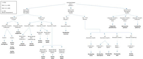
Perform bacterial identifications using different biochemical tests and 16S rDNA sequencing. Tests used to identify the bacteria within the class Bacilli are shown in the
Figure 1, as described by T. W. Fisher and Garczynski
[28][34].
-
Figure 1. Different biochemical tests for the identification of Bacilli species. The figure was adapted and redrawn after modifications from T.W. Fisher and Garczynski [17]. Some details of the tests presented include VP (Voges–Proskauer test (Barritt’s method)), Gelatin (proteolysis of gelatin), ADH (presence of the amino acid arginine dihydrolase), Glucose (fermentation) and Mannitol (fermentation); Starch (hydrolysis), Nitrate (nitrate reduction to nitrite), and Urea (Urease test).
Different biochemical tests for the identification of Bacilli species. The figure was adapted and redrawn after modifications from T.W. Fisher and Garczynski [23]. Some details of the tests presented include VP (Voges–Proskauer test (Barritt’s method)), Gelatin (proteolysis of gelatin), ADH (presence of the amino acid arginine dihydrolase), Glucose (fermentation) and Mannitol (fermentation); Starch (hydrolysis), Nitrate (nitrate reduction to nitrite), and Urea (Urease test).
3. Isolation of Entomopathogenic Fungi
Fungal entomopathogens can directly be isolated from insect cadavers in the case of visible mycosis
[29][35]. Moreover, they can also be isolated from soils or phylloplane as they spend a considerable part of their life as saprophytes in soils or as plant endophytes. However, to
the our
esearchers' knowledge, their survival as soil saprophytes has not been proven yet
[4][5][6][7][8][29][30][4,5,6,7,8,35,36]. In either case, the material can be cultured directly onto a medium selective for an EPF or the material can be baited with an infection-sensitive insect
[31][37]. In case of the isolation of EPF as endophyte, proper disinfection of the material is needed. Nonetheless, different antibacterial and fungal saprophyte-inhibiting chemicals are added in the selective medium, as per the research interest. Here, different culture media used to isolate fungal entomopathogens, especially those belonging to the order Hypocreales are discussed.
3.1. Isolations from Naturally Mycosed Insect Cadavers
This method is applied to study the natural EPF infections in the fields as it relies on the collection of the dead insects from the fields. The protocol described below is similar to that employed by Sharma et al.
[7].
- (a)
-
Insect cadavers are brought to the laboratory as separate entities in sterile tubes.
-
- (b)
-
Insects are observed under a stereomicroscope (40×) for probable mycosis.
-
- (c)
-
In case of a visible mycosis, the insects are surface sterilized using 70% ethanol or 1% NaOCl, for 3 min, followed by 3 distinct washes with 100 mL of sterilized water. Then, the sporulating EPF from the insect cadaver is plated directly.
-
- (d)
-
Cadavers are then cultured on a selective medium at 22 °C for up to 3 weeks, depending on the time taken by the fungi for germination and proliferation. In case of no germination, the cadavers can be homogenized and plated on the selective medium. Details of the different selective medium are provided later in the text.
-
- (e)
-
Obtained fungi are subcultured on potato dextrose agar (PDA) or Sabouraud dextrose agar (SDA) until pure culture is obtained.
-
- (f)
-
Fungi are identified by comparing morphological characteristics using light microscopy (400×), described in several fungal identification keys, such as Domsch et al.
[32][38] and Humber
[33][39].
-
- (g)
-
Molecular identifications can be done by extracting the DNA and performing PCR for the amplification and subsequent sequencing of the nuclear internal transcribed spacer (nrITS) region of the fungal nuclear ribosomal DNA, as described in Yurkov et al.
[34][40].
-
Note: If the objective of the work is to study the diversity of the fungal entomopathogens, irrespective of the genus of interest, a few media can be used: (a) SDA with 0.2% yeast extract (
w/v), i.e., SDAY further supplemented with 0.08% (
w/v) streptomycin-sulphate and 0.03% (
w/v) penicillin
[35][41]; (b) SDA supplemented with 0.05% (
w/v) streptomycin-sulphate and 0.025% (
w/v) chloramphenicol
[36][42]; (c) PDA supplemented with either 0.01% (
w/v) streptomycin-sulphate and 0.005% (
w/v) tetracycline
[37][43], 0.01% (
w/v) chloramphenicol
[38][39][44,45], or 0.01% (
w/v) penicillin, 0.02% (
w/v) streptomycin-sulphate and 0.005% (
w/v) tetracycline
[40][46]; (d) oatmeal agar supplemented with 0.06% (
w/v) cetyl trimethyl ammonium bromide and 0.05 % (
w/v) chloramphenicol (OM-CTAB)
[41][47]; (e) Dichloran Rose Bengal chloramphenicol agar (DRBCA)
[4][42][4,48], or DRBCA supplemented with 0.05% (
w/v) streptomycin-sulphate
[31][37]. It is always advisable to use more than one selective medium pertaining to the susceptibility of a few EPF species to a particular concentration of the inhibitory chemical used.
3.2. Isolations from Soils
Isolations of fungal entomopathogens from soils can be done in 2 ways, i.e., either by culturing the soil inoculums or by employing bait insects. In any of the cases, after visible mycosis. If the research objective is to isolate a particular EPF genus, then the relevant selective medium described below can be used.
3.3. Isolation from Phyllosphere
Some studies have also isolated EPF from the phylloplane and other parts of the plant phyllosphere, as these fungi can also be present as plant epiphytes or endophytes
[35][41]. Meyling et al. suggested a leaf imprinting methodology where the leaf is cultured onto a selective agar medium
[43][64]. Petri dishes with partitions are used and the upper (adaxial), and the lower (abaxial) surface of the leaf are pressed on the separate sides of the petri plate. Henceforth, the same leaf is put on a paper sheet and photocopied to estimate its surface area using image analysis software at a later stage. The petri plates are incubated in the dark at 23 °C to count fungal colony forming units (CFUs)
[43][64]. Surface sterilization is quite important in isolating hypocrealean fungi as endophytes. This can be done by dipping the plant part in either 70% ethanol and/or 1–5% NaOCl for 3 min. In case of the leaves, the petiole can be first kept out of the sanitizer to avoid the chemical reaching inside the leaf, and then it can be cut to culture the sterilized part of the leaf on either of the selective mediums described above. It is always recommended to sanitize the intact plant part and then cut it into pieces for further culturing, as this avoids the sterilization of the endophytic fungi
[44][111]. Different studies have isolated EPF from the phyllosphere, such as bark and branch samples
[45][46][56,112] and leaves
[47][48][59,113].
3.4. Molecular Identifications of the Isolated Entomopathogenic Fungi
After obtaining a single spore fungal culture on a PDA or SDA, the species can be resolved or identified by amplifying the regions of nuclear ribosomal DNA, such as
nrITS, large (28S) subunit (
nrLSU), or small (18S) subunit (
nrSSU). Another, nuclear ribosomal DNA region, i.e., the intergenic spacer region between
nrSSU and
nrLSU or
IGS, has also been used to understand
Beauveria and
Metarhizium speciation
[48][49][50][51][113,114,115,116]. The resolution of the molecular identification can be increased by amplifying other nuclear DNA regions of interest, e.g., for Bloc for
Beauveria [48][49][50][113,114,115] and the 5′ intron-containing region of translation elongation factor 1-alpha subunit (5′-
tef1α) for
Metarhizium [51][52][116,117]. Other nuclear DNA markers, such as the regions of the gene encoding for the largest subunit of RNA polymerase II (
rpb1), the second largest subunit of RNA polymerase II (
rpb2); β-tublin (
β-tub), and the coding region of Tef1-α, can also be employed, in general, for any EPF
[53][54][118,119].
Moreover, in the last decades, researchers have been constantly developing and validating the use of several microsatellite markers for the genotyping of
Beauveria [50][55][56][57][58][59][93,115,120,121,122,123] and
Metarhizium [60][61][124,125] isolates. For example, Oulevey et al.
[61][125] described 18 small single repeats or microsatellite marker sets for
Metarhizium, i.e., Ma145, Ma325, Ma307, Ma2049, Ma2054, Ma2055, Ma2056, Ma2057, Ma2060, Ma2063, Ma2069, Ma2070, Ma2077, Ma2089, Ma2283, Ma2287, Ma2292, and Ma2296. Similarly, Meyling et al.
[55][93] and Goble et al.
[59][123] validated the use of 17 to 18 microsatellite marker sets for
Beauveria, i.e., Ba06, Ba08, and Ba12-Ba29. This methodology enables enhanced resolution among very closely related isolates which may otherwise be rendered as clones. Recently, Kepler and Rehner
[54][119] developed primers for the amplification and sequencing of nuclear intergenic spacer markers for the resolution of
Metarhizium isolates, i.e., BTIGS, MzFG543, MzFG546, MzIGS2, MzIGS3, MzIGS5, and MzIGS7, and Kepler et al.
[62][99] successfully validated the use of MzIGS3 and MzFG543 on the
Metarhizium isolated from agricultural soils.