Modulation of Tumor Immunogenicity: Comparison
Please note this is a comparison between Version 2 by Fanny Huang and Version 1 by M. WALID QORONFLEH.

Immunotherapy is now established as a potent therapeutic paradigm engendering antitumor immune response against a wide range of malignancies and other diseases by modulating the immune system either through the stimulation or suppression of immune components such as CD4+ T cells, CD8+ T cells, B cells, monocytes, macrophages, dendritic cells, and natural killer cells. By targeting several immune checkpoint inhibitors or blockers (e.g., PD-1, PD-L1, PD-L2, CTLA-4, LAG3, and TIM-3) expressed on the surface of immune cells, several monoclonal antibodies and polyclonal antibodies have been developed and already translated clinically. In addition, natural killer cell-based, dendritic cell-based, and CAR T cell therapies have been also shown to be promising and effective immunotherapeutic approaches. In particular, CAR T cell therapy has benefited from advancements in CRISPR-Cas9 genome editing technology, allowing the generation of several modified CAR T cells with enhanced antitumor immunity.

  • cancer
  • immunogenicity
  • immunotherapy
  • immune checkpoint
  • CAR T cell therapy
  • CRISPR-Cas9

1. Introduction

Immunotherapy is an important therapeutic strategy where extrinsic therapeutic substances either stimulate or repress the immune system to combat tumors, infections, as well as other disease ailments [1]. Immunotherapy has transpired to be the touchstone in the treatment of cancer, unlike other traditional treatment options such as targeted therapy, radiation therapy, and chemotherapy, which are emerging dynamic fields in the biopharmaceutical industry [2,3][2][3]. Cancer immunotherapy is a promising and advantageous tool for tumor treatment which acts by boosting the immune system to generate antitumor effects against tumor cells [4]. Nowadays, it gains more traction than other cancer treatments due to its ability to enhance patients’ overall survival as well as their quality of life [5]. The mechanism of antitumor immunity involves the presentation of tumor-specific antigens (TSAs) and tumor-associated antigens (TAAs) by antigen-presenting cells (APCs) such as dendritic cells and macrophages to cytotoxic T lymphocytes (CTLs) and helper T cells, thereby exerting its antitumor effects [6]. There exist various types of immunotherapy approaches in treating cancer of any origin through T-cell transfer therapy, monoclonal antibodies (mAbs), natural killer (NK) cell therapy, dendritic cell (DC)-based vaccines, gene-editing using clustered regularly interspaced short palindromic repeats-CRISPR- and its associated protein-9 (CRISPR-Cas9) technology, immune checkpoint inhibitors, as well as other cancer vaccines [7,8][7][8]. In the case of advanced tumors of various origins, the accepted treatment regime employs immune checkpoint inhibitors (ICPs or ICIs), which are well known to target programmed cell death protein 1 (PD-1)/programmed cell death-ligand 1 (PD-L1) and cytotoxic T-lymphocyte associated antigen 4 (CTLA-4) [9]. More recently, in 2020, globally active chimeric antigen receptor (CAR) T cell therapies were clinically employed to treat patients, which also encompassed advanced precision medicine immunotherapy measures like the use of checkpoint inhibitors [10,11][10][11]. The mechanism of CAR T cells involves the adoptive transfer of modified T cells, i.e., chimeric antigen receptor (CAR) T cells intended towards the cluster of differentiation 19 (CD19), which is currently authorized to cure patients with advanced B cell lymphoma and refractory B cell acute lymphoblastic leukemia-ALL [12]. In cancer immunotherapy, the DC-based therapy is specifically used to treat prostate cancer. The NK-cell-based therapy involves the modification of NK cells and enables the clinical treatment of different cancer types like breast cancer, ALL, neuroblastoma, gastrointestinal (GI)-tract cancer, etc. [13]. Nowadays, CRISPR Cas-9 technology is widely used to inactivate oncogenes by editing tumor genes with the Cas-9 endonuclease (RNA-guided mechanism) [14]. Some of the FDA-approved immunotherapies for the treatment of different types of cancer conditions, along with their mechanisms, are illustrated in Figure 1.
Figure 1.
List of FDA-approved immunotherapies and their mechanism of action.
Despite the progress in cancer immunotherapy, there is a challenge as cancer cells can develop resistance either initially or in response to subsequent treatments, including primary, secondary, or acquired resistance. The drawback of immunotherapy resistance is more intricate because tumors subsist in a progressive microenvironment [15]. The dynamic microenvironment of tumors encompasses different types of malignant cancer cells, innate and adaptive immune components, extracellular matrix, signaling molecules, and blood vessels that act separately as well as in combination to increase the sensitivity and efficacy of immunotherapy [16,17][16][17]. In this fashion, multiple stratagems are adopted to transform the niche with the aim of improving their response to immunotherapy against cancer. These are widely classified into two types, namely, direct and indirect modulation of tumor immunogenicity. Direct modulation involves the alteration of the tumor by itself, whereas the indirect modulation of immunogenicity acts on the tumor niche. 

2. Modulation of Tumor Immunogenicity

Immunogenicity is the ability of a substance to provoke a protective immune response. Most preferable is eliciting the adaptive immune response of the host immune system. The immunogenicity of an antigen can be determined by the following three aspects: immunological defense (the capability to eliminate the antigens and combat pathogenic infection), immunological homeostasis (maintaining a stable homeostasis upon the recognition and elimination of damaged cells or tissue), and immunological surveillance (the capacity to recognize and kill the mutated, abnormally behaving cells, and prevent malignant growth in the body) [18].
Tumor immunogenicity is basically nothing but a tumor antigen, which elicits tumor immune response to restrict tumor growth. Several modulators have already been discovered to modify the tumor microenvironment to generate an antitumor immune response.

2.1. Direct Modulation of Tumor Immunogenicity

The direct modulation of immunogenicity against tumors involves radiation, chemotherapy, targeted therapy, and metabolic modifiers [19].
In chemotherapy, several cytotoxic drugs, besides their direct killing of cancer cells and preventing the cancer burden, have been used in combination with immunotherapy due to their immunomodulatory role. In destroying tumor cells, cytotoxic drugs elicit a potent antitumor immune response by releasing myeloid-derived tumor-associated antigens. Additionally, chemotherapy can reduce immunosuppressive cells such as regulatory T cells (Treg) and myeloid-derived suppressor cells (MDSCs). Gemcitabine (a deoxycytidine analog) and Cyclophosphamide (an alkylating agent) are examples of widely used chemotherapeutic drugs with immunotherapeutic drugs due to cross-priming and antigen cross-presentation; mechanistically, this occurs because of the suppression of Treg cells, and the stimulation of the proliferation of Teff cells, respectively [20]. The FDA approved the first chemotherapy combination including ICI in 2018. Carboplatin, Pemetrexed, and Pembrolizumab are now regularly used as first-line treatments for non-small-cell lung cancer (NSCLC) [21].
Radiation therapy, similar to chemotherapy, causes the direct killing of tumor cells; it also releases tumor antigens to provoke immune response, stimulate antigen presentation, and induce the infiltration of tumor-infiltrating lymphocytes (TILs) via inflammation. The innate immune systems recognize radiation-mediated DNA damage and induce the migration of Teff cells to the tumor microenvironment [22].
Targeted therapy, like chemotherapy, can induce cytoreduction; additionally, it modulates immune cells, promotes the infiltration of T cells and NK cells, alters the tumor endothelium, and reduces tolerogenic cell infiltration (a heterogeneous pool of dendritic cells capable of producing immunological tolerance) [23]. The benefits of targeted therapy are that it improves the defensive ability of the immune system to fight against various types of tumors, inhibits the growth of tumors, provides inhibitory signals to the angiogenesis process of tumor cells, and initiates apoptosis or inhibits metastasis, ultimately leading to cancer cell death. Targeted therapy in the form of mAb targets specific overexpressed antigens on tumors and it can be administered intravenously [24]. The main mechanism of the induction of tumor cell death in targeted therapies is the blocking of growth factor receptor signaling. mAbs play a very significant role in blocking target receptors expressed on cancer cells. Studies have revealed that EGFR- and HER2-targeting monoclonal antibody Cetuximab causes the induction of the apoptosis of tumor cells by blocking the binding of the ligand to the particular receptor, thereby inhibiting the receptor dimerization [25,26,27][25][26][27].
The combined use of BRAF and MEK inhibitors against melanoma has been found to upregulate MHC levels and melanoma differentiation antigens, such as gp-100 and melanoma-associated antigen recognized by T cells (MART-1) [28]. Axitinib (vascular endothelial growth factor receptor-VEGFR-kinase) and Pembrolizumab (a humanized anti-PD-L1 mAb) have shown improved overall survival (OS) and progression-free survival (PFS) in advanced renal cell carcinoma patients [29]. Similarly, Lenvatinib (a VEGFR kinase inhibitor) in combination with Pembrolizumab was also shown to improve the health of patients with advanced endometrial cancer [30]. Combination therapy is becoming the norm in cancer treatment. Pembrolizumab and Lenvatinib were first approved by the FDA in 2014 and 2015, respectively. The combination has shown significant overall survival among patients with advanced endometrial cancer in a phase III clinical trial. The FDA also granted accelerated drug approval for Pembrolizumab for advanced endometrial cancer as a single treatment in March 2022 [31].
The intake and digestion of nutrients, as well as the removal of cellular waste, are critical aspects of every tumor’s interaction with its local milieu. Many of the substances that fuel tumor cells are also important to immune cells, complicating efforts to target metabolic pathways therapeutically. Glucose, lactate, fatty acids, and amino acids are the most significant target molecules of the cancer cells. Several therapeutic aspects have demonstrated in vitro success in modulating the tumor’s interaction with microenvironmental glucose, such as reducing glucose levels, modifying glycolytic pathways, and/or altering lactic acid metabolism [32]. Metformin and Phenformin are two important anti-diabetic drugs that have been widely used against breast cancer [33]. In a preclinical-level study, Phenformin has shown antitumor efficacy against hematological cancer and solid tumors [34]. Phenformin drugs suppress the MDSCs and enhance the ICPs in inhibitory-efficacy melanoma models [35].

2.2. Indirect Modulation of Tumor Immunogenicity

The indirect modulation of immunogenicity involves markers of the immunogenic microenvironment, ICPs blockers, molecules that stimulate T cells response to the tumor microenvironment, compounds that trigger stimulatory pathways, molecular vaccines (i.e., cell-based vaccines, protein/peptide vaccines, and genetic vaccines), oncolytic viruses, and epigenetic modifications [19].
Three main kinds of T cell therapy modulate the T cell response to tumor microenvironment including TILs infusion/treatment [36], CAR T cell therapy, and T-cell receptor (TCR)-engineered cell therapy. Immunogenic markers of the tumor niche are effector CD8+ T cells (Teff), regulatory T cells (Treg), and myeloid-derived suppressor cells. In many types of tumors, the Teff:Treg ratio has been used as an important prognostic and predictive marker [37,38,39,40][37][38][39][40]. Additionally, tumor intrinsic factors that include PD-L1 expression and tumor mutation burden (TMB), and mismatch repair deficiency have emerged as potential biomarkers with mixed success, as clinical predictors of multiple types of cancer in response to ICP inhibitor therapy. Immunotherapies targeting PD-1/PD-L1 have yielded a noticeable clinical response against a subset of patients with melanoma, NSCLC, and urothelial cancer [41,42,43][41][42][43]. ICI therapy targeting TMB within the tumor genome has also shown a remarkable clinical response against multiple cancers such as NSCLC [44[44][45],45], small-cell lung cancer (SCLC) [46], melanoma [47], and colorectal cancer [48].
Activating stimulatory pathways may involve Theralizumab (TGN1412), a mAb agonist of CD28 known to interact with B7, which is a co-stimulatory molecule that activates T cells; hence, proliferated and differentiated T cells improve the antitumor immune response [49]. Moreover, inducible T-cell co-stimulator (ICOS, i.e., CD28 superfamily), Toll-like receptors (TLRs, i.e., CD40), and OX40 receptor (CD134) are some of the secondary co-stimulatory immune checkpoint targets [50]. Vaccines like cell-based vaccines, protein/peptide-based vaccines, and genetic vaccines (DNA/RNA-based vaccines) regulate the immune niche and are known to amplify the immune reaction by giving direct antigen injection or else through the lysis of tumor cells to render intratumoral immunogen. The first FDA-approved cell-based vaccine was Sipuleucel–T, which is a DC vaccine [51]. In 2018, Pan, RY et al. illustrated the power of personalized cancer vaccines mainly by concentrating on neoantigens/tumor-specific antigens, which are fabricated by non-synonymous mutation or errors in transcription in tumor cells. By utilizing next-generation sequencing, neoantigens can be characterized by evaluating cancer cells from peripheral blood mononuclear cells since neoantigens are restricted to cancer cells [52]. Tumor-specific epitopes were therefore used to assess the antigenic landscape of tumors, which allowed researchers to study the relationship between immunogenic peptides binding to the major histocompatibility complex (MHC) molecule and the presentation on the cancer cell surface to engage T cells [53,54][53][54]. Since not all neoantigens are immunogenic and due to the high degree of polymorphism in TCRs, studies have focused on developing bioinformatic tools to predict the binding ability of neoantigens to TCRs [55]. Intracellular short ~9-mer peptides bind to a protein complex consisting of β2-microglobulin (β2M) and human leukocyte antigen (HLA) proteins (HLA-A, HLA-B, and HLA-C) that are presented on the cell surface to engage CD8+ T cells [56]. Several processes were reported as impairing MHC function, such as somatic mutations affecting the MHC complex and its dysregulation [57]. In this process, the integrity of the MHC complex holds excellent importance for immune surveillance in relieving the potential of neoantigens to induce immune response [58,59,60][58][59][60]

2.3. Existing Immunotherapies for the Indirect Modulation of Immunogenicity

Fundamentally, immunotherapy is a type of cancer treatment strategy that works by boosting the immune system against cancer cells with the help of modulators (body-synthesized molecules) or biologics made in the laboratory [61]. Different immunotherapy approaches have been advanced, including the administration of therapeutic vaccines or exogenous cytokines to enhance the T cell number against tumor cells, the adoptive transfer of immune effector cells specific to tumor antigens, and nowadays, a variety of agonists of co-stimulatory receptors and ICPs that are applied to alleviate tumor-induced immunosuppressive action [62]. Here follows a description of some existing immunotherapeutic approaches.

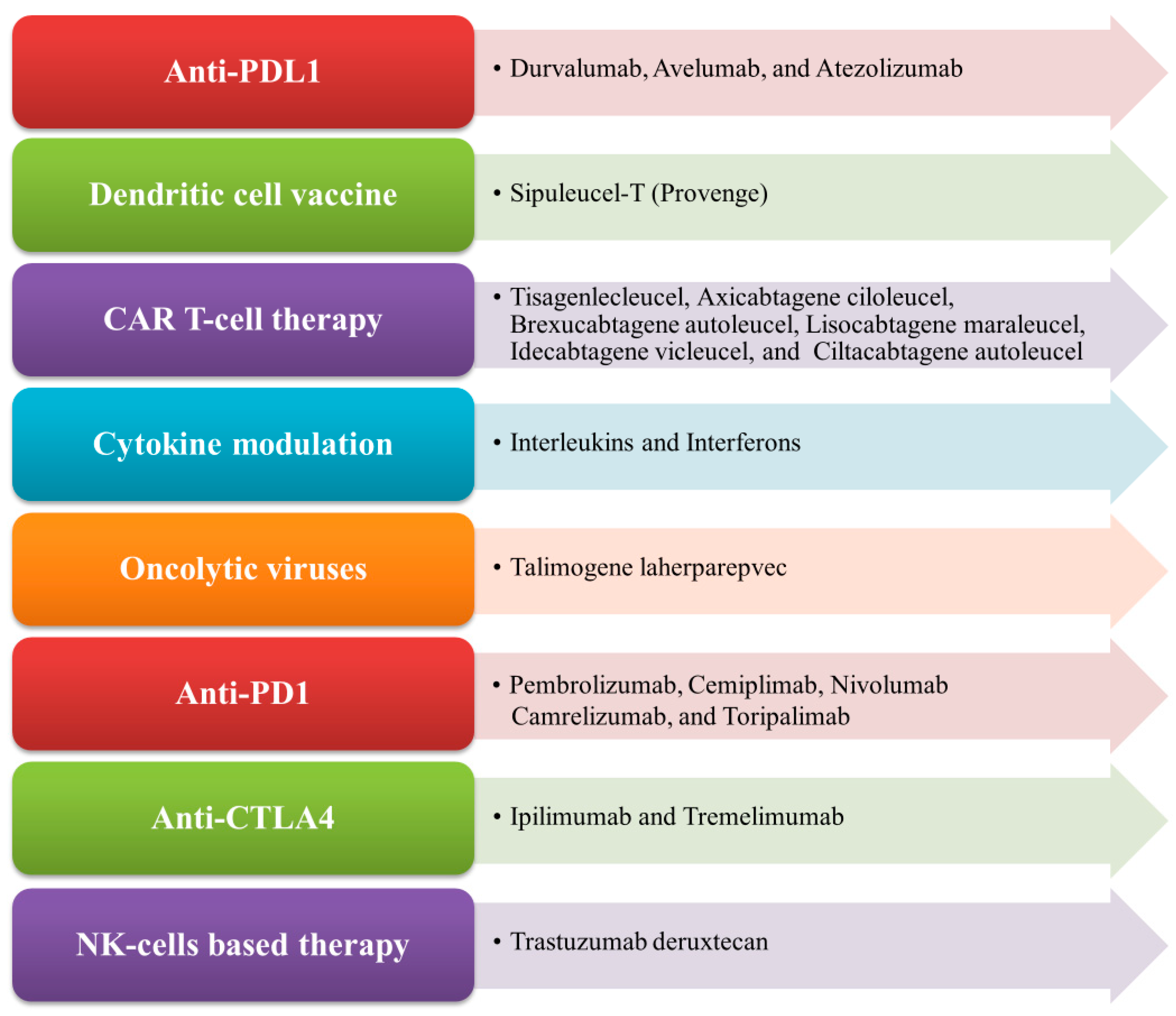
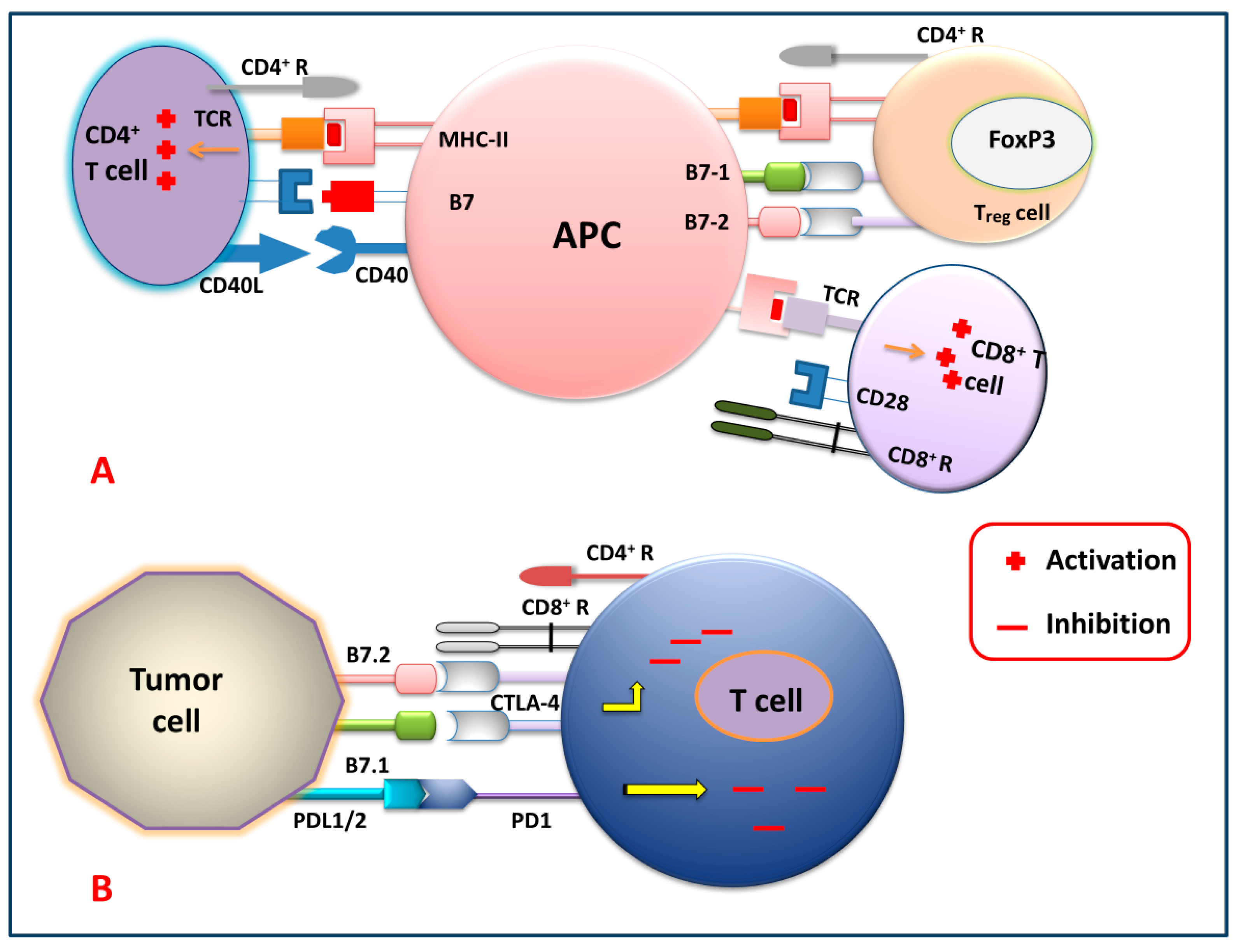
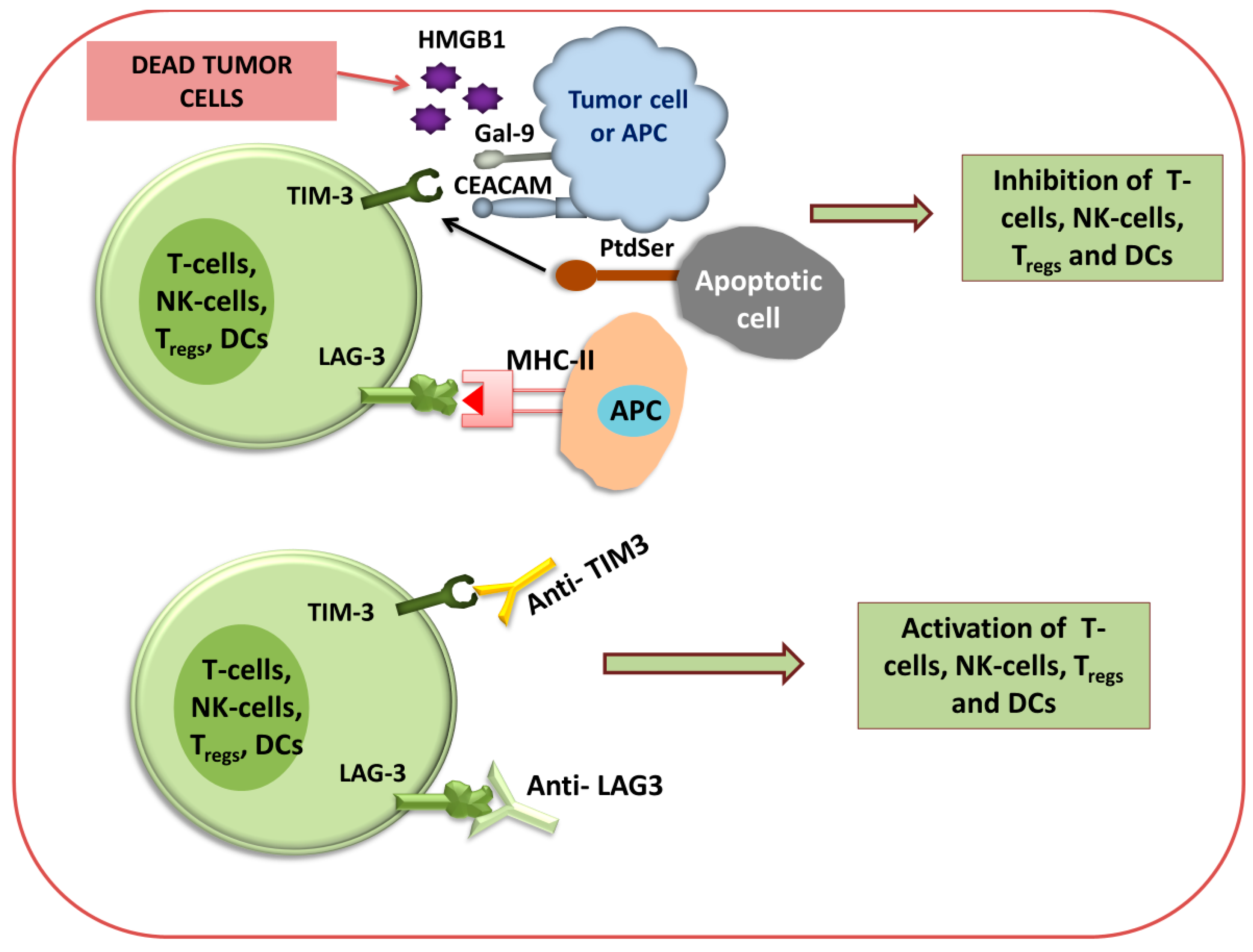
2.3.1. Checkpoint Blockade Therapy

Figure 2 shows different immune checkpoints through which the tumor cells send inhibitory signals to the T cells, thereby inhibiting the generation of the antitumor immune response against tumor cells by suppressing the activation, proliferation, and differentiation of T cells. By blocking these checkpoints, the cancer cells cannot express their receptor against killer T cells. So, generations of these checkpoint-specific antibodies are commonly used for the signal blockade. For that reason, checkpoint-specific antibodies that are administered to the patients eventually block the cancer cells and mediate the killer T cells to target and degrade the cancerous cells precisely.
Figure 2. Immune checkpoint signaling pathways. (A) T cell (CD4+, Treg and CD8+) activation by APC, and (B) T cell inhibition by tumor cells via targeting several immune checkpoint signaling pathways.
Figure 3 and Figure 4 show that different immune checkpoints are inhibited by different antibodies such as anti-PDL1, anti-PDL2, anti-PD1, anti-CTLA4, anti-lymphocyte-activation gene 3 (anti-LAG3), and anti-T cell immunoglobulin and mucin domain-containing protein 3 (anti-TIM-3), which are involved in the generation of an antitumor immune response against cancerous cells.
Figure 3. Immune checkpoint inhibition (antibody-mediated immunotherapeutic strategy). Diagram for anti-PDL-1, anti-PDL-2, anti-PD-1, and anti-CTLA-4 immunotherapy.
Figure 4. Immune checkpoint inhibition of TIM3 and LAG3 using antibodies. Diagram for anti-TIM3 and anti-LAG3 immunotherapy via the activation or inhibition of T cells, NK cells, T-reg cells, and DCs.
Among various types of immune checkpoints, two receptor classes are actively expressed on activated T cells, CTLA-4 and PD-1, which are targeted by cancer cells to negatively regulate the T cells’ function from killing the tumors [63]. mAb-based therapies have been used against these immune checkpoints, where they have produced a long-term durable immune response against different types of malignancies [64,65,66,67][64][65][66][67]. Furthermore, in mAb-based therapies, a specific mAb has been designed that particularly targets either CTLA-4 (such as Ipilimumab and Tremelimumab) or PD-1 (such as Nivolumab and Pembrolizumab), resulting in significant clinical advantages, including a durable immune response against different malignancies [68,69,70][68][69][70].

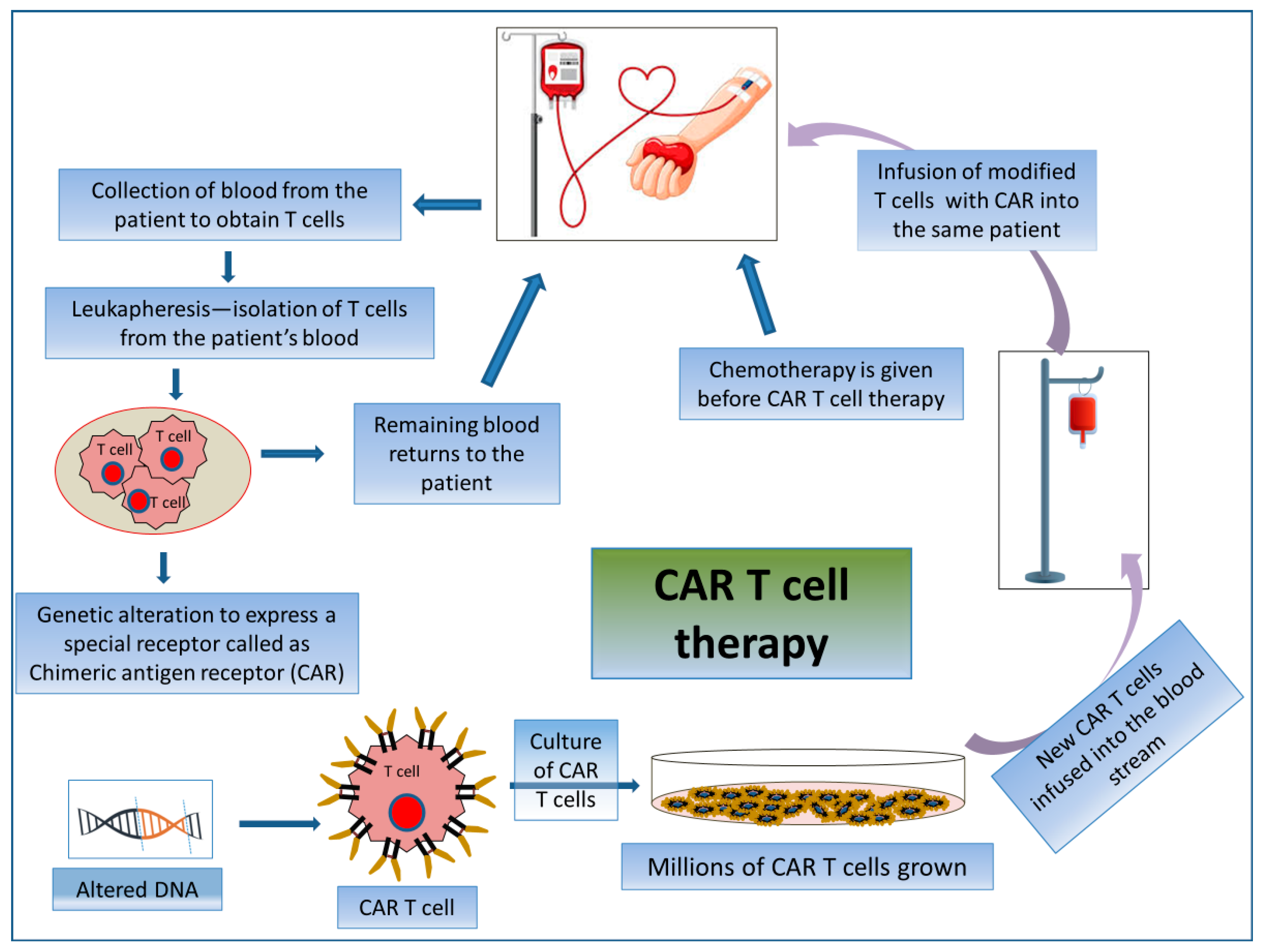
2.3.2. CAR T Cell Therapy

CAR T cell therapy is an adoptive T cell transfer (ACT) therapy whereby killing cancerous cells does not involve MHC molecules but rather the patient’s T-lymphocytes. CAR T cell therapy encompasses the exogenous selection, modification, expansion, and re-infusion of modified T cells into the patient’s body (Figure 5). CARs are generated by collecting patient T cells via a process known as apheresis, and engineered cells are reintroduced after a preparative regimen. Here, T-lymphocytes are collected from their peripheral blood and then are genetically modified ex vivo via the single-chain variable fragment (scFv) antigen recognition domain to express CD19-specific CARs. Thus, the reinfused modified T cells seek out and destroy the tumor cells expressing CD19 in the patient. By this mechanism, modified T cells raise an immunogenic niche inside the patient’s body [127][71]. These are used specifically for treating patients with advanced-stage B-cell malignancies [128][72].
Figure 5. Mechanism of action involved in CAR T cell therapy by acquiring patient’s own T cells. Schematic representation of the process of CAR T cell therapy.
First-generation CAR T cells bind to a receptor derived from an extracellular antibody mainly designed for a tumor antigen with a CD3-based intracellular activating domain [129][73]. Second- and third-generation CAR T cells are formed based on different molecular components of co-stimulatory molecules, including CD28, ICOS, OX40 (CD134), and 4-1BB (CD137), to generate durable immune response even after repeated times of antigenic stimulation [130,131,132][74][75][76]. Fourth-generation CAR T cells contain signaling domains from an inducible expression of different inflammatory cytokines, e.g., IL-12 or IL-18, or cytokine receptors [133,134][77][78]. The most successful FDA-approved CAR T cell therapy, Tisagenlecleucel, Brexucabtagene autoleucel, and Axicabtagene ciloleucel (Figure 1), targets the B-cell lineage antigen CD19. The CD-19-targeted CAR T cells have been used as an important option to treat patients with certain incursive B-cell non-Hodgkin lymphomas (NHLs) and/or acute B-cell lymphoblastic leukemia (B-ALL) [130][74]. Recently, the FDA approved several CAR T cell therapeutic regimens, such as Lisocabtagene maraleucel, which targets CD19 antigen and is used to treat relapsed or refractory large B-cell lymphoma; and Idecabtagene vicleucel and Ciltacabtagene autoleucel both of which target B cell maturation antigen (BCMA) and are used against relapsed or refractory multiple myeloma [135][79].

2.3.3. Natural Killer (NK) Cell-Based Immunotherapy (Alternative to CAR T Cell Therapy)

NK cell immunotherapy for cancer has been extensively reviewed, presenting a perspective on NK cell biology and function, therapy types, and clinical trials [136,137][80][81]. NK cells play a very specialized role in innate immune defense and have a potent activity in the killing of abnormal cells (virus-infected or cancer cells) in an MHC-unrestricted manner [138,139][82][83]. NK cells have the ability to recognize metastatic cells and kill these cells by releasing either lysing enzymes or through ADCC. For that, NK cells do not need any prior sensitization of any particular antigen. Upon activation in the presence of malignant cells, NK cells mimic the activated cytotoxic T cell activity against antigens by secreting cytotoxic molecules containing perforin and granzyme granules to lyse these malignant cells directly. NK cells also have a role in modulating adaptive immunity by producing cytokines and chemokines such as TNF-α and IFN-γ. For many decades, NK cells have been investigated to be used against cancer [138][82]. The efficacy of NK cells can be increased by using some immune stimulants, particularly various types of antibodies and cytokines during antitumor immunotherapy [140][84] including adoptive transfer (direct transfer into the patients) of ex vivo cultured NK cells [141][85]. CAR NK cells are constructed by genetic modification of NK cells from different sources, including hematopoietic pluripotent stem cells (HPSCs), primary NK cells, as well as NK cell lines, to express CARs in order to enhance the recognition of a particular surface marker of cancer cells such as CD19, CD20, and ErbB2. After recognition of the target, the downstream signaling domain (such as CD3ζ and CD28) of CAR causes the activation of PI3K and ultimately influences the release of IFN-γ and stimulates cytotoxicity [142,143][86][87].
CAR NK cells have both CAR-dependent and CAR-independent potential cytotoxic effects against cancer cells [144][88]. Some preclinical studies suggested that NK-cell-derived extracellular vesicles (EVs) have potent antitumor activity. NK EVs bear the CD56 marker and contain some lytic proteins like perforin-granzyme and FasL [145][89]. Other studies have reported that NK EVs showed potent cytotoxic effects of killing against various cancer cell lines together with breast carcinoma, ALL, and neuroblastomas [145][89]. In human epidermal growth factor receptor 2 (HER2)-positive breast cancer patients, Trastuzumab deruxtecan (see Figure 1) was used as a monotherapy and was found to stimulate the activation of NK cells against HER2. Dual HER2 blockage with Trastuzumab and Pertuzumab is currently the mainstay of therapy in early and advanced cancer illness, since this combination may have an additive impact on ADCC [146][90].

2.3.4. Dendritic Cell Vaccine Therapy (a Cross between a Vaccine and a Cell Therapy)

DC vaccines elicit a specific immune response that can selectively eliminate cancer cells. Progress in this area has been recently reviewed [147][91]. DCs have been used in clinical trials to test their application in boosting antitumor immunity [148][92]. DCs are a heterogeneous population of different types of dendritic cells comprising conventional dendritic cells 1 (cDC1), conventional dendritic cells 2 (cDC2), monocyte-derived DCs (MoDCs), and pDCs developed from hematopoietic cells. They are involved in maintaining the connection between innate and adaptive immunity. DCs are used in vaccine production due to their ability to express TAAs primarily on CD4+ T cells, and cross-presentation (i.e., cross-priming) to CD8+ T cells [149,150][93][94]. As DCs are found across the skin, mucosa, blood, and as well as lymphoid tissues, they have the capacity of antigen processing and presentation to naïve T-lymphocyte cells, consequently inducing them to convert into tolerogenic subsets or effector subsets. Therefore, they can orchestrate an adaptive immune response [151,152,153][95][96][97]. DCs have the capacity to cross the representation of exogenous tumor antigens by class-I MHC molecules to recently matured CD8+ T lymphocyte cells from the thymus. In addition, they have the ability to polarize the CD4+ T cells towards the Th-1 subset effectively and to activate the NK cells [154,155][98][99]. Moreover, activated and proliferated CTLs trigger the process of elimination of tumor cells by recognizing the antigenic peptide complex with the MHC-I molecule presented on the tumor cells [156][100].
DC-based vaccines induce the activity of CTLs expressing low levels of CTLA-4 and PD-1 to increase their cytolytic ability and to enhance the expression of different molecules (CXCR3 and CD103/CD49a) to empower the migration of CTLs towards the tumor microenvironment [157][101]. Currently, the FDA has approved Sipuleucel-T (Figure 1) as the first DC-based autologous cellular immunotherapeutic drug to treat prostate cancer. The major component of Sipuleucel-T is the fusion protein (PA2024) composed of two constituents which are cancer antigen-prostatic acid phosphatase (PAP) conjugated to adjuvant GM-CSF. Some clinical studies have reported that under ex vivo conditions, this fusion protein (PAP-GMCSF) undergoes activation when incubated with the isolated APCs. These activated APCs can now fight against prostate cancer cells once re-infused into the patients [158,159][102][103].

2.3.5. CRISPR-Cas9-Based Immunotherapy

Few reviews tackled the cutting-edge application of gene editing in cancer immunotherapy [14,160][14][104]. CRISPR-Cas9 is a genome-editing technology tool that uses mRNA nuclease to cleave genomic DNA at a target sequence of interest with efficiency, precision, and specificity. In cancer, excessive accumulation of mutations leads to the activation of different oncogenes via the gain of proto-oncogene function and the inactivation of tumor suppressor genes via the loss of gene function. Hence, CRISPR-Cas9 can be used as an important therapeutic tool for oncogene inactivation by tumor genome editing and the restoration of tumor suppressor and apoptotic functions [161][105]. The CRISPR-Cas9 system has two essential components, single guide RNA (sgRNA) and Cas-9 endonuclease, which cut the site-specific double-stranded DNA with the help of sgRNA [162][106]. Recently, the CRISPR-Cas9 system has emerged as a therapeutic technology for generating CAR T cells in the cancer immunotherapy field. In 2017, one study reported that the CRISPR-Cas9 system could disrupt several genomic sites simultaneously to make universal CAR T cells, which have defective TCR and MHC-I expression, showing potent antitumor activity [163][107]. The Fas receptor, known as CD95, causes the apoptosis of expanded T cells and can damage CAR T cell functions when it interacts with the Fas ligand (FasL). Thus, using CRIPSR-Cas9 technology, Fas knockout CAR T cells can be formed, which have a better ability to eliminate tumor cells [164][108]. Apart from constructing universal CAR T cells, CRISPR-Cas9-mediated genome editing eliminates multiple genes which encode inhibitory T-cell surface receptor PD-1 and CTLA-4 to improve the efficacy of T cell-based antitumor immunotherapy [165][109].

References

  1. Esfahani, K.; Elkrief, A.; Calabrese, C.; Lapointe, R.; Hudson, M.; Routy, B.; Miller, W.H., Jr.; Calabrese, L. Moving towards personalized treatments of immune-related adverse events. Nat. Rev. Clin. Oncol. 2020, 17, 504–515.
  2. Wang, Y.; Wang, M.; Wu, H.-X.; Xu, R.-H. Advancing to the era of cancer immunotherapy. Cancer Commun. 2021, 41, 803–829.
  3. Sharmiladevi, P.; Girigoswami, K.; Haribabu, V.; Girigoswami, A. Nano-enabled theranostics for cancer. Mater. Adv. 2021, 2, 2876–2891.
  4. Yang, K.; Halima, A.; Chan, T.A. Antigen presentation in cancer—Mechanisms and clinical implications for immunotherapy. Nat. Rev. Clin. Oncol. 2023, 20, 604–623.
  5. Kruger, S.; Ilmer, M.; Kobold, S.; Cadilha, B.L.; Endres, S.; Ormanns, S.; Schuebbe, G.; Renz, B.W.; D’Haese, J.G.; Schloesser, H.; et al. Advances in cancer immunotherapy 2019—Latest trends. J. Exp. Clin. Cancer Res. 2019, 38, 268.
  6. Alatrash, G.; Jakher, H.; Stafford, P.D.; Mittendorf, E.A. Cancer immunotherapies, their safety and toxicity. Expert Opin. Drug Saf. 2013, 12, 631–645.
  7. Pilard, C.; Ancion, M.; Delvenne, P.; Jerusalem, G.; Hubert, P.; Herfs, M. Cancer immunotherapy: It’s time to better predict patients’ response. Br. J. Cancer 2021, 125, 927–938.
  8. Sahin, U.; Türeci, Ö. Personalized vaccines for cancer immunotherapy. Science 2018, 359, 1355–1360.
  9. Robert, C. A decade of immune-checkpoint inhibitors in cancer therapy. Nat. Commun. 2020, 11, 3801.
  10. June, C.H.; O’Connor, R.S.; Kawalekar, O.U.; Ghassemi, S.; Milone, M.C. CAR T cell immunotherapy for human cancer. Science 2018, 359, 1361–1365.
  11. Eno, J. Immunotherapy through the years. J. Adv. Pract. Oncol. 2017, 8, 747–753.
  12. Yun, S.; Vincelette, N.D.; Green, M.R.; Wahner Hendrickson, A.E.; Abraham, I. Targeting immune checkpoints in unresectable metastatic cutaneous melanoma: A systematic review and meta-analysis of anti-CTLA-4 and anti-PD-1 agents trials. Cancer Med. 2016, 5, 1481–1491.
  13. Bachanova, V.; Miller, J.S. NK cells in therapy of cancer. Crit. Rev. Oncog. 2014, 19, 133–141.
  14. Khalaf, K.; Janowicz, K.; Dyszkiewicz-Konwińska, M.; Hutchings, G.; Dompe, C.; Moncrieff, L.; Jankowski, M.; Machnik, M.; Oleksiewicz, U.; Kocherova, I.; et al. CRISPR/Cas9 in Cancer Immunotherapy: Animal Models and Human Clinical Trials. Genes 2020, 11, 921.
  15. O’Donnell, J.S.; Teng, M.W.L.; Smyth, M.J. Cancer immunoediting and resistance to T cell-based immunotherapy. Nat. Rev. Clin. Oncol. 2019, 16, 151–167.
  16. Sadeghi Rad, H.; Monkman, J.; Warkiani, M.E.; Ladwa, R.; O’Byrne, K.; Rezaei, N.; Kulasinghe, A. Understanding the tumor microenvironment for effective immunotherapy. Med. Res. Rev. 2021, 41, 1474–1498.
  17. Yalcin, G.D.; Danisik, N.; Baygin, R.C.; Acar, A. Systems Biology and Experimental Model Systems of Cancer. J. Pers. Med. 2020, 10, 180.
  18. Zhang, J.; Tao, A. Antigenicity, immunogenicity, allergenicity. In Allergy Bioinformatics; Springer Nature B.V.: Dordrecht, The Netherlands, 2015; pp. 175–186.
  19. Murciano-Goroff, Y.R.; Warner, A.B.; Wolchok, J.D. The future of cancer immunotherapy: Microenvironment-targeting combinations. Cell Res. 2020, 30, 507–519.
  20. Brode, S.; Cooke, A. Immune-potentiating effects of the chemotherapeutic drug cyclophosphamide. Crit. Rev. Immunol. 2008, 28, 109–126.
  21. Gandhi, L.; Rodríguez-Abreu, D.; Gadgeel, S.; Esteban, E.; Felip, E.; De Angelis, F.; Domine, M.; Clingan, P.; Hochmair, M.J.; Powell, S.F.; et al. Pembrolizumab plus chemotherapy in metastatic non–small-cell lung cancer. N. Engl. J. Med. 2018, 378, 2078–2092.
  22. Campbell, A.M.; Decker, R.H. Harnessing the Immunomodulatory Effects of Radiation Therapy. Oncology 2018, 32, 370–374.
  23. Atkins, M.B.; Plimack, E.R.; Puzanov, I.; Fishman, M.N.; McDermott, D.F.; Cho, D.C.; Vaishampayan, U.; George, S.; Olencki, T.E.; Tarazi, J.C.; et al. Axitinib in combination with pembrolizumab in patients with advanced renal cell cancer: A non-randomised, open-label, dose-finding, and dose-expansion phase 1b trial. Lancet Oncol. 2018, 19, 405–415.
  24. Stanculeanu, D.L.; Daniela, Z.; Lazescu, A.; Bunghez, R.; Anghel, R. Development of new immunotherapy treatments in different cancer types. J. Med. Life 2016, 9, 240–248.
  25. Slamon, D.J.; Godolphin, W.; Jones, L.A.; Holt, J.A.; Wong, S.G.; Keith, D.E.; Levin, W.J.; Stuart, S.G.; Udove, J.; Ullrich, A.; et al. Studies of the HER-2/neu proto-oncogene in human breast and ovarian cancer. Science 1989, 244, 707–712.
  26. Li, S.; Schmitz, K.R.; Jeffrey, P.D.; Wiltzius, J.J.; Kussie, P.; Ferguson, K.M. Structural basis for inhibition of the epidermal growth factor receptor by cetuximab. Cancer Cell 2005, 7, 301–311.
  27. Patel, D.; Bassi, R.; Hooper, A.; Prewett, M.; Hicklin, D.J.; Kang, X. Anti-epidermal growth factor receptor monoclonal antibody cetuximab inhibits EGFR/HER-2 heterodimerization and activation. Int. J. Oncol. 2009, 34, 25–32.
  28. Pelster, M.S.; Amaria, R.N. Combined targeted therapy and immunotherapy in melanoma: A review of the impact on the tumor microenvironment and outcomes of early clinical trials. Ther. Adv. Med. Oncol. 2019, 11, 1758835919830826.
  29. Rini, B.I.; Plimack, E.R.; Stus, V.; Gafanov, R.; Hawkins, R.; Nosov, D.; Pouliot, F.; Alekseev, B.; Soulières, D.; Melichar, B.; et al. Pembrolizumab plus axitinib versus sunitinib for advanced renal-cell carcinoma. N. Engl. J. Med. 2019, 380, 1116–1127.
  30. Makker, V.; Rasco, D.; Vogelzang, N.J.; Brose, M.S.; Cohn, A.L.; Mier, J.; Di Simone, C.; Hyman, D.M.; Stepan, D.E.; Dutcus, C.E.; et al. Lenvatinib plus pembrolizumab in patients with advanced endometrial cancer: An interim analysis of a multicentre, open-label, single-arm, phase 2 trial. Lancet Oncol. 2019, 20, 711–718.
  31. Makker, V.; Colombo, N.; Casado Herráez, A.; Santin, A.D.; Colomba, E.; Miller, D.S.; Fujiwara, K.; Pignata, S.; Baron-Hay, S.; Ray-Coquard, I.; et al. Lenvatinib plus Pembrolizumab for Advanced Endometrial Cancer. N. Engl. J. Med. 2022, 386, 437–448.
  32. García Rubiño, M.E.; Carrillo, E.; Ruiz Alcalá, G.; Domínguez-Martín, A.; Marchal, J.A.; Boulaiz, H. Phenformin as an anticancer agent: Challenges and prospects. Int. J. Mol. Sci. 2019, 20, 3316.
  33. Orecchioni, S.; Reggiani, F.; Talarico, G.; Mancuso, P.; Calleri, A.; Gregato, G.; Labanca, V.; Noonan, D.M.; Dallaglio, K.; Albini, A.; et al. The biguanides metformin and phenformin inhibit angiogenesis, local and metastatic growth of breast cancer by targeting both neoplastic and microenvironment cells. Int. J. Cancer Res. 2015, 136, E534–E544.
  34. Vara-Ciruelos, D.; Dandapani, M.; Russell, F.M.; Grzes, K.M.; Atrih, A.; Foretz, M.; Viollet, B.; Lamont, D.J.; Cantrell, D.A.; Hardie, D.G. Phenformin, but not metformin, delays development of T cell acute lymphoblastic leukemia/lymphoma via cell-autonomous AMPK activation. Cell Rep. 2019, 27, 690–698.
  35. Kim, S.H.; Li, M.; Trousil, S.; Zhang, Y.; di Magliano, M.P.; Swanson, K.D.; Zheng, B. Phenformin inhibits myeloid-derived suppressor cells and enhances the anti-tumor activity of PD-1 blockade in melanoma. J. Investig. Dermatol. 2017, 137, 1740–1748.
  36. Divyapriya, C.; Kannan, A.; Raghavan, V. Expression of CD4, CD8 Biomarkers in Invasive Carcinoma of Breast with Clinicopathological Correlation. J. Pharm. Res. Int. 2021, 33, 65–73.
  37. Baras, A.S.; Drake, C.; Liu, J.J.; Gandhi, N.; Kates, M.; Hoque, M.O.; Meeker, A.; Hahn, N.; Taube, J.M.; Schoenberg, M.P.; et al. The ratio of CD8 to Treg tumor-infiltrating lymphocytes is associated with response to cisplatin-based neoadjuvant chemotherapy in patients with muscle invasive urothelial carcinoma of the bladder. Oncoimmunology 2016, 5, e1134412.
  38. Sasidharan Nair, V.; Elkord, E. Immune checkpoint inhibitors in cancer therapy: A focus on T-regulatory cells. Immunol. Cell. Biol. 2018, 96, 21–33.
  39. Fumet, J.D.; Richard, C.; Ledys, F.; Klopfenstein, Q.; Joubert, P.; Routy, B.; Truntzer, C.; Gagné, A.; Hamel, M.A.; Figueiredo Guimaraes, C.; et al. Prognostic and predictive role of CD8 and PD-L1 determination in lung tumor tissue of patients under anti-PD-1 therapy. Br. J. Cancer 2018, 119, 950–960.
  40. Huang, L.; Guo, Y.; Liu, S.; Wang, H.; Zhu, J.; Ou, L.; Xu, X. Targeting regulatory T cells for immunotherapy in melanoma. Mol. Biomed. 2021, 2, 11.
  41. Taube, J.M.; Klein, A.; Brahmer, J.R.; Xu, H.; Pan, X.; Kim, J.H.; Chen, L.; Pardoll, D.M.; Topalian, S.L.; Anders, R.A. Association of PD-1, PD-1 ligands, and other features of the tumor immune microenvironment with response to anti-PD-1 therapy. Clin. Cancer Res. 2014, 20, 5064–5074.
  42. Reck, M.; Rodríguez-Abreu, D.; Robinson, A.G.; Hui, R.; Csőszi, T.; Fülöp, A.; Gottfried, M.; Peled, N.; Tafreshi, A.; Cuffe, S.; et al. Pembrolizumab versus Chemotherapy for PD-L1-Positive Non-Small-Cell Lung Cancer. N. Engl. J Med. 2016, 375, 1823–1833.
  43. Rosenberg, J.E.; Hoffman-Censits, J.; Powles, T.; Van Der Heijden, M.S.; Balar, A.V.; Necchi, A.; Dawson, N.; O’Donnell, P.H.; Balmanoukian, A.; Loriot, Y.; et al. Atezolizumab in patients with locally advanced and metastatic urothelial carcinoma who have progressed following treatment with platinum-based chemotherapy: A single-arm, multicentre, phase 2 trial. Lancet 2016, 387, 1909–1920.
  44. Rizvi, N.A.; Hellmann, M.D.; Snyder, A.; Kvistborg, P.; Makarov, V.; Havel, J.J.; Lee, W.; Yuan, J.; Wong, P.; Ho, T.S.; et al. Cancer immunology. Mutational landscape determines sensitivity to PD-1 blockade in non-small cell lung cancer. Science 2015, 348, 124–128.
  45. Hellmann, M.D.; Ciuleanu, T.E.; Pluzanski, A.; Lee, J.S.; Otterson, G.A.; Audigier-Valette, C.; Minenza, E.; Linardou, H.; Burgers, S.; Salman, P.; et al. Nivolumab plus Ipilimumab in Lung Cancer with a High Tumor Mutational Burden. N. Engl. J. Med. 2018, 378, 2093–2104.
  46. Hellmann, M.D.; Nathanson, T.; Rizvi, H.; Creelan, B.C.; Sanchez-Vega, F.; Ahuja, A.; Ni, A.; Novik, J.B.; Mangarin, L.M.; Abu-Akeel, M.; et al. Genomic features of response to combination immunotherapy in patients with advanced non-small-cell lung cancer. Cancer Cell 2018, 33, 843–852.
  47. Van Allen, E.M.; Miao, D.; Schilling, B.; Shukla, S.A.; Blank, C.; Zimmer, L.; Sucker, A.; Hillen, U.; Geukes Foppen, M.H.; Goldinger, S.M.; et al. Genomic correlates of response to CTLA-4 blockade in metastatic melanoma. Science 2015, 350, 207–211.
  48. Le, D.T.; Uram, J.N.; Wang, H.; Bartlett, B.R.; Kemberling, H.; Eyring, A.D.; Skora, A.D.; Luber, B.S.; Azad, N.S.; Laheru, D.; et al. PD-1 Blockade in Tumors with Mismatch-Repair Deficiency. N. Engl. J. Med. 2015, 372, 2509–2520.
  49. Suntharalingam, G.; Perry, M.R.; Ward, S.; Brett, S.J.; Castello-Cortes, A.; Brunner, M.D.; Panoskaltsis, N. Cytokine storm in a phase 1 trial of the anti-CD28 monoclonal antibody TGN1412. N. Engl. J. Med. 2006, 355, 1018–1028.
  50. Omar, H.A.; Tolba, M.F. Tackling molecular targets beyond PD-1/PD-L1: Novel approaches to boost patients’ response to cancer immunotherapy. Crit. Rev. Oncol. Hematol. 2019, 135, 21–29.
  51. Kantoff, P.W.; Higano, C.S.; Shore, N.D.; Berger, E.R.; Small, E.J.; Penson, D.F.; Redfern, C.H.; Ferrari, A.C.; Dreicer, R.; Sims, R.B.; et al. Sipuleucel-T immunotherapy for castration-resistant prostate cancer. N. Engl. J. Med. 2010, 363, 411–422.
  52. Pan, R.Y.; Chung, W.H.; Chu, M.T.; Chen, S.J.; Chen, H.C.; Zheng, L.; Hung, S.I. Recent Development and Clinical Application of Cancer Vaccine: Targeting Neoantigens. J. Immunol. Res. 2018, 2018, 4325874.
  53. Chong, C.; Coukos, G.; Bassani-Sternberg, M. Identification of tumor antigens with immunopeptidomics. Nat. Biotechnol. 2022, 40, 175–188.
  54. Thomas, C.; Tampé, R. MHC I assembly and peptide editing—Chaperones, clients, and molecular plasticity in immunity. Curr. Opin. Immunol. 2021, 70, 48–56.
  55. Liu, C.C.; Steen, C.B.; Newman, A.M. Computational approaches for characterizing the tumor immune microenvironment. Immunology 2019, 158, 70–84.
  56. Garcia-Garijo, A.; Fajardo, C.A.; Gros, A. Determinants for Neoantigen Identification. Front. Immunol. 2019, 10, 1392.
  57. Dersh, D.; Hollý, J.; Yewdell, J.W. A few good peptides: MHC class I-based cancer immunosurveillance and immunoevasion. Nat. Rev. Immunol. 2021, 21, 116–128.
  58. Balachandran, V.P.; Łuksza, M.; Zhao, J.N.; Makarov, V.; Moral, J.A.; Remark, R.; Herbst, B.; Askan, G.; Bhanot, U. Identification of unique neoantigen qualities in long-term survivors of pancreatic cancer. Nature 2017, 551, 512–516.
  59. Łuksza, M.; Riaz, N.; Makarov, V.; Balachandran, V.P.; Hellmann, M.D.; Solovyov, A.; Rizvi, N.A.; Merghoub, T.; Levine, A.J.; Chan, T.A.; et al. A neoantigen fitness model predicts tumour response to checkpoint blockade immunotherapy. Nature 2017, 551, 517–520.
  60. McGranahan, N.; Swanton, C. Neoantigen quality, not quantity. Sci. Transl. Med. 2019, 11, eaax7918.
  61. Kim, T.K.; Herbst, R.S.; Chen, L. Defining and understanding adaptive resistance in cancer immunotherapy. Trends Immunol. 2018, 39, 624–631.
  62. Disis, M.L. Mechanism of action of immunotherapy. Semin. Oncol. 2014, 41 (Suppl. 5), S3–S13.
  63. Pardoll, D. The blockade of immune checkpoints in cancer immunotherapy. Nat. Rev. Cancer 2012, 12, 252–264.
  64. Wolchok, J.D.; Kluger, H.; Callahan, M.K.; Postow, M.A.; Rizvi, N.A.; Lesokhin, A.M.; Segal, N.H.; Ariyan, C.E.; Gordon, R.-A.; Reed, K.; et al. Nivolumab plus ipilimumab in advanced melanoma. N. Engl. J. Med. 2013, 369, 122–133.
  65. Hamid, O.; Robert, C.; Daud, A.; Hodi, F.S.; Hwu, W.J.; Kefford, R.; Wolchok, J.D.; Hersey, P.; Joseph, R.W.; Webber, J.S.; et al. Safety and tumor responses with lambrolizumab (anti-PD-1) in melanoma. N. Engl. J. Med. 2013, 369, 134–144.
  66. Topalian, S.L.; Hodi, F.S.; Brahmer, J.R.; Gettinger, S.N.; Smith, D.C.; McDermott, D.F.; Powderly, J.D.; Carvajal, R.D.; Sosman, J.A.; Atkins, M.B.; et al. Safety, activity, and immune correlates of anti–PD-1 antibody in cancer. N. Engl. J. Med. 2012, 366, 2443–2454.
  67. Hodi, F.S.; O’Day, S.J.; McDermott, D.F.; Weber, R.W.; Sosman, J.A.; Haanen, J.B.; Gonzalez, R.; Robert, C.; Schadendorf, D.; Hassel, J.C.; et al. Improved survival with ipilimumab in patients with metastatic melanoma. N. Engl. J. Med. 2010, 363, 711–723.
  68. Savoia, P.; Astrua, C.; Fava, P. Ipilimumab (Anti-Ctla-4 Mab) in the treatment of metastatic melanoma: Effectiveness and toxicity management. Hum. Vaccin. Immunother. 2016, 12, 1092–1101.
  69. Tartari, F.; Santoni, M.; Burattini, L.; Mazzanti, P.; Onofri, A.; Berardi, R. Economic sustainability of anti-PD-1 agents nivolumab and pembrolizumab in cancer patients: Recent insights and future challenges. Cancer Treat. Rev. 2016, 48, 20–24.
  70. Fumet, J.D.; Isambert, N.; Hervieu, A.; Zanetta, S.; Guion, J.F.; Hennequin, A.; Rederstorff, E.; Bertaut, A.; Ghiringhelli, F. Phase Ib/II trial evaluating the safety, tolerability and immunological activity of durvalumab (MEDI4736) (anti-PD-L1) plus tremelimumab (anti-CTLA-4) combined with FOLFOX in patients with metastatic colorectal cancer. ESMO Open 2018, 3, e000375.
  71. Zhang, C.; Liu, J.; Zhong, J.F.; Zhang, X. Engineering car-t cells. Biomark. Res. 2017, 5, 1–6.
  72. Wagner, D.L.; Fritsche, E.; Pulsipher, M.A.; Ahmed, N.; Hamieh, M.; Hegde, M.; Ruella, M.; Savoldo, B.; Shah, N.N.; Turtle, C.J.; et al. Immunogenicity of CAR T cells in cancer therapy. Nat. Rev. Clin. Oncol. 2021, 18, 379–393.
  73. June, C.H.; Sadelain, M. Chimeric Antigen Receptor Therapy. N. Engl. J. Med. 2018, 379, 64–73.
  74. Krause, A.; Guo, H.F.; Latouche, J.B.; Tan, C.; Cheung, N.K.; Sadelain, M. Antigen-dependent CD28 signaling selectively enhances survival and proliferation in genetically modified activated human primary T lymphocytes. J. Exp. Med. 1998, 188, 619–626.
  75. Guedan, S.; Chen, X.; Madar, A.; Carpenito, C.; McGettigan, S.E.; Frigault, M.J.; Lee, J.; Posey, A.D., Jr.; Scholler, J.; Scholler, N.; et al. ICOS-based chimeric antigen receptors program bipolar TH17/TH1 cells. Blood 2014, 124, 1070–1080.
  76. Rosado-Sánchez, I.; Herrero-Fernández, I.; Genebat, M.; Del Romero, J.; Riera, M.; Podzamczer, D.; Olalla, J.; Vidal, F.; Muñoz-Fernández, M.; Leal, M.; et al. HIV-infected subjects with Poor CD4 T-cell recovery despite effective therapy express high levels of OX40 and α4β7 on CD4 T-cells prior therapy initiation. Front. Immunol. 2018, 9, 1673.
  77. Kerkar, S.P.; Muranski, P.; Kaiser, A.; Boni, A.; Sanchez-Perez, L.; Yu, Z.; Palmer, D.C.; Reger, R.N.; Borman, Z.A.; Zhang, L.; et al. Tumor-specific CD8+ T cells expressing interleukin-12 eradicate established cancers in lymphodepleted hosts. Cancer Res. 2010, 70, 6725–6734.
  78. Chmielewski, M.; Hombach, A.A.; Abken, H. Of CARs and TRUCKs: Chimeric antigen receptor (CAR) T cells engineered with an inducible cytokine to modulate the tumor stroma. Immunol. Rev. 2014, 257, 83–90.
  79. Li, Y.; Ming, Y.; Fu, R.; Li, C.; Wu, Y.; Jiang, T.; Li, Z.; Ni, R.; Li, L.; Su, H.; et al. The pathogenesis, diagnosis, prevention, and treatment of CAR-T cell therapy-related adverse reactions. Front. Pharmacol. 2022, 13, 950923.
  80. Chu, J.; Gao, F.; Yan, M.; Zhao, S.; Yan, Z.; Shi, B.; Liu, Y. Natural killer cells: A promising immunotherapy for cancer. J. Transl. Med. 2022, 20, 240.
  81. Myers, J.A.; Miller, J.S. Exploring the NK cell platform for cancer immunotherapy. Nat. Rev. Clin. Oncol. 2021, 18, 85–100.
  82. Liu, S.; Galat, V.; Galat4, Y.; Lee, Y.K.A.; Wainwright, D.; Wu, J. NK cell-based cancer immunotherapy: From basic biology to clinical development. J. Hematol. Oncol. 2021, 14, 7.
  83. Rees, R.C. MHC restricted and non-restricted killer lymphocytes. Blood Rev. 1990, 4, 204–210.
  84. Sanmamed, M.F.; Chen, L. A paradigm shift in cancer immunotherapy: From enhancement to normalization. Cell 2018, 175, 313–326.
  85. Parkhurst, M.R.; Riley, J.P.; Dudley, M.E.; Rosenberg, S.A. Adoptive transfer of autologous natural killer cells leads to high levels of circulating natural killer cells but does not mediate tumor regression. Clin. Cancer Res. 2011, 17, 6287–6297.
  86. Zhang, C.; Oberoi, P.; Oelsner, S.; Waldmann, A.; Lindner, A.; Tonn, T.; Wels, W.S. Chimeric antigen receptor-engineered NK-92 cells: An off-the-shelf cellular therapeutic for targeted elimination of cancer cells and induction of protective antitumor immunity. Front. Immunol. 2017, 8, 533.
  87. Oberschmidt, O.; Kloess, S.; Koehl, U. Redirected primary human chimeric antigen receptor natural killer cells as an “off-the-shelf immunotherapy” for improvement in cancer treatment. Front. Immunol. 2017, 8, 654.
  88. Khawar, M.B.; Sun, H. CAR-NK Cells: From Natural Basis to Design for Kill. Front. Immunol. 2021, 12, 707542.
  89. Jong, A.Y.; Wu, C.H.; Li, J.; Sun, J.; Fabbri, M.; Wayne, A.S.; Seeger, R.C. Large-scale isolation and cytotoxicity of extracellular vesicles derived from activated human natural killer cells. J. Extracell. Vesicles 2017, 6, 1294368.
  90. Mandó, P.; Rivero, S.G.; Rizzo, M.M.; Pinkasz, M.; Levy, E.M. Targeting ADCC: A different approach to HER2 breast cancer in the immunotherapy era. Breast 2021, 60, 15–25.
  91. Yu, J.; Sun, H.; Cao, W.; Song, Y.; Jiang, Z. Research progress on dendritic cell vaccines in cancer immunotherapy. Exp. Hematol. Oncol. 2022, 11, 3.
  92. Constantino, J.; Gomes, C.; Falcão, A.; Cruz, M.T.; Neves, B.M. Antitumor dendritic cell-based vaccines: Lessons from 20 years of clinical trials and future perspectives. Transl. Res. 2016, 168, 74–95.
  93. Huber, A.; Dammeijer, F.; Aerts, J.G.J.V.; Vroman, H. Current State of Dendritic Cell-Based Immunotherapy: Opportunities for in vitro Antigen Loading of Different DC Subsets? Front. Immunol. 2018, 9, 2804.
  94. Wimmers, F.; Schreibelt, G.; Sköld, A.E.; Figdor, C.G.; De Vries, I.J. Paradigm shift in dendritic cell-based immunotherapy: From in vitro generated monocyte-derived DCs to naturally circulating DC subsets. Front. Immunol. 2014, 5, 165.
  95. Okamoto, M.; Kobayashi, M.; Yonemitsu, Y.; Koido, S.; Homma, S. Dendritic cell-based vaccine for pancreatic cancer in Japan. World J. Gastrointest. Pharmacol. Ther. 2016, 7, 133–138.
  96. Sabado, R.L.; Balan, S.; Bhardwaj, N. Dendritic cell-based immunotherapy. Cell Res. 2017, 27, 74–95.
  97. Steinman, R.M. Decisions about dendritic cells: Past, present, and future. Annu. Rev. Immunol. 2012, 30, 1–22.
  98. Palucka, K.; Banchereau, J. Dendritic-cell-based therapeutic cancer vaccines. Immunity 2013, 39, 38–48.
  99. Melief, C.J.; Kast, W.M. Cytotoxic T lymphocyte therapy of cancer and tumor escape mechanisms. Semin. Cancer Biol. 1991, 2, 347–354.
  100. Martínez-Lostao, L.; Anel, A.; Pardo, J. How Do Cytotoxic Lymphocytes Kill Cancer Cells? Clin. Cancer Res. 2015, 21, 5047–5056.
  101. Zhang, N.; Bevan, M.J. CD8+ T cells: Foot soldiers of the immune system. Immunity 2011, 35, 161–168.
  102. Sutherland, S.I.; Ju, X.; Horvath, L.G.; Clark, G.J. Moving on From Sipuleucel-T: New Dendritic Cell Vaccine Strategies for Prostate Cancer. Front. Immunol. 2021, 12, 641307.
  103. Anassi, E.; Ndefo, U.A. Sipuleucel-T (provenge) injection: The first immunotherapy agent (vaccine) for hormone-refractory prostate cancer. Pharmacol. Ther. 2011, 36, 197–202.
  104. Ou, X.; Ma, Q.; Yin, W.; Ma, X.; He, Z. CRISPR/Cas9 Gene-Editing in Cancer Immunotherapy: Promoting the Present Revolution in Cancer Therapy and Exploring More. Front. Cell Dev. Biol. 2021, 9, 674467.
  105. Azangou-Khyavy, M.; Ghasemi, M.; Khanali, J.; Boroomand-Saboor, M.; Jamalkhah, M.; Soleimani, M.; Kiani, J. CRISPR/Cas: From Tumor Gene Editing to T Cell-Based Immunotherapy of Cancer. Front. Immunol. 2020, 11, 2062.
  106. Xia, A.L.; He, Q.F.; Wang, J.C.; Zhu, J.; Sha, Y.Q.; Sun, B.; Lu, X.J. Applications and advances of CRISPR-Cas9 in cancer immunotherapy. J. Med. Genet. 2019, 56, 4–9.
  107. Ren, J.; Zhang, X.; Liu, X.; Fang, C.; Jiang, S.; June, C.H.; Zhao, Y. A versatile system for rapid multiplex genome-edited CAR T cell generation. Oncotarget 2017, 8, 17002–17011.
  108. Künkele, A.; Johnson, A.J.; Rolczynski, L.S.; Chang, C.A.; Hoglund, V.; Kelly-Spratt, K.S.; Jensen, M.C. Functional Tuning of CARs Reveals Signaling Threshold above Which CD8+ CTL Antitumor Potency Is Attenuated due to Cell Fas-FasL-Dependent AICD. Cancer Immunol. Res. 2015, 3, 368–379.
  109. Ren, J.; Liu, X.; Fang, C.; Jiang, S.; June, C.H.; Zhao, Y. Multiplex genome editing to generate universal CAR T cells resistant to PD1 inhibition. Clin. Cancer Res. 2017, 23, 2255–2266.
More
Video Production Service