NETs Affect the Outcome of Cancer Therapy: Comparison
Please note this is a comparison between Version 2 by Catherine Yang and Version 1 by Adelina Vlad.

Neutrophil extracellular traps (NETs) were originally discovered as a part of the innate immune response of the host to bacteria. They form a web-like structure that can immobilize microorganisms or exhibit direct antimicrobial properties, such as releasing reactive oxygen species (ROS). Resistance to cancer therapy is an important prognostic factor that influences the survival rates of patients. As neutrophil activation and recruitment are present in most solid tumors, it is important to establish if and how the presence of NETs in the tumor microenvironment (TME) might influence the outcome of cancer therapy. In the past, low levels of circulating neutrophils were associated with higher survival rates for patients who underwent different cancer treatments, which was initially considered coincidental.

  • neutrophil extracellular traps (NETs)
  • NETosis
  • cancer therapy resistance
  • epithelioma

1. NETs Can Provide Resistance to Chemotherapy

Clinical studies reveal a significant association between high NET serum levels and reduced chemotherapy efficacy [12][1]. For example, Ramachandran et al. [46][2] reported a chemoprotective role of NETs in multiple myeloma patients treated with doxorubicin. By using confocal microscopy and flow cytometry, the study proposed a mechanism whereby cancer cells could internalize the web-like structure of NETs, which acts as a strong antioxidant and binds with doxorubicin [47][3]. The researchers found that in MM-bearing mice, myeloid-derived suppressor cells (MDSCs), especially polymorphonuclear MDSCs (PMN-MDSCs), were significantly more prevalent in the bone marrow compared to tumor-free mice. Intriguingly, both MDSCs from MM-bearing mice and similar cells from tumor-free mice notably diminished the effectiveness of chemotherapy agents, doxorubicin and melphalan, on mouse MM cell lines. This suggests a protective role of these cells against chemotherapy-induced cytotoxicity. Further, the study extended these findings to human cells, demonstrating that both PMN-MDSCs and mature neutrophils from the bone marrow of MM patients significantly reduced the cytotoxic effects of these chemotherapy drugs on human MM cell lines. The protective mechanism of these cells was found to be distinct from other bone marrow cells, relying on soluble factors rather than direct cell contact. This indicates a unique protective pathway that may involve a range of cytokines and growth factors known to modulate tumor cell chemosensitivity. These findings have significant implications for cancer treatment, highlighting the potential benefit of targeting MDSCs to enhance the efficacy of combined chemo- and immunotherapy treatments [46][2]. Notably, when the web-like structure is dissolved using DNase, a potent cytotoxic effect is restored.

2. NETs Can Provide Resistance to Immunotherapy

Several molecules have been identified and used in the last decade to promote the anti-cancer activity of certain immune cells. Immunotherapy focuses on the endogenous immune capacities of the host, which increase dramatically after inhibiting checkpoint molecules, such as PD-L1 and PD-1, known to exert a physiological role in preventing auto-immune responses. Cancer cells use these molecules to escape the immune response and continue to develop [48][4]. However, an amplification in the recruitment of neutrophils, objectified by the total count in the bloodstream, is associated with a significant decrease in the effectiveness of checkpoint blockade immunotherapies [49][5].
Zhang et al. [50][6] recently demonstrated that neutrophil extracellular traps derived from interleukin-17-activated neutrophils can mediate checkpoint blockade immunotherapy in pancreatic cancer. The study found that there was an elevated production of IL-17 in mice and patients with pancreatic cancer, while IL-17 is known for playing a crucial role in the progression and initiation of premalignant pancreatic lesions. This production led to the recruitment of neutrophils and an upregulation of PD-1 and PD-L1 expression in CD8+ T-cells [50][6]. The finding was further validated by comparing the results with a second study group that received pharmacological inhibitors of IL-17 signaling pathway, who have shown an increase in CD8+ activity, as well as a reduction in their circulating blood count. IL17 plays a critical role in modulating the tumor microenvironment, particularly influencing the recruitment and function of neutrophils. IL17 neutralization was shown to reduce myeloid cell recruitment and increase activation and exhaustion markers in CD8+ T cells. This remodeling of the pancreatic tumor microenvironment by IL17/IL17R signaling affects the spatial distribution and activation of CD8+ T cells, favoring their exclusion and inactivation in the tumor. Furthermore, the study explores the pharmacological and genetic blockade of IL17 signaling as a method to overcome resistance to immune checkpoint inhibition [50][6]. Despite the IL17 blockade’s positive immunomodulatory effects, single-agent therapy did not yield significant antitumor efficacy. However, a synergistic antitumoral effect was observed when the IL17 blockade was combined with PD-1 inhibition. This combination was effective in different preclinical models of PDAC and showed dependency on CD8+ T cell activation. The study also identifies metabolic changes, particularly lactate levels, as potential biomarkers for the activity of IL17 and PD-1 blockade combination therapy. Moreover, it was demonstrated that IL17 enhances its immunosuppressive effects by promoting neutrophil infiltration and NETosis in pancreatic tumors. The blockade of neutrophils or Padi4-dependent NETosis, in combination with PD-1 inhibition, led to a significant reduction in tumor growth. These findings indicate that IL17 plays a pivotal role in PDAC immunosuppression and resistance to immune checkpoint blockade through its effects on neutrophils and NETosis [50][6].
Similarly, a study conducted by Alvaro Teijeira et al. [51][7] proposed that neutrophil extracellular traps create a physical barrier able to impede the contact between cancer cells and T-cells or natural killer cells, thereby reducing the cytotoxic effect of immunotherapy. By using DNase, NETs were removed from the surface of cancer cells and the effector-target contact between them and the immune cells was restored. A proposed role of how NETs can influence cancer therapy is showed in Figure 21.
Figure 21. The role of NETs in cancer therapy. A: Cancer cells can internalize the web-like structure which allows them to use it as a strong antioxidant; this causes the chemotherapeutic drug to bind to the structure, resulting in decreased efficacy of the treatment. B: NETs can also organize at the exterior of the tumoral mass, acting as a mechanical barrier to radiation treatment and thus lowering its efficiency. C: NETs can negatively impact the mediation of checkpoint blockade molecules, which in turn lowers the efficiency of immunotherapy.

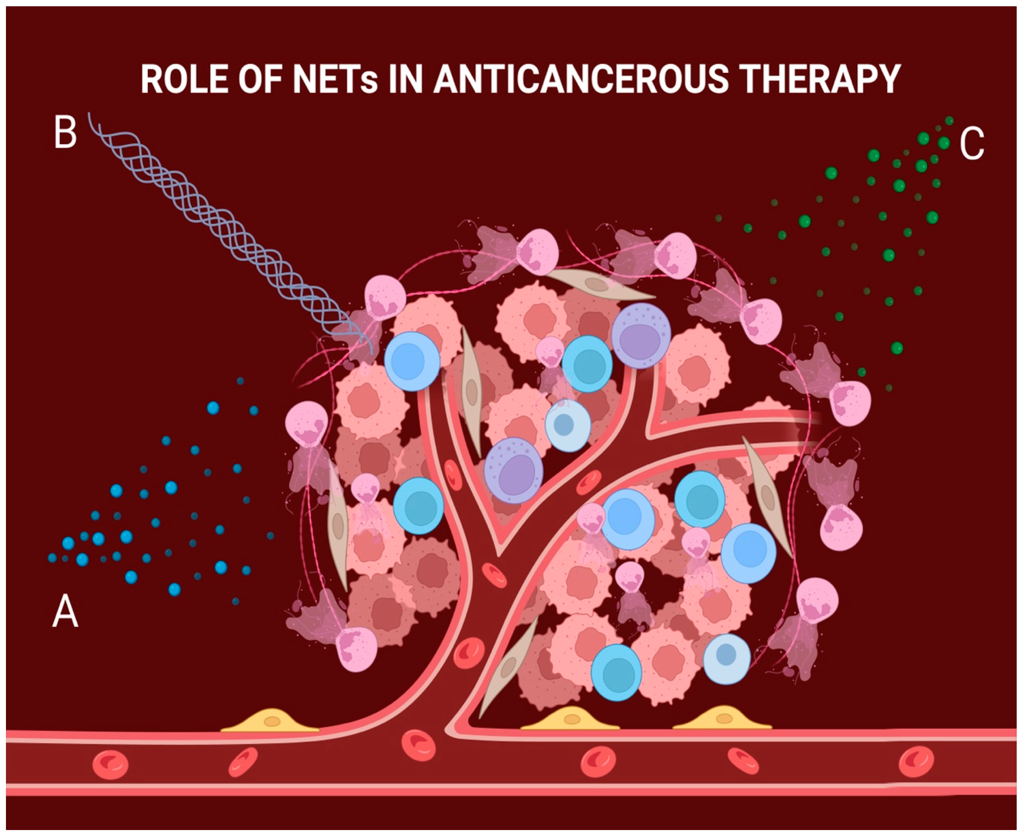
3. NETs Can Provide Resistance to Radiotherapy

Radiotherapy, either alone or in combination with other treatments, is a common curative approach for patients with various types of cancers, and resistance to it is a major obstacle in improving oncologic treatment outcomes. Consequently, increasing efforts have been focused on understanding the mechanisms that cause cancer to become resistant to radiation [52,53,54][8][9][10].
When studying the tissue’s normal response to irradiation, an initial fast influx of neutrophils can be observed. This is a primary response aimed at reducing inflammation. However, some studies suggest that tissues may develop resistance to radiotherapy as the immune infiltrate of neutrophils comes into direct contact with soluble factors that can stimulate NETosis [53][9].
A group of researchers led by Shinde-Jadhav conducted experiments on mice with invasive bladder cancer to understand how the production of NETs can affect cancer’s resistance to radiation [55][11]. They proposed a mechanism where NETs, induced by irradiation or other factors dependent on TME, can coat the surface of cancer cells, acting as a mechanical barrier and lowering the efficiency of the treatment [55][11]. The study showed a significant increase in NETs in irradiated tumors compared to non-irradiated ones, leading to a diminishing radiotherapy sensitivity over time. Clinically, the relevance of these findings was evaluated in a cohort of human MIBC patients treated with RT. The study found that NETs were present in the tumor immune microenvironment (TIME) of these patients, particularly in those who did not respond to RT. A high ratio of intratumoral PMNs to CD8 T-cells, which correlates with the presence of NETs, was associated with poorer overall survival. These findings suggest that NETs can impede RT effectiveness by hindering intratumoral CD8 T-cell infiltration, thereby promoting tumor radioresistance [55][11]. However, when DNase was added to the therapeutic protocol, the response to irradiation was dramatically restored [55][11]. It was reported also that a protein called high mobility group box protein-1 (HMGB1), which is produced excessively in several types of cancers and acts as a trigger of inflammation, has a selective affinity for the Toll-like receptors known to induce NETosis when stimulated. Thus, it can influence radiotherapy resistance by increasing NET production [55,56,57][11][12][13]. The most important ways that NETs can influence cancer regulation or cancer therapies are summarized in Table 1.
Table 1.
Different functions of NETs in cancer and cancer therapy.
Role of NETs in Cancer References
Regulate EndMT [15,16,17][14][15][16]
Positive effect on tumor progression, invasion, and growth [18,19,20,21][17][18][19][20]
Positive effect on angiogenesis [22,23,24,25][21][22][23][24]
Provide resistance to chemotherapy [26,27,28][25][26][27]
Provide resistance to immunotherapy [31,32][28][29]
Provide resistance to radiotherapy [33,,42][30]34,[31]35,[32]40[33][34]

References

  1. Jaillon, S.; Ponzetta, A.; Di Mitri, D.; Santoni, A.; Bonecchi, R.; Mantovani, A. Neutrophil Diversity and Plasticity in Tumour Progression and Therapy. Nat. Rev. Cancer 2020, 20, 485–503.
  2. Ramachandran, I.R.; Condamine, T.; Lin, C.; Herlihy, S.E.; Garfall, A.; Vogl, D.T.; Gabrilovich, D.I.; Nefedova, Y. Bone Marrow PMN-MDSCs and Neutrophils Are Functionally Similar in Protection of Multiple Myeloma from Chemotherapy. Cancer Lett. 2016, 371, 117–124.
  3. Lin, C.; Herlihy, S.E.; Li, M.; Deng, H.; Bernabei, L.; Gabrilovich, D.I.; Vogl, D.T.; Nefedova, Y. NETs Promote Tumor Resistance to Anthracyclines. Cancer Res. 2019, 79, 2103.
  4. Heinhuis, K.M.; Ros, W.; Kok, M.; Steeghs, N.; Beijnen, J.H.; Schellens, J.H.M. Enhancing Anti-Tumor Response by Combining Immune Checkpoint Inhibitors with Chemotherapy in Solid Tumors. Ann. Oncol. 2019, 30, 219–235.
  5. Zhang, Y.; Liu, G.; Sun, M.; Lu, X. Targeting and Exploitation of Tumor-Associated Neutrophils to Enhance Immunotherapy and Drug Delivery for Cancer Treatment. Cancer Biol. Med. 2020, 17, 32–43.
  6. Zhang, Y.; Chandra, V.; Sanchez, E.R.; Dutta, P.; Quesada, P.R.; Rakoski, A.; Zoltan, M.; Arora, N.; Baydogan, S.; Horne, W.; et al. Interleukin-17-Induced Neutrophil Extracellular Traps Mediate Resistance to Checkpoint Blockade in Pancreatic Cancer. J. Exp. Med. 2020, 217, e20190354.
  7. Teijeira, Á.; Garasa, S.; Gato, M.; Alfaro, C.; Migueliz, I.; Cirella, A.; de Andrea, C.; Ochoa, M.C.; Otano, I.; Etxeberria, I.; et al. CXCR1 and CXCR2 Chemokine Receptor Agonists Produced by Tumors Induce Neutrophil Extracellular Traps That Interfere with Immune Cytotoxicity. Immunity 2020, 52, 856–871.
  8. Moding, E.J.; Kastan, M.B.; Kirsch, D.G. Strategies for Optimizing the Response of Cancer and Normal Tissues to Radiation. Nat. Rev. Drug Discov. 2013, 12, 526–542.
  9. Wisdom, A.J.; Hong, C.S.; Lin, A.J.; Xiang, Y.; Cooper, D.E.; Zhang, J.; Xu, E.S.; Kuo, H.C.; Mowery, Y.M.; Carpenter, D.J.; et al. Neutrophils Promote Tumor Resistance to Radiation Therapy. Proc. Natl. Acad. Sci. USA 2019, 116, 18584–18589.
  10. Shaul, M.E.; Fridlender, Z.G. Tumour-Associated Neutrophils in Patients with Cancer. Nat. Rev. Clin. Oncol. 2019, 16, 601–620.
  11. Shinde-Jadhav, S.; Mansure, J.J.; Rayes, R.F.; Marcq, G.; Ayoub, M.; Skowronski, R.; Kool, R.; Bourdeau, F.; Brimo, F.; Spicer, J.; et al. Role of Neutrophil Extracellular Traps in Radiation Resistance of Invasive Bladder Cancer. Nat. Commun. 2021, 12, 1–14.
  12. Tadie, J.M.; Bae, H.B.; Jiang, S.; Park, D.W.; Bell, C.P.; Yang, H.; Pittet, J.F.; Tracey, K.; Thannickal, V.J.; Abraham, E.; et al. HMGB1 Promotes Neutrophil Extracellular Trap Formation through Interactions with Toll-like Receptor 4. Am. J. Physiol. Lung Cell. Mol. Physiol. 2013, 304, 8–12.
  13. Kim, S.W.; Lee, H.; Lee, H.K.; Kim, I.D.; Lee, J.K. Neutrophil Extracellular Trap Induced by HMGB1 Exacerbates Damages in the Ischemic Brain. Acta Neuropathol. Commun. 2019, 7, 94.
  14. Park, J.; Wysocki, R.W.; Amoozgar, Z.; Maiorino, L.; Fein, M.R.; Jorns, J.; Schott, A.F.; Kinugasa-Katayama, Y.; Lee, Y.; Won, N.H.; et al. Cancer Cells Induce Metastasis-Supporting Neutrophil Extracellular DNA Traps. Sci. Transl. Med. 2016, 8, 361ra138.
  15. Ronchetti, L.; Boubaker, N.S.; Barba, M.; Vici, P.; Gurtner, A.; Piaggio, G. Neutrophil Extracellular Traps in Cancer: Not Only Catching Microbes. J. Exp. Clin. Cancer Res. 2021, 40, 231.
  16. Veglia, F.; Tyurin, V.A.; Blasi, M.; De Leo, A.; Kossenkov, A.V.; Donthireddy, L.; To, T.K.J.; Schug, Z.; Basu, S.; Wang, F.; et al. Fatty Acid Transport Protein 2 Reprograms Neutrophils in Cancer. Nature 2019, 569, 73–78.
  17. Cedervall, J.; Zhang, Y.; Huang, H.; Zhang, L.; Femel, J.; Dimberg, A.; Olsson, A.K. Neutrophil Extracellular Traps Accumulate in Peripheral Blood Vessels and Compromise Organ Function in Tumor-Bearing Animals. Cancer Res. 2015, 75, 2653–2662.
  18. Hidalgo, A.; Libby, P.; Soehnlein, O.; Aramburu, I.V.; Papayannopoulos, V.; Silvestre-Roig, C. Neutrophil Extracellular Traps: From Physiology to Pathology. Cardiovasc. Res. 2022, 118, 2737–2753.
  19. Snoderly, H.T.; Boone, B.A.; Bennewitz, M.F. Neutrophil Extracellular Traps in Breast Cancer and beyond: Current Perspectives on NET Stimuli, Thrombosis and Metastasis, and Clinical Utility for Diagnosis and Treatment. Breast Cancer Res. 2019, 21, 145.
  20. Tohme, S.; Yazdani, H.O.; Al-Khafaji, A.B.; Chidi, A.P.; Loughran, P.; Mowen, K.; Wang, Y.; Simmons, R.L.; Huang, H.; Tsung, A. Neutrophil Extracellular Traps Promote the Development and Progression of Liver Metastases after Surgical Stress. Cancer Res. 2016, 76, 1367–1380.
  21. Cristinziano, L.; Modestino, L.; Antonelli, A.; Marone, G.; Simon, H.U.; Varricchi, G.; Galdiero, M.R. Neutrophil Extracellular Traps in Cancer. Semin. Cancer Biol. 2022, 79, 91–104.
  22. Kaltenmeier, C.; Simmons, R.L.; Tohme, S.; Yazdani, H.O. Neutrophil Extracellular Traps (Nets) in Cancer Metastasis. Cancers 2021, 13, 6131.
  23. Söderberg, D.; Segelmark, M. Neutrophil Extracellular Traps in Vasculitis, Friend or Foe? Curr. Opin. Rheumatol. 2018, 30, 16–23.
  24. Kaltenmeier, C.; Yazdani, H.O.; Morder, K.; Geller, D.A.; Simmons, R.L.; Tohme, S. Neutrophil Extracellular Traps Promote T Cell Exhaustion in the Tumor Microenvironment. Front. Immunol. 2021, 12, 785222.
  25. Li, P.; Lu, M.; Shi, J.; Hua, L.; Gong, Z.; Li, Q.; Shultz, L.D.; Ren, G. Dual Roles of Neutrophils in Metastatic Colonization Are Governed by the Host NK Cell Status. Nat. Commun. 2020, 11, 4387.
  26. Ribatti, D.; Tamma, R.; Annese, T. Epithelial-Mesenchymal Transition in Cancer: A Historical Overview. Transl. Oncol. 2020, 13, 100773.
  27. Pieterse, E.; Rother, N.; Garsen, M.; Hofstra, J.M.; Satchell, S.C.; Hoffmann, M.; Loeven, M.A.; Knaapen, H.K.; Van Der Heijden, O.W.H.; Berden, J.H.M.; et al. Neutrophil Extracellular Traps Drive Endothelial-to-Mesenchymal Transition. Arterioscler. Thromb. Vasc. Biol. 2017, 37, 1371–1379.
  28. Demers, M.; Wong, S.L.; Martinod, K.; Gallant, M.; Cabral, J.E.; Wang, Y.; Wagner, D.D. Priming of Neutrophils toward NETosis Promotes Tumor Growth. Oncoimmunology 2016, 5, e1134073.
  29. Miller-Ocuin, J.L.; Liang, X.; Boone, B.A.; Doerfler, W.R.; Singhi, A.D.; Tang, D.; Kang, R.; Lotze, M.T.; Zeh, H.J. DNA Released from Neutrophil Extracellular Traps (NETs) Activates Pancreatic Stellate Cells and Enhances Pancreatic Tumor Growth. Oncoimmunology 2019, 8, e1605822.
  30. Yang, L.Y.; Luo, Q.; Lu, L.; Zhu, W.W.; Sun, H.T.; Wei, R.; Lin, Z.F.; Wang, X.Y.; Wang, C.Q.; Lu, M.; et al. Increased Neutrophil Extracellular Traps Promote Metastasis Potential of Hepatocellular Carcinoma via Provoking Tumorous Inflammatory Response. J. Hematol. Oncol. 2020, 13, 3.
  31. Milette, S.; Quail, D.F.; Spicer, J.D. Neutrophil DNA Webs Untangled. Cancer Cell 2020, 38, 164–166.
  32. Yang, L.; Liu, Q.; Zhang, X.; Liu, X.; Zhou, B.; Chen, J.; Huang, D.; Li, J.; Li, H.; Chen, F.; et al. DNA of Neutrophil Extracellular Traps Promotes Cancer Metastasis via CCDC25. Nature 2020, 583, 133–138.
  33. Webb, N.J.A.; Myers, C.R.; Watson, C.J.; Bottomley, M.J.; Brenchley, P.E.C. Activated Human Neutrophils Express Vascular Endothelial Growth Factor (VEGF). Cytokine 1998, 10, 254–257.
  34. Scapini, P.; Calzetti, F.; Cassatella, M.A. On the Detection of Neutrophil-Derived Vascular Endothelial Growth Factor (VEGF). J. Immunol. Methods 1999, 232, 121–129.
More
Video Production Service