Your browser does not fully support modern features. Please upgrade for a smoother experience.
Please note this is a comparison between Version 1 by Karolina Kryczka and Version 2 by Rita Xu.
Peripartum cardiomyopathy (PPCM) is a form of heart failure, often severe, that occurs in previously healthy women at the end of their pregnancy or in the first few months after delivery. In PPCM, the recovery of heart muscle function reaches 45–50%.
- peripartum cardiomyopathy
- heart disease in pregnancy
- heart failure
- cardiac biomarkers
1. Introduction
Peripartum cardiomyopathy (PPCM) is a life-threatening disease leading to a deterioration in the systolic function of the left ventricle (LV) associated with pregnancy. The onset of heart failure (HF) is usually observed a few weeks before or in the first few months after delivery in women with no previous cardiac history [1]. PPCM is almost always characterized by a left ventricular ejection fraction (LVEF) under 45% on echocardiography with or without LV enlargement [2][3][2,3].
Cardiomyopathies are diseases of the heart muscle that include dilated, hypertrophic, restrictive, and arrhythmogenic cardiomyopathies and PPCM [4]. Recently, a new type of cardiomyopathy with a preserved LVEF has been distinguished [4]. The pathophysiology and morphology of different types of cardiomyopathies have been elucidated elsewhere [5]. Most types of cardiomyopathies differ morphologically and are easy to differentiate through echocardiography or cardiac magnetic resonance imaging (CMRI). PPCM may mimic dilated cardiomyopathy (DCM) in the case of a reduced LVEF with dilated LV. However, DCM usually occurs later in life due to the slow and irreversible process of heart muscle damage [4]. In the case of genetic mutations, an overlapping phenomenon may be observed, which means that different cardiomyopathies may share mutations in the same gene [5]. This phenomenon is observed in PPCM and DCM, most frequently in the titin gene (TTN) [5]. PPCM is characterized by an approximately 45–50% recovery rate, depending on the studied population, and a wide range of phenotypes and courses of the disease [1][2][6][1,2,6]. However, the mortality from PPCM remains high in long-term observation, and in some registries even reached 24% within three years of observation [2][7][2,7]. PPCM may relapse in future pregnancies, especially in patients who did not improve an LVEF of ≥50% [2][7][2,7]. Even in patients who recovered from PPCM, the subsequent pregnancies may again decrease LV contractility. Although the deterioration of LV function associated with subsequent pregnancy is greater in PPCM patients who do not show improved LV function, the mortality rate during eight years of observation is similar, reaching 20% irrespective of the value of the LVEF before subsequent pregnancies [8].
The etiology of the disease is complex and not fully recognized, including unbalanced oxidative stress leading to the formation of 16-kDa prolactin (PRL) with antiangiogenic and cardiotoxic properties. PPCM pathophysiology includes also extracellular matrix dysfunction, fibrosis, and genetic mutations. Bromocriptine, which blocks PRL being released from the pituitary gland, is currently the most specific PPCM treatment. However, not all patients respond to this treatment. This may be due to other mechanisms beyond 16-kDa PRL or delayed diagnosis. Therefore, novel pathophysiological pathways and biomarkers need further examination, particularly those engaged in microcirculatory, cardiac muscle, and extracellular matrix dysfunction. Currently, there is a deficiency in specific diagnostic and prognostic biomarkers that can be widely used in clinical practice to distinguish the symptoms observed in physiological pregnancy and puerperium from those pathological signs associated with PPCM. According to the International Programme on Chemical Safety, on behalf of the World Health Organization, biomarkers are defined as “any substance, structure, or process that can be measured in the body or its products and influence or predict the incidence of outcome or disease” [9]. This definition indicates the investigation of a broad range of body tissues and genes. Most of the biomarkers already known to be associated with PPCM may be classified according to their role in the pathophysiology of the disease or their diagnostic and prognostic utility (Figure 1). Their role in PPCM pathophysiology, diagnosis and prognosis is discussed in this review.
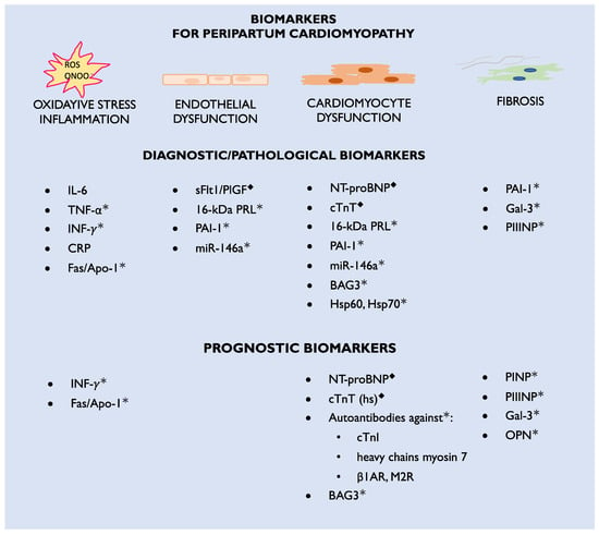
Figure 1. Diagnostic and prognostic biomarkers for peripartum cardiomyopathy; β1AR—beta 1-adrenergic receptors; CRP—C-reactive protein; cTnI—cardiac troponin I; cTnT (hs)—cardiac troponin T (high specific); Fas/Apo1—apoptosis antigen-1; Gal-3—galectin-3; Hsp—heat shock protein; IL-6—interleukin-6; INF-γ—interferon gamma; M2R—M2-muscarinic receptors; miR—microRNA; NT-proBNP—N-terminal pro-Brain-type natriuretic peptide; ONOO•—peroxynitrite; OPN—osteopontin; PAI-1—plasminogen activator inhibitor-1; PINP—procollagen type-I N-terminal propeptide; PIIINP—procollagen type-III N-terminal propeptide; PlGF—placental growth factor; PRL—prolactin; ROS—reactive oxygen species; sFlt1—soluble Fms-like tyrosine kinase-1; TNF-α—tumor necrosis factor alpha; ◆—biomarkers currently used in clinical practice; *—biomarkers which are candidates for future clinical practice.
1.1. Epidemiology, Risk Factors, and Outcomes
As PPCM is a rare disease, the sources of data on different biomarkers are limited. Moreover, the study population’s number of participants did not exceeded 151 patients. It is not uncommon for a certain biomarker to be investigated in only one study with a limited number of patients. For this Sreview, studies available from online medical databases on the topic of biomarkers in PPCM were used. In addition, some representative case reports were presented to highlight new ideas.
The exact and up-to-date statistics on PPCM epidemiology are limited. So far, it has been reported that the disease is most frequent in Nigeria (1:100 deliveries), Haiti (1:300 deliveries), and South Africa (1:1000 deliveries). In the United States, among Caucasians, the frequency increased from 1:2500 in 2004 to 1:1316 deliveries in 2011 [10][11][12][13][14][15][10,11,12,13,14,15]. This process is associated with the older age of and concomitant diseases affecting mothers, such as hypertension or diabetes [1][16][1,16]. The data from the six-month observation of PPCM women in the EuroObservationRegistry Project indicate that PPCM occurs globally, and the frequency in Europe may be comparable to that in Africa [17].
The risk factors for PPCM include the mother’s older or younger age (>30 years old and <18 years old, respectively), multiparity, twin pregnancies, hypertension, preeclampsia, smoking, diabetes, and race [1][2][1,2]. The disease may be underdiagnosed, since signs of HF usually mimic those associated with normal pregnancy and puerperium, such as fatigue or leg edema. Data from the registry validated the main risk factors for PPCM, with preeclampsia being observed in almost one-fourth of patients. The registry highlighted the importance of not only relying on physical examination, as over 40% of PPCM patients did not present with peripheral edema or pulmonary congestion [6][17][6,17].
In most patients, the onset of PPCM was mainly observed in the first month postpartum with sever impairment of LVEF, <35%. The mortality of mothers and neonates was high, reaching 6% and 5%, respectively. In mothers, sudden cardiac death was the main cause of death. The frequency of thrombosis and rehospitalization reached 7 and 10%, respectively. LVEF recovery was observed in less than half of the patients, calling for the improvement of treatment [6].
1.2. Different Phenotypes and Courses of Peripartum Cardiomyopathy
The first case concerns the acute onset of HF in a 26-year-old woman on the third day after delivery of her first pregnancy. Figure 2 demonstrates an enlarged LV with an LV end-diastolic diameter of 68 mm and a severe decrease in LVEF, up to 17%, assessed by CMRI. Apart from pharmacological treatment, also with bromocriptine, the patient required interventional treatment with a biventricular assist device (BiVAD). As a result, the LVEF increased to 35% with BiVAD treatment (Figure 2B) and further to 40–45% in the six-month follow-up period [18]. The effect of the treatment was monitored with biomarkers. NT-proBNP decreased approximately 10 times during treatment, from baseline 10,275 pg/mL (N < 125) to 1019 pg/mL at the six-month observation. Cardiac troponin T was 52.78 ng/L (N < 14.00) at baseline and 7.18 after six months.
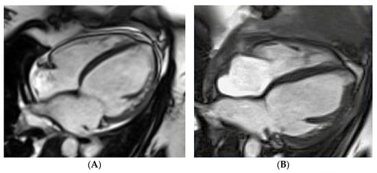
Figure 2. Peripartum cardiomyopathy in a gravida para26-year-old woman in cardiac magnetic resonance imaging: (A) fourth day postpartum, left ventricular ejection fraction (LVEF) of 17%, (B) after bi-ventricle assist device treatment, LVEF of 35%.
By contrast, rwesearchers hospitalized a 35-year-old woman on the fourth day after her third cesarean section, who presented with atrial fibrillation, a severe decrease in LVEF to 20%, an enlarged LV end-diastolic diameter of 62 mm, and severe mitral insufficiency. The global longitudinal strain (GLS) was impaired up to −11.5% (norm from −26% to −18%) (Figure 3). According to the literature, a GLS > −11.4% is recognized as a predictor of increased cardiovascular events, death, and a lack of LVEF improvement ≥50% [19]. The LV insufficiency was still present after successful cardioversion. The pharmacological treatment of HF with bromocriptine was introduced, and the patient showed an improved LVEF of over 50% at the six-month observation. The biomarkers were significantly elevated: NT-proBNP equaled 6776.00 pg/mL (N < 125.00) and cardiac troponin T 33.38 ng/L (N < 14.00). The biomarkers’ levels decreased during the six-month follow-up to 170.40 pg/mL for NT-proBNP and to 4.99 for TnT.
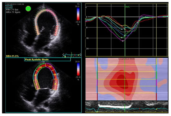
Figure 3. Global strain in echocardiography of 35-year-old woman with peripartum cardiomyopathy with severe impairment of left ventricular (LV) heart muscle functions. Four-chamber apical view: diastolic (upper left); systolic (bottom left); LV systolic and level and asynchrony of maximal contractility of LV segments illustrated by lines of different colors (upper right); longitudinal strain map of LV (bottom right); FR—frame rate, fps—frames per second; GS—global strain longitudinal; HR—heart rate; bpm—beats per minute.
2. Therapy for Peripartum Cardiomyopathy
Currently, rwesearchers are lacking a specifically targeted therapy for PPCM. Bromocriptine, the D2 receptor agonist that inhibits the secretion of PRL from the pituitary gland, in addition to standard HF pharmacotherapy, currently appears to be the most specific drug for PPCM [1][20][1,42]. The pathophysiology of PPCM and the effect of bromocriptine treatment were first validated on a STAT 3 CKO mouse model that developed PPCM during pregnancy and postpartum [21]. Mice with PPCM were characterized by increased cathepsin D levels, the presence of 16 kDa PRL, decreased levels of capillaries and cardiomyocytes in the heart, an increased level of MMP3, and fibrosis, which resulted in decreased survival correlated with an increased number of pregnancies [21].
Bromocriptine treatment was associated with an increased number of capillaries and cardiomyocytes in the heart, a decreased MMP3 level, and fibrosis [21]. The bromocriptine-treated mice had a normal shortening fraction and LV end-diastolic and end-systolic diameters in contrast to the untreated mice with PPCM [21]. Currently, PPCM treatment is based on the BOARD (Bromocriptine, Oral heart failure therapies, Anticoagulants, vaso-Relaxing agents, and Diuretics) concept that recommends using bromocriptine and anticoagulants on top of standard HF treatment (Figure 4) [22][60]. This concept originates from two randomized studies that suggested the beneficial role of bromocriptine treatment [20][23][42,103]. The outcome of the last randomized study highlights the necessity for further studies on bromocriptine, other pathophysiological aspects of the disease, and new drug targets.
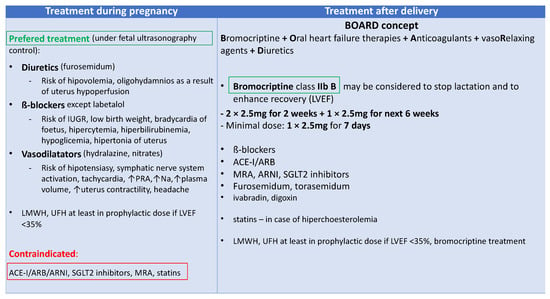
Figure 4. Peripartum cardiomyopathy (PPCM) treatment; ACE-I—angiotensin convertase enzyme inhibitor; ARB—angiotensin receptor blockers; ARNI—angiotensin receptor neprilysin inhibitor; IUGR—intrauterine growth retardation; LMWH—low molecular weight heparin; LVEF—left ventricular ejection fraction; MRA—mineral corticosteroid receptor agonists; Na—natrium; PRA—plasma renin activity; SGLT2—sodium-glucose cotransporter-2; UFH—unfractionated heparin.
2.1. New Biomarker-Based Therapies
Biomarkers associated with certain diseases may serve as a potential target for new therapies. The first reported target therapy for PPCM involved treating a mother with anti-miRNA-146a. One of the potential disadvantages of this treatment is that it may enable mothers to nurse neonates. However, studies on STAT 3 CKO mice have demonstrated that, in contrast to bromocriptine treatment, despite improvement in LV function, the LV remains dilated, suggesting that other pathological pathways have not been assessed with this treatment [24][22]. Other new therapies for microcirculatory dysfunction include the anti-sFlt-1 monoclonal antibody (mAb), which has been successful in the treatment of bronchopulmonary dysplasia in infants of mothers with preeclampsia in a rat model [25][104]. VEGF-modified RNA encoding VEGF (AZD-8601) was useful for the induction of therapeutic revascularization in the heart. In preclinical studies, it has been shown to regulate endothelial cells and cardiomyocyte survival and proliferation [26][105]. Pro-angiogenic therapy with recombinant VEGF was found to ameliorate PPCM [24][22]. However, VEGF treatment of PGC-1α HKO mice with sFlt-1-induced HF did not cause a full recovery from PPCM [24][22]. Therefore, treatment with anti-sFlt-1 mAb may improve results. The glucose-uptake-enhancing drug Perhexiline was found to decrease the cardiotoxic side effects of β-AR stimulation in CKO mice [27][106]. The cardioprotective property of this drug appears to be promising in patients with PPCM and cardiogenic shock when β-AR stimulation cannot be avoided [27][106].
The targets and biomarkers under investigation include proteins, such as Gal-3, proteoglycans, and miRNAs, which have been reviewed previously [28][107]. miRs that act as upstream regulators or downstream effectors of the fibrotic process may be useful in biomarker profiling for the identification of patients most likely to respond to the treatment with these agents. Some data demonstrate that fibrosis may be a reversible process. Therefore, as fibrosis is associated with an inferior outcome, more effort should be engaged in identifying therapeutic targets and developing new direct therapies [29][30][108,109]. New therapeutic targets in PPCM should include MPO and Gal-3. A novel, covalent, irreversible MPO inhibitor that decreases inflammation and improves microvascular function in preclinical models is currently being tested in a phase II clinical study (NCT03611153). The authors are investigating whether a single dose of 30 mg of AZD4831 given orally influences hemodynamic processes in patients with preserved LVEF ≥ 50% and with elevated filling pressures at rest or during exercise which can be assessed by pulmonary capillary wedge pressure during catheterization of the right heart. This is currently the most advanced clinical study on MPO inhibitors [31][110]. The available clinical data from phases I and II support further clinical development of AZD4831 for patients with HF with preserved ejection fraction. Anti-gal-3 therapy includes novel small-molecule gal-3 inhibitors, successful in the treatment of fibrosis in preclinical models, and modified citrus pectin multibranched polysaccharide, which ameliorated cardiac dysfunction, decreased myocardial injury, and decreased collagen deposition in rat HF models. It is worth mentioning that eplerenone and spironolactone downregulate gal-3 expression and therefore decrease the levels of collagen type I, collagen III, and TNF-α, preventing fibrosis after acute myocardial infarction in rats. Some molecules targeting Hsps are known to have a beneficial effect on improving HF. These include geranylgeranylacetone for increased Hsp70 expression, which was cardioprotective in cardiomyopathy models, as well as functional inhibitors that decrease the inflammatory effects of Hsps on cardiac tissue, such as an anti-Hsp70 antibody, polymixin B, colistin sulfate, and epigallocatechin-3-gallate [32][102].
2.2. Biomarker-Guided Therapy
Guiding HF therapy with biomarkers such as NT-proBNP and cardiac troponins can be helpful in clinical practice [5][8][9][10][11][12][13][14][15][16][17][18][19][20][21][22][23][24][25][26][27][28][29][30][31][32][33][34][35][36][37][38][39][40][41][42][43][44][45][46][47][48][49][50][51][52][53][54][55][56][57][58][59][60][61][62][63][64][65][66][67][68][69][70][71][72][73][74][75][76][77][78][79][80][81][82][83][84][85][86][87][88][89][90][91][92][93][94][95][96][97][98][99][100][101][102][103][104][105][106][107][108][109][110][111][5,8,9,10,11,12,13,14,15,16,17,18,19,20,21,22,23,24,25,26,27,28,29,30,31,32,33,34,35,36,37,38,39,40,41,42,43,44,45,46,47,48,49,50,51,52,53,54,55,56,57,58,59,60,61,62,63,64,65,66,67,68,69,70,71,72,73,74,75,76,77,78,79,80,81,82,83,84,85,86,87,88,89,90,91,92,93,94,95,96,97,98,99,100,101,102,103,104,105,106,107,108,109,110,111]. However, randomized trials on guiding therapy with natriuretic peptides have shown contradictory results. Some of them demonstrated the superiority of natriuretic peptide-guided HF treatment over traditional treatment based on clinical experience and guidelines. In these studies, the decrease in hospitalizations and mortality was lower in natriuretic peptide-guided therapy [112][113][112,113]. This was particularly true for patients ≤75 years of age [114]. However, some studies demonstrated no benefits from natriuretic peptide-guided therapy compared with clinically guided management, especially in older patients >60 years of age [115][116][115,116]. Metanalyses were found to have beneficial effects on natriuretic peptide-guided therapy according to a decrease in all-cause mortality compared with usual management, especially in younger patients. In addition, one demonstrated benefits such as a decrease in cardiovascular hospitalizations [117][118][117,118].
Studies on biomarker-managed therapy in PPCM are lacking. However, in patients with an improved LVEF in the six-month observation period, the levels of different biomarkers, including NT-proBNP, Fas/Apo1, IFN-γ, and prolactin, decreased more than in patients with no LVEF improvement [22][60]. A published example of one of theour PPCM patients demonstrated that monitoring treatment with 23-kDa PRL may be beneficial in treating this disease, as an increase in PRL level after bromocriptine discontinuation was associated with the exacerbation of symptoms. Prolonged bromocriptine treatment for up to 12 months was particularly beneficial for this patient, with an increase in LVEF >50% [45][34].